

doi: 10.1038/nchembio.1231.
Epub 2013 Apr 14.
Structure of a class II preQ1 riboswitch reveals ligand recognition by a new fold
Affiliations
- PMID: 23584677
- PMCID: PMC3661761
- DOI: 10.1038/nchembio.1231
Item in Clipboard
Structure of a class II preQ1 riboswitch reveals ligand recognition by a new fold
Nat Chem Biol.
2013 Jun.
Abstract
PreQ1 riboswitches regulate genes by binding the pyrrolopyrimidine intermediate preQ1 during the biosynthesis of the essential tRNA base queuosine. We report what is to our knowledge the first preQ1-II riboswitch structure at 2.3-Å resolution, which uses a previously uncharacterized fold to achieve effector recognition at the confluence of a three-way helical junction flanking a pseudoknotted ribosome-binding site. The results account for translational control mediated by the preQ1-II riboswitch class and expand the known repertoire of ligand-binding modes used by regulatory RNAs.
Figures
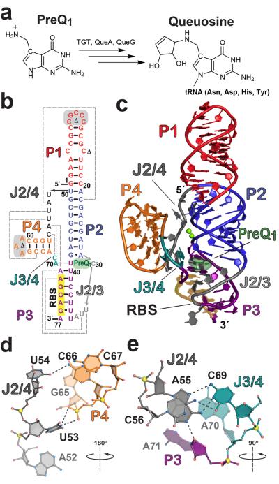
(a) Queuosine biosynthesis from preQ1 with known enzymes shown. Although animals must obtain Q from dietary sources or gut flora, bacteria can produce it by de novo synthesis (reviewed in 21, 22). TGT, tRNA:guanine transglycosylase; QueA, epoxyqueuosine synthase; and QueG, oQ (epoxyqueuosine) reductase (b) Secondary structure of the wild type L. rhamnosus preQ1-II riboswitch used in this investigation based on the crystal structure. PreQ1 is dark green; various pairing regions, P, are color coded with long-range interactions indicated by dashed gray lines; junctions are labeled J. Sites modified for crystallization are highlighted in gray or marked with a Δ. See Supplementary Fig. 1 for the modified construct (MC) used in crystallization and isothermal titration calorimetry (ITC); numbering is based on the MC 77-mer sequence. The consensus RBS sequence 5′-AGGAG-3′ is highlighted in yellow. (c) Cartoon depiction of the preQ1-bound crystal structure. Coloring is the same as b with the preQ1 effector depicted as a semitransparent surface model. The RBS is labeled and highlighted in yellow. (d) Hydrogen-bond tertiary interactions (dashed lines) between P4 and J2/4 that stabilize the core fold; the view is rotated ~180° about the axis shown, relative to the orientation in c. (e) Tertiary interactions that knit together J2/4, J3/4, and A71 of the three-way helical junction; the view is rotated ~90° about the indicated axis relative to c.
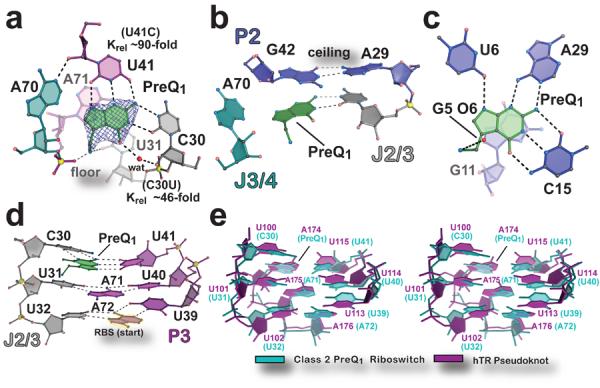
(a) View of the preQ1 ligand-binding site. The final refined ligand is covered by an unbiased Fo-Fc omit electron density map, contoured at the 3.0 σ level, that was calculated prior to inclusion of preQ1 in the model. The “floor” of the binding pocket is formed by a Hoogsteen base pair between A71•U31. Krel (KD mutant / KD wild type) of binding site mutants, determined by ITC, is shown next to the respective base. (b) The “ceiling” of the preQ1 binding pocket is formed by a cis-Watson-Crick/Watson-Crick base pair between G42 and A29. (c) The preQ1-I translational riboswitch in complex with preQ1 (PDB ID 3Q50). (d) Major-groove base triples that stack on the RBS to facilitate formation of the P3 pseudoknot. (e) Stereo view of an all-atom superposition between the eight nucleotides of the preQ1-II riboswitch base triples in d and equivalent base triples from the hTR pseudoknot (PDB ID 1YMO). The average rmsd was 1.46 Å (excluding hTR A174, which spatially overlaps preQ1).
References
Publication types
MeSH terms
Substances
Associated data
- Actions
Grants and funding
LinkOut - more resources
Full Text Sources
Other Literature Sources
