

Topical diquafosol for patients with obstructive meibomian gland dysfunction
- PMID: 23584719
- PMCID: PMC3664386
- DOI: 10.1136/bjophthalmol-2012-302668
Topical diquafosol for patients with obstructive meibomian gland dysfunction
Abstract
Aims: To evaluate the effect of topical diquafosol in patients with meibomian gland dysfunction (MGD) using tear film parameters and quantitatively analyse the meibomian gland morphology.
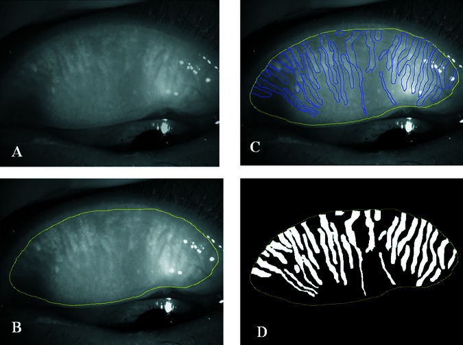
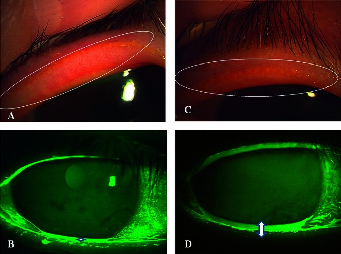
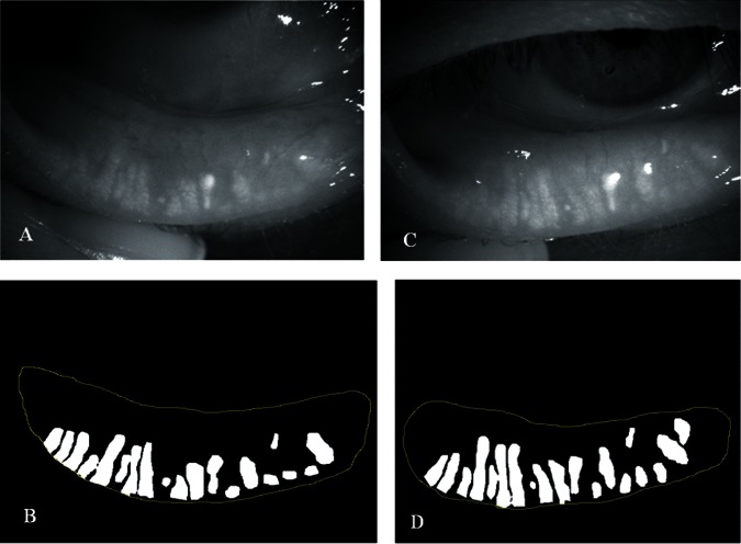
Subjects and methods: The subjects were 19 eyes of 10 patients diagnosed with obstructive MGD. All subjects were given 3% diquafosol ophthalmic solution with instructions to use one drop four times a day. Ocular symptoms were scored from 0 to 14. Lid margin abnormalities were scored from 0 to 4. Changes in the meibomian glands were scored using non-contact meibography (meiboscore). Superficial punctate keratopathy (SPK) was scored from 0 to 3. Meibum was graded from 0 to 3. Tear film production was evaluated by Schirmer's test. Quantitative image analysis of the meibomian glands was performed using the original software.
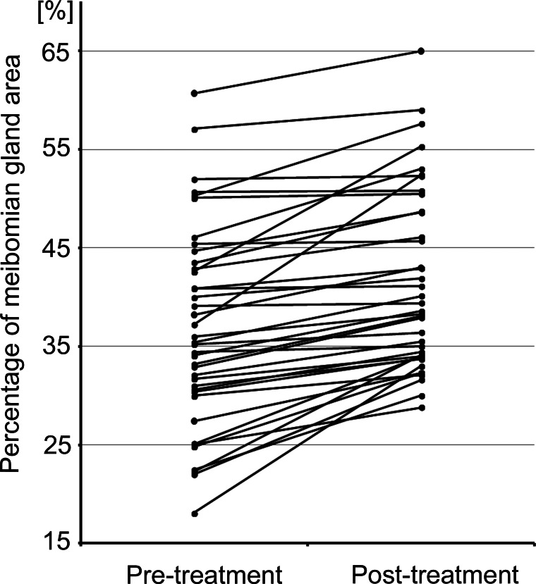
Results: 10 patients completed more than 4 months of therapy. Ocular symptoms, lid margin abnormalities, SPK score and meibum grade were decreased. Break-up time and tear film meniscus were increased. Mean ratio of the meibomian gland area was significantly increased after treatment (p<0.0001).
Conclusions: Quantitative image analysis was useful for evaluating the morphological changes of the meibomian glands. Topical diquafosol therapy was effective for patients with obstructive MGD.
Figures
References
-
- Mishima S, Maurice DM. The oily layer of the tear film and evaporation from the corneal surface. Exp Eye Res 1961;1:39–45 - PubMed
-
- McCulley JP. Meibomitis. In: Kaufman HE, Barron BA, McDonald MB, Waltman SR, eds. The Cornea. New York, NY: Churchill Livingstone Inc., 1988:125–38
-
- Lemp MA. Report of the National Eye Institute/Industry Workshop on Clinical Trials in Dry Eyes. CLAO J 1995;21:221–32 - PubMed
-
- Mathers WD. Ocular evaporation in meibomian gland dysfunction and dry eye. Ophthalmology 1993;100:347–51 - PubMed
-
- Shimazaki J, Sakata M, Tsubota K. Ocular surface changes and discomfort in patients with meibomian gland dysfunction. Arch Ophthalmol 1995;113:1266–70 - PubMed
Publication types
MeSH terms
Substances
LinkOut - more resources
Full Text Sources
Other Literature Sources
Medical