. 2013 Jun 1;304(11):H1525-37.
doi: 10.1152/ajpheart.00874.2012. Epub 2013 Apr 12.

Inotropic and lusitropic effects of calcitonin gene-related peptide in the heart

Affiliations

Inotropic and lusitropic effects of calcitonin gene-related peptide in the heart

Mustafa Al-Rubaiee et al. Am J Physiol Heart Circ Physiol. .

Abstract

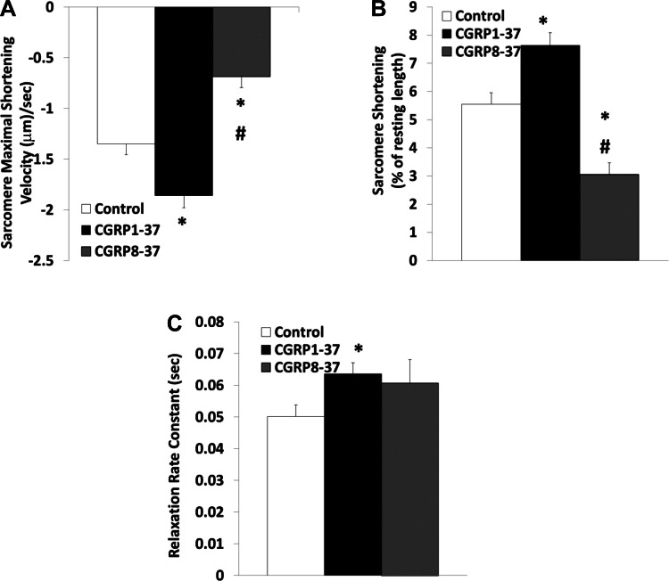
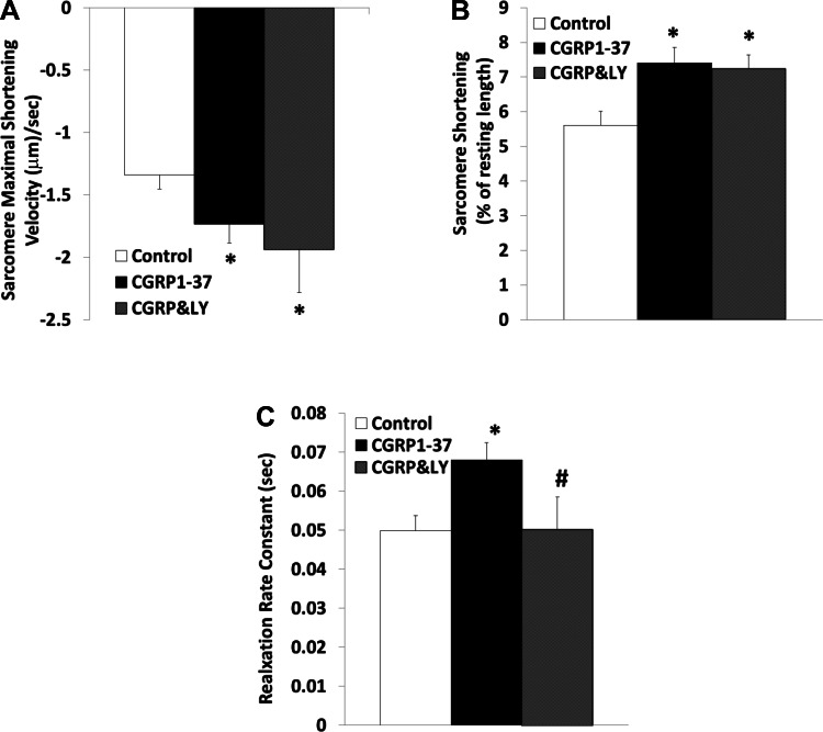
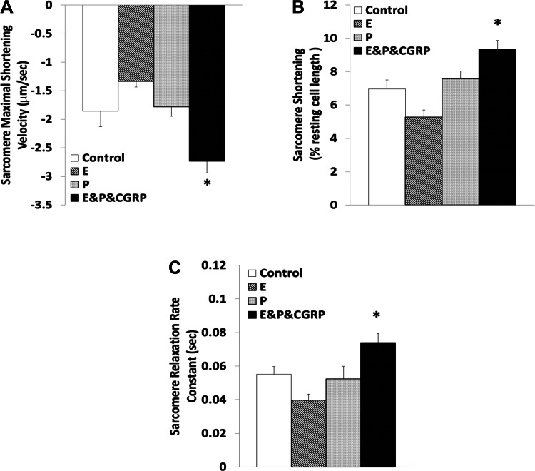
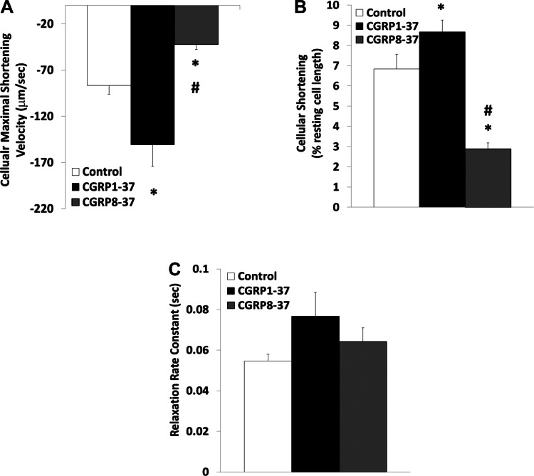
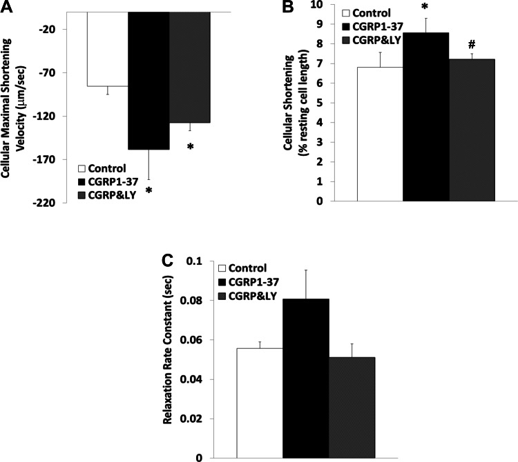
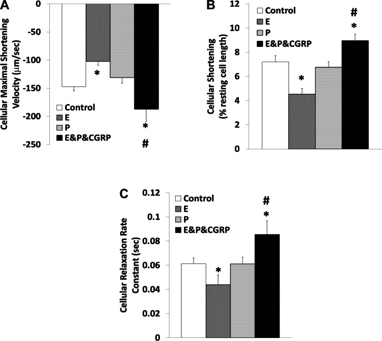
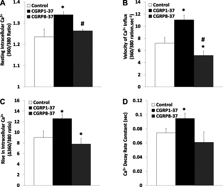
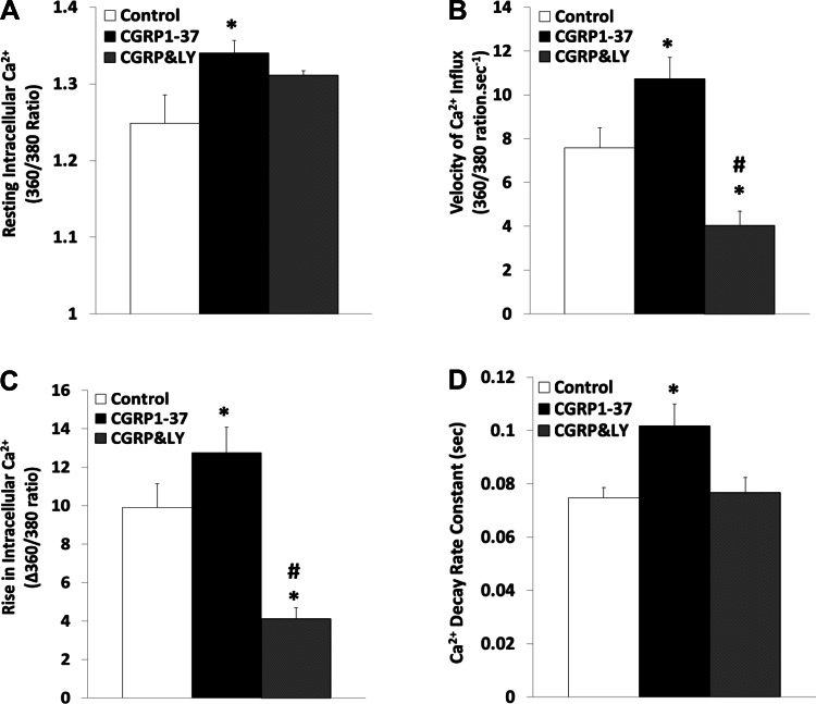
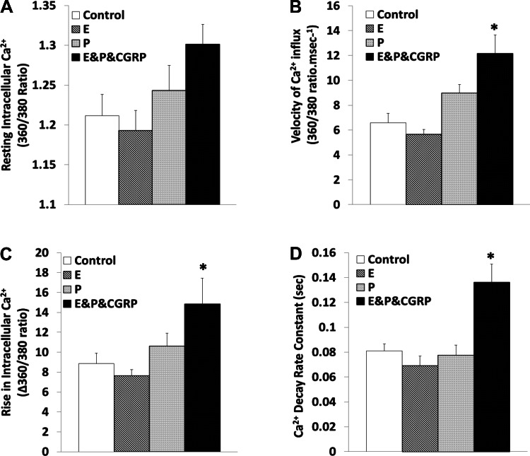
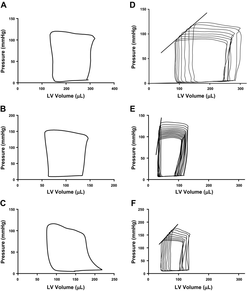
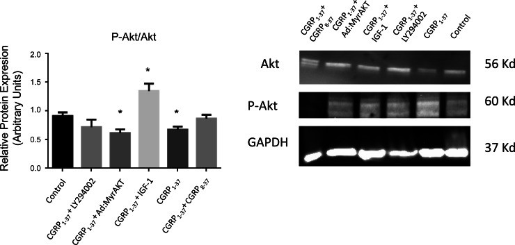
Previous studies have demonstrated positive-inotropic effects of calcitonin gene-related peptide (CGRP), but the mechanisms remain unclear. Therefore, two experiments were performed to determine the physiological correlates of the positive-inotropic effects of CGRP. Treatments designed to antagonize the effects of physiologically active CGRP₁₋₃₇ included posttreatment with CGRP₈₋₃₇ and pretreatment with LY-294002 (LY, an inhibitor of phosphatidylinositol 3-kinase), 17β-estradiol (E), and progesterone (P) were also used to modulate the effects of CGRP₁₋₃₇. Experiment 1 was in vitro studies on sarcomeres and cells of isolated adult rat cardiac myocytes. CGRP₁₋₃₇, alone and in combination with E and P, decreased sarcomere shortening velocities and increased shortening percentages, effects that were antagonized by CGRP₈₋₃₇, but not by LY. CGRP₁₋₃₇ increased resting intracellular calcium ion concentrations and Ca(2+) influxes, effects that were also antagonized by both CGRP₈₋₃₇ and LY. Experiment 2 was in vivo studies on left ventricular pressure-volume (PV) loops. CGRP₁₋₃₇ increased end-systolic pressure, ejection fraction, and velocities of contraction and relaxation while decreasing stroke volume, cardiac output, stroke work, PV area, and compliance. After partial occlusion of the vena cava, CGRP₁₋₃₇ increased the slope of the end-systolic PV relationship. CGRP₈₋₃₇ and LY attenuated most of the CGRP-induced changes. These findings suggest that CGRP-induced positive-inotropic effects may be increased by treatments with estradiol and progesterone and inhibited by LY. The physiological correlates of CGRP-induced positive inotropy observed in rat sarcomeres, cells, and intact hearts are likely to reveal novel mechanisms of heart failure in humans.

Keywords: PI3K; calcitonin gene-related peptide; cardiac; estrogen; progesterone.


Figures

Fig. 1.
Fig. 1.
Effects of calcitonin gene-related peptide (CGRP)1–37 and CGRP8–37 on cardiomyocyte sarcomere contractile dynamics. Bars show effects of control, 1 nM CGRP1–37 treatments, and 1 nM CGRP8–37 posttreatments on maximal shortening velocity of sarcomeres during contraction (A), sarcomere shortening as a percentage of resting length (B), and sarcomere relaxation rate constant (C). Values are means ± SE; n = 20 from 4–5 animals for each group. *Significantly different from controls at P < 0.05. #Significantly different from CGRP1–37 treatments at P < 0.05.
Fig. 2.
Fig. 2.
Effects of CGRP1–37 and LY-294002 (LY) on cardiomyocyte sarcomere contractile dynamics. Bars show effects of control, 1 nM CGRP1–37 treatments, and 1 μM LY (CGRP&LY) posttreatments on maximal shortening velocity of sarcomeres during contraction (A), sarcomere shortening as a percentage of resting length (B), and sarcomere relaxation rate constant (C). Values are means ± SE; n = 20 from 4–5 animals for each group. *Significantly different from controls at P < 0.05. #Significantly different from CGRP1–37 treatments at P < 0.05.
Fig. 3.
Fig. 3.
Modulation of the contractile effects of CGRP1–37 by 17β-estradiol (E) and progesterone (P) at the sarcomere level. Bars show effects of control, 10 nM E, 10 nM P, and 1 nM CGRP1–37 with 10 nM of E and 10 nM of P (E&P&CGRP) treatments on maximal shortening velocity of sarcomeres during contraction (A), sarcomere shortening as a percentage of resting length (B), and sarcomere relaxation rate constant (C). Values are means ± SE; n = 20 from 4–5 animals for each group. *Significantly different from controls at P < 0.05.
Fig. 4.
Fig. 4.
Effects of CGRP1–37 and CGRP8–37 on cardiomyocyte contractile dynamics at the cellular level. Bars show effects of control, 1 nM CGRP1–37 treatments, and 1 nM CGRP8–37 posttreatments on maximal shortening velocity of cells during contraction (A), cell shortening as a percentage of resting length (B), and cell relaxation rate constant (C). Values are means ± SE; n = 20 from 4–5 animals for each group. *Significantly different from controls at P < 0.05. #Significantly different from CGRP1–37 treatments at P < 0.05.
Fig. 5.
Fig. 5.
Effects of CGRP1–37 and LY on cardiomyocyte contractile dynamics at the cellular level. Bars show effects of control, 1 nM CGRP1–37 treatments, and 1 μM LY (CGRP&LY) posttreatments on maximal shortening velocity of cells during contraction (A), cell shortening as a percentage of resting length (B), and cell relaxation rate constant (C). Values are means ± SE; n = 20 from 4–5 animals for each group. *Significantly different from controls at P < 0.05. #Significantly different from CGRP1–37 treatments at P < 0.05.
Fig. 6.
Fig. 6.
Modulation of cardiomyocyte contractile effects of CGRP1–37 by E and P at the cellular level. Bars show effects of control, 10 nM E, 10 nM P, and 1 nM CGRP1–37 with 10 nM of E and 10 nM of P (E&P&CGRP) on maximal shortening velocity of cells during contraction (A), cell shortening as a percentage of resting length (B), and cell relaxation rate constant (C). Values are means ± SE; n = 20 from 4–5 animals for each group. *Significantly different from controls at P < 0.05. #Significantly different from CGRP1–37 treatments at P < 0.05.
Fig. 7.
Fig. 7.
Effects of CGRP1–37 and CGRP8–37 on cardiomyocyte calcium dynamics. Bars show effects of control, 1 nM CGRP1–37 treatments, and 1 nM CGRP8–37 posttreatments on resting intracellular calcium ion concentration ([Ca2+]i; A), Ca2+ influx during cell contraction (B), [Ca2+]i increment during cell contraction (C), and Ca2+ decay rate constant during cell relaxation (D). Values are means ± SE; n = 20 from 4–5 animals for each group. *Significantly different from controls at P < 0.05. #Significantly different from CGRP1–37 treatments at P < 0.05.
Fig. 8.
Fig. 8.
Effects of CGRP1–37 and LY on cardiomyocyte calcium dynamics. Bars show effects of control, 1 nM CGRP1–37 pretreatments, and 1 μM LY posttreatments (CGRP&LY) on resting [Ca2+]i (A), Ca2+ influx during cell contraction (B), [Ca2+]i increment during cell contraction (C), and Ca2+ decay rate constant during cell relaxation (D). Values are means ± SE; n = 20 from 4–5 animals for each group. *Significantly different from controls at P < 0.05. #Significantly different from CGRP1–37 treatments at P < 0.05.
Fig. 9.
Fig. 9.
Modulation of cardiomyocyte calcium effects of CGRP1–37 by E and P. Bars show effects of control, 10 nM E, 10 nM P, and 1 nM CGRP1–37 with 10 nM of E and 10 nM of P (E&P&CGRP) on resting [Ca2+]i (A), Ca2+ influx during cell contraction (B), [Ca2+]i increment during cell contraction (C), and Ca2+ decay rate constant during cell relaxation (D). Values are means ± SE; n = 20 from 4–5 animals for each group. *Significantly different from controls at P < 0.05 on the rate of Ca2+ decay during relaxation.
Fig. 10.
Fig. 10.
Effects of CGRP1–37 and CGRP8–37 on pressure-volume (PV) relationships of the left ventricle (LV) of adult male rats. Steady-state PV loop for control conditions (A), treatment with 1 nM CGRP1–37 (B), and pretreatment with 1 nM CGRP1–37 and posttreatment with CGRP8–37 (C) are shown. Regression of PV loop following occlusion of the vena cava where the slope of the regression line depicts the end-systolic PV relationship for control conditions (D), treatment with 1 nM CGRP1–37 (E), and pretreatment with 1 nM CGRP 1–37 and posttreatment with 1 nM CGRP8–37 (F) are shown.
Fig. 11.
Fig. 11.
Effects of CGRP1–37, CGRP8–37, and LY on the end-systolic PV relationship associated with vena cava occlusion. Regression lines of the end-systolic PV relationship during jugular intravenous infusions in control treatment, pretreatment with 1 nM CGRP1–37, and posttreatment with 1 nM CGRP8–37 (A) and in control, 1 μM LY, and 1 nM CGRP1–37 treatments (B), the latter with a 1 μM LY (CGRP+LY) posttreatment, are shown.
Fig. 12.
Fig. 12.
Effects of CGRP1–37 on the expression and activation level of Akt in the LV of adult male rats. Right: representative Western blot gels demonstrating the effects of CGRP1–37 alone and with cotreatment using either the CGRP1–37 antagonist CGRP8–37, the phosphatidylinositol 3-kinase inhibitor LY, adenoviral transfection with negative Akt (Ad.myrAkt), or the phosphatidylinositol 3-kinase agonist insulin-like growth factor (IGF)-I, compared with control (untreated) ventricles. Left: bar graph showing the average effects of the aforementioned treatments on the activation level of Akt represented as the p-Akt-to-Akt ratio. Values are means ± SE; n = 6 for each group. *P < 0.05, compared with the control.

References

    1. Alvin Z, Laurence GG, Coleman BR, Zhao A, Hajj-Moussa M, Haddad GE. Regulation of L-type inward calcium channel activity by captopril and angiotensin II via the phosphatidyl inositol 3-kinase pathway in cardiomyocytes from volume-overload hypertrophied rat hearts. Can J Physiol Pharmacol 89: 206–215, 2011 - PMC - PubMed
    1. Bell D, McDermott BJ. Calcitonin gene-related peptide stimulates a positive contractile response in rat ventricular cardiomyocytes. J Cardiovasc Pharmacol 23: 1011–1021, 1994 - PubMed
    1. Bell D, Schlnter KD, Zhou XJ, McDermott BJ, Piper MH. Hypertrophic effects of calcitonin gene-related peptide (CGRP) and amylin on adult mammalian ventricular cardiomyocytes. J Mol Cell Cardiol 27: 2433–2443, 1995 - PubMed
    1. Bell D, Tamamori M, Marumo F, Hiroe M, McDermott BJ, Ito H. Calcitonin gene-related peptide (CGRP) increases cell surface area and induces expression of skeletal [alpha]-actin and ANP mRNA in hypertrophying neonatal cardiomyocytes. Regul Pept 71: 1–7, 1997 - PubMed
    1. Bick RJ, Poindexter BJ, Davis RA, Schiess MC. Determination of the site of action of calcitonin gene-related peptide in the alteration of intracellular calcium levels in adult and neonatal rodent myocytes. Peptides 26: 2231–2238, 2005 - PubMed

Publication types

MeSH terms

LinkOut - more resources