. 2013 May 1;41(10):5457-68.
doi: 10.1093/nar/gkt212. Epub 2013 Apr 12.

Zebrafish AID is capable of deaminating methylated deoxycytidines

Affiliations

Zebrafish AID is capable of deaminating methylated deoxycytidines

Hala Abdouni et al. Nucleic Acids Res. .

Abstract

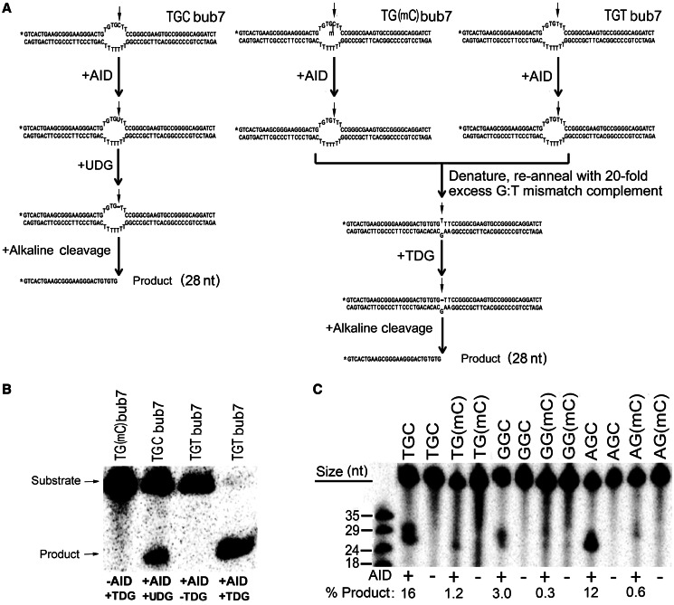
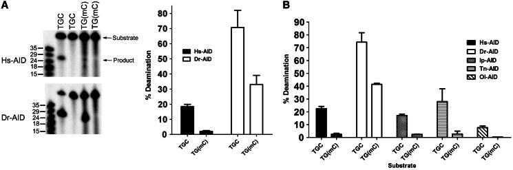
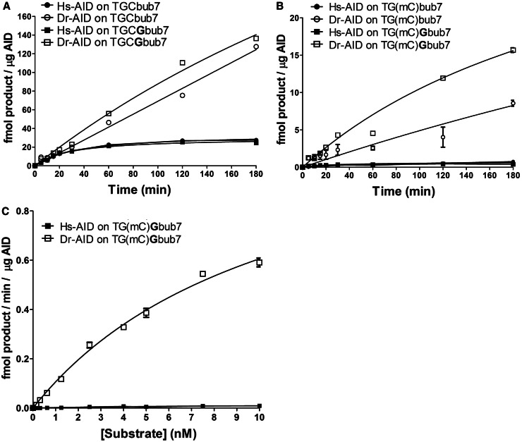
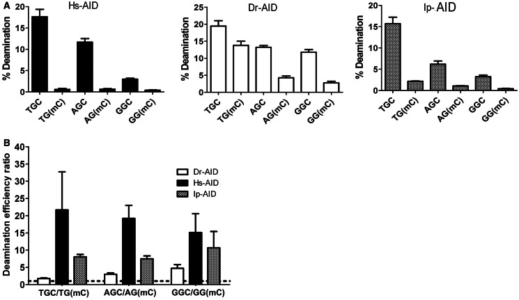
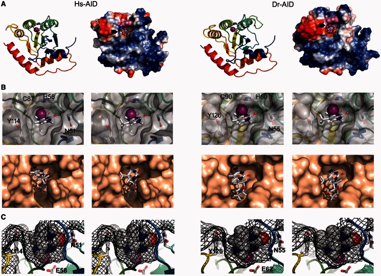
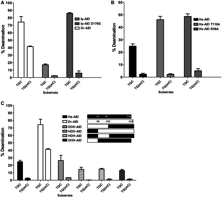
Activation-induced cytidine deaminase (AID) deaminates deoxycytidine (dC) to deoxyuracil (dU) at immunoglobulin loci in B lymphocytes to mediate secondary antibody diversification. Recently, AID has been proposed to also mediate epigenetic reprogramming by demethylating methylated cytidines (mC) possibly through deamination. AID overexpression in zebrafish embryos was shown to promote genome demethylation through G:T lesions, implicating a deamination-dependent mechanism. We and others have previously shown that mC is a poor substrate for human AID. Here, we examined the ability of bony fish AID to deaminate mC. We report that zebrafish AID was unique among all orthologs in that it efficiently deaminates mC. Analysis of domain-swapped and mutant AID revealed that mC specificity is independent of the overall high-catalytic efficiency of zebrafish AID. Structural modeling with or without bound DNA suggests that efficient deamination of mC by zebrafish AID is likely not due to a larger catalytic pocket allowing for better fit of mC, but rather because of subtle differences in the flexibility of its structure.


Figures

Figure 1.
Figure 1.
Experimental scheme for measurement of deamination activity on 5-methylcytosine. (A) Typical bubble type substrates used in this study are shown. TGCbub7 denotes a substrate bearing the WRC motif TGC located in a seven-nucleotide-long bubble region. Left shows the scheme for the UDG-based alkaline cleavage assay on the WRC bearing substrate TGCbub7. The middle shows the scheme for the TDG-based alkaline cleavage assay on the WR(mC) bearing substrate TG(mC)bub7. Right shows a control reaction for the TDG-based alkaline cleavage assay to detect the activity of AID on mC. The substrate is TGTbub7, which is the expected product of TG(mC)bub7 after deamination by AID. (B) Control reactions for the efficiency and specificity of TDG. In all, 50 fmol substrate was incubated with AID, TDG or both (as shown in A). This control was included in each subsequent experiment to ensure 100% TDG efficiency. (C) Comparison of Hs-AID activity on dC versus mC located either in a WRC motif (TGC, AGC) or non-WRC motif (GGC). In all, 50 fmol substrate was incubated with AID. A typical alkaline cleavage gel used to measure the generation of deaminated product is shown and percentage of product formation is shown below each lane.
Figure 2.
Figure 2.
Dr-AID exhibits high activity on mC. (A) Left shows alkaline cleavage assay gels showing the deamination of TGCbub7 or TG(mC)bub7 by of Hs-AID (top) and Dr-AID (bottom). Right shows the graphed percentage of deaminated product. Hs- and Dr-AID were incubated overnight with 50 fmol substrate at 37°C and 25°C, respectively. (B) Deamination of TGCbub7 and TG(mC)bub7 by AID of three other bony fish (Medaka: Ol-AID, tetradon: Tn-AID and Channel catfish: Ip-AID) compared with the activities of Hs- and Dr-AID. Each AID was incubated with 50 mol substrate overnight at optimal temperature (37°C and 25°C, respectively, for Hs- and fish AIDs).
Figure 3.
Figure 3.
Deamination kinetics comparing the activities of Hs-AID and Dr-AID. (A) Comparison of activity kinetics between Hs-AID and Dr-AID on seven-nucleotide bubble substrates bearing dC in a WRC or a WRCG motif. In all, 50 fmol substrate was incubated with AID for various times. Product formation for 1 µg AID is shown as a function of incubation time. (B) Comparison of activity kinetics between Hs-AID and Dr-AID on seven-nucleotide bubble substrates bearing mC in a WRC or a WRCG motif. (C) Deamination kinetics to compare the deamination rates of dC and mC in a WRCG motif by Hs-AID and Dr-AID. Various concentrations of the substrate TG(mC)Gbub7 ranging from 0.1 to 10 nM were incubated with AID. Velocity was calculated as the amount of deaminated product generated by a given amount of AID in a unit of time and plotted against substrate concentration.
Figure 4.
Figure 4.
The influence of WRC sequence specificity of AID on mC activity. (A) Left, middle and right show the activities of Hs-, Dr- and Ip-AID, respectively, on C and mC, located in two WRC motifs (TGC, AGC) or a non-WRC motif (GGC). AID was incubated with 40 fmol substrate for 1 h. Percentage of deamination product was quantitated and graphed. (B) The ratio of percentage of deamination product for C/mC was calculated for each AID and graphed to show the fold preference for C over mC, in each of the WRC and non-WRC sequence motifs. Dotted line represents a ratio of 1.
Figure 5.
Figure 5.
Models of the putative catalytic pockets of Hs- and Dr-AID with docked C or mC. (A) Models of the AID monomer were generated based on the solved structure of the APOBEC3G catalytic region. Left shows a ribbon and surface charge diagram of Hs-AID and right shows the same for Dr-AID. N- to C-terminus progression is shown in color from blue to red. The ribbon diagram shows the putative catalytic (Zn-coordinating) residues are shown in stick (Hs-AID: H56, E58, C87, C90; Dr-AID: H60, E62, C99, C102). The surface topology and charge models show positively and negatively charged residues are blue and red, respectively. The putative catalytic pockets containing the Zn-coordinating residues are shown in magenta, with a docked cytidine base inside the pocket as marked by black arrows. (B) Modeled top down view of the surface topology of the putative catalytic pocket of Hs-AID (left) or Dr-AID (right), with either C or mC bases (top row) using manual docking, or by using Autodock software to dock dC or mC nucleoside versions (bottom row). Zn is shown as a magenta sphere. Residues that surround the catalytic pocket and affect the fit of C or mC are shown. (C) Mesh diagram of the modeled side view of the catalytic pockets of Hs-AID (left), or Dr-AID (right), showing the fit of C (left side) or mC (right side). The mesh diagram illustrates the spaces occupied by the atomic radii of both substrate and AID. Zn is shown as a magenta sphere. Residues that surround the catalytic pocket and affect the fit of C or mC are shown.
Figure 6.
Figure 6.
The activity of AID mutants and domain-swapped AIDs on mC. (A) Comparison of the deamination activities of Dr-AID, Ip-AID and Ip-AID (D176G) on TGCbub7 and TGC(mC)bub7. (B) Comparison of the deamination activities of Hs-AID, Hs-AID (R36A) and Hs-AID (T110A) on TGCbub7 and TG(mC)bub7. (C) Comparison of the deamination activities of Hs-AID, Dr-AID and hybrid enzymes with domains swapped between Hs- and Dr-AID on TGCbub7 and TGC(mC)bub7. AID was incubated with 50 fmol substrate for 3 h, and percentage of deamination product was graphed.

References

    1. Muramatsu M, Sankaranand VS, Anant S, Sugai M, Kinoshita K, Davidson NO, Honjo T. Specific expression of activation-induced cytidine deaminase (AID), a novel member of the RNA-editing deaminase family in germinal center B cells. J. Biol. Chem. 1999;274:18470–18476. - PubMed
    1. Conticello SG. The AID/APOBEC family of nucleic acid mutators. Genome Biol. 2008;9:229. - PMC - PubMed
    1. Pham P, Bransteitter R, Petruska J, Goodman MF. Processive AID-catalysed cytosine deamination on single-stranded DNA simulates somatic hypermutation. Nature. 2003;424:103–107. - PubMed
    1. Larijani M, Petrov AP, Kolenchenko O, Berru M, Krylov SN, Martin A. AID associates with single-stranded DNA with high affinity and a long complex half-life in a sequence-independent manner. Mol. Cell. Biol. 2007;27:20–30. - PMC - PubMed
    1. Larijani M, Frieder D, Basit W, Martin A. The mutation spectrum of purified AID is similar to the mutability index in Ramos cells and in ung(−/−)msh2(−/−) mice. Immunogenetics. 2005;56:840–845. - PubMed

Publication types