. 2013 Jun 1;73(11):3402-11.
doi: 10.1158/0008-5472.CAN-12-4619. Epub 2013 Apr 12.

Elevation of receptor tyrosine kinases by small molecule AKT inhibitors in prostate cancer is mediated by Pim-1

Affiliations

Elevation of receptor tyrosine kinases by small molecule AKT inhibitors in prostate cancer is mediated by Pim-1

Bo Cen et al. Cancer Res. .

Abstract

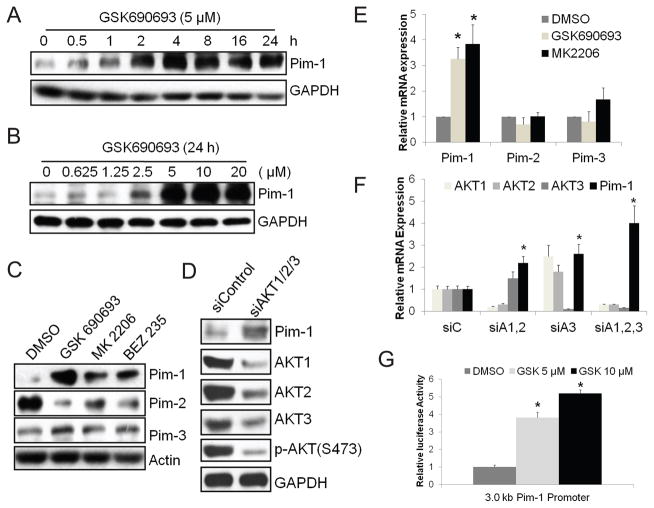
The PI3K/AKT pathway is hyperactivated in prostate cancer but its effective therapeutic targeting has proven difficult. In particular, the antitumor activity of AKT inhibitors is attenuated by upregulation of receptor tyrosine kinases (RTK) through an uncharacterized feedback mechanism. In this report, we show that RNA interference-mediated silencing or pharmacologic inhibition of Pim-1 activity curtails AKT inhibitor-induced upregulation of RTKs in prostate cancer cells. Although Pim kinases have been implicated in cap-dependent translational control, we find that in the context of AKT inhibition, the expression of RTKs is controlled by Pim-1 in a cap-independent manner by controlling internal ribosome entry. Combination of Pim and AKT inhibitors resulted in synergistic inhibition of prostate tumor growth in vitro and in vivo. Together, our results show that Pim-1 mediates resistance to AKT inhibition and suggest its targeting to improve the efficacy of AKT inhibitors in anticancer therapy.


Conflict of interest statement

The authors declare no potential conflicts of interest.

Figures

Fig. 1
Fig. 1
AKT inhibition induces expression of Pim-1. PC3-LN4 cells were treated with (A) 5 μM GSK690693 for the times indicated, (B) increasing doses of GSK690693 as indicated for 24 h, (C) 5 μM GSK690693, 2 μM MK2206, or 0.5 μM BEZ235 for 24 h, and (D) siRNAs against AKT1, AKT2, and AKT3 or a negative control siRNA for 72 h. Whole cell lysates were subjected to immunoblot analyses with the indicated antibodies. (E) Cells as in (C) were harvested and total RNA was isolated. Real-time qPCR analyses were performed with Pim-1, Pim-2, Pim-3-specific primers. Results were normalized to the expression of β-actin. (F) PC3-LN4 cells were treated with siRNAs against AKT1,2 (siA1,2), AKT3 (siA3), AKT1,2,3 (siA1,2,3), or a nontargeting control siRNA (siC) for 72 h and then RNA isolated, and real-time qPCR with indicated primers performed. (G) PC3-LN4 cells were transfected with a luciferase reporter containing a 3.0 kb human Pim-1 promoter. After 24 h, cells were treated with DMSO or two different doses of GSK690693 (GSK) as indicated for additional 24 h before harvesting for luciferase assays. Results were normalized to Renilla luciferase activity by a co-transfected plasmid carrying this enzyme. Data in E, F, and G are mean ±SD of three independent experiments. *p<0.05 compared to the corresponding negative control.
Fig. 2
Fig. 2
Pim-1 is required for elevated expression of RTKs induced by AKT inhibition. Immunoblot analyses were carried out with the indicated antibodies in (A) PC3-LN4 cells were treated with two different siRNAs (1 & 2) against Pim-1 as well as a nontargeting control siRNA (two left lanes) for 48 h followed by the addition of GSK690693 (5 μM) for an additional 24 h, (B) PC3-LN4 cells were treated with GSK690693 (GSK, 5 μM), or SMI-4a (4a, 10 μM), or the combination of the two compounds for 24 h, (C) Wild-type (WT), Pim kinase triple-knockout (TKO) murine embryonic fibroblast cells were treated with 5 μM GSK690693 for 24 h, (D) PC3-LN4 cells were treated with three different Pim inhibitors, SMI-4a (4a, 10 μM), SMI-16a (16a, 10 μM), or K00135 (K, 5 μM) for 24 h, (E) PC3-LN4 cells were transfected with a nontargeting control siRNA, siRNA against Pim-1, an empty vector, or a Pim-1 expressing plasmid for 72 h.
Fig. 3
Fig. 3
AKT inhibition increases cap-independent translation. (A) PC3-LN4 cells were treated with GSK690693 (5 μM) alone, or in combination with PP242 (2 μM) or AZD8055 (1 μM) for 24 h, and immunoblotting performed. (B) Dicistronic luciferase plasmids containing viral (CrPV and HCV) or cellular (HIF1α, VEGF, and Myc) IRESs were transfected into PC3-LN4 cells. GSK690693 (5 μM) was added 6 h after transfection for additional 24 h and luciferase activities were determined. Data are mean ±SD of four independent experiments. *p<0.05 compared to the corresponding DMSO control. (C) PC3-LN4 cells were treated with increasing doses of GSK690693 as indicated for 24 h and lysates examined by Western blotting.
Fig. 4
Fig. 4
Pim-1 regulates RTK translation by controlling IRES activity. (A) Dicistronic plasmids pRF, pR-EMCV-F, pR-HIF-F, pR-VEGF-F, and pR-MET-F were transfected into PC3-LN4 cells. A Pim-1 expressing plasmid was cotransfected with pR-MET-F as indicated. GSK690693 (GSK, 5 μM) was added 6 h after transfection and luciferase activities were determined 24 h after transfection. Data are mean ±SD of four independent experiments. *p<0.05 compared to the MET. (B) Capped, polyadenylated dicistronic mRNAs were transfected into PC3-LN4 cells. The ratios of firefly/Renilla activities are shown relative to the ratio for RF, which was given a value of 1. (C) A dicistronic plasmid containing IGF1R IRES was transfected into PC3-LN4 cells with or without either a Pim-1 expressing plasmid or siRNA targeted at Pim-1. GSK690693 (GSK, 5 μM) was added 6 h after transfection and at 48 h luciferase activities were determined. (D) Dicistronic luciferase plasmids containing viral (CrPV and HCV) or cellular (HIF1α and Myc) IRESs were transfected into PC3-LN4 cells together with siRNA against Pim-1 or a nontargeting control siRNA, and luciferase activities were determined 48h after transfection. (E) PC3-LN4 cells were treated with two different siRNAs (1 & 2) against Pim-1 as well as a nontargeting control siRNA (two left lanes) for 48h followed by adding GSK690693 (5 μM) for additional 24h as indicated. Whole cell lysates were subjected to immunoblot analyses with the indicated antibodies. Data in B, C, D, and E reflect the mean ±SD of four independent experiments. *p<0.05 compared to corresponding negative controls.
Fig. 5
Fig. 5
Ribosomal stress abrogates RTK upregulation induced by GSK690693. (A) PC3-LN4 cells were treated for 48 h with siRNAs against Pim-1, ribosomal protein S19, S6 as well as a nontargeting control siRNA (two left lanes) followed by adding GSK690693 (5 μM) for an additional 24 h. (B) PC3-LN4 cells were treated with increasing dose of actinomycin D (ActD) with and without 5 μM GSK690693 for 24 h. (C) PC3-LN4 cells were transfected with a Pim-1 expressing plasmid or a control vector. ActD (5 nM) was added 24h after transfection for an additional 16 h. Whole cell lysates were subjected to immunoblot analyses with the indicated antibodies.
Fig. 6
Fig. 6
Combined inhibition of AKT and Pim demonstrates synergistic antitumor activity. (A) PC3-LN4 cells were treated with increasing doses of GSK690693 and SMI-4a (4a) as indicated in media containing 0.2% serum for 72 h followed by a MTT assay. Similar results were obtained from three independent experiments. One representative experiment is shown. (B) PC3-LN4 cells were plated in 10% serum and 0.7% agarose-containing medium with 10 μM of GSK690693 or SMI-4a alone or in combination. Colonies were stained with crystal violet and counted after 21 days and the data are mean ±SD of three independent experiments. Bar, 200 μM. (C) Nu/Nu mice bearing PC3-LN4 tumors were randomized into four groups: vehicle, GSK690693 (30 mg/kg i.p. daily), SMI-4a (60 mg/kg oral twice/day), and the combination. Tumor size was measured every three days. The results are presented as the mean tumor volume ±SEM (n = 6 mice/group). **P < 0.02 for the GSK + SMI-4a group vs all other treatment groups. (D) Immunoblot analyses of tumors in (C) with the indicated antibodies. Tumors were harvested on Day 36 6 h after the last dose of therapy. The numbers above each lane represent individual tumors in that treatment group. (E) A model for the feedback upregulation of RTK expression mediated by Pim-1 kinase.

Comment in

References

    1. Vivanco I, Sawyers CL. The phosphatidylinositol 3-Kinase AKT pathway in human cancer. Nat Rev Cancer. 2002;2:489–501. - PubMed
    1. Yoshimoto M, Cunha IW, Coudry RA, Fonseca FP, Torres CH, Soares FA, et al. FISH analysis of 107 prostate cancers shows that PTEN genomic deletion is associated with poor clinical outcome. Br J Cancer. 2007;97:678–85. - PMC - PubMed
    1. Suzuki H, Freije D, Nusskern DR, Okami K, Cairns P, Sidransky D, et al. Interfocal heterogeneity of PTEN/MMAC1 gene alterations in multiple metastatic prostate cancer tissues. Cancer Res. 1998;58:204–9. - PubMed
    1. Bellacosa A, Kumar CC, Di Cristofano A, Testa JR. Activation of AKT kinases in cancer: implications for therapeutic targeting. Adv Cancer Res. 2005;94:29–86. - PubMed
    1. Hynes NE, Lane HA. ERBB receptors and cancer: the complexity of targeted inhibitors. Nat Rev Cancer. 2005;5:341–54. - PubMed

Publication types

MeSH terms

Substances