. 2013 May;123(5):2317-31.
doi: 10.1172/JCI67356. Epub 2013 Apr 15.

GM-CSF contributes to aortic aneurysms resulting from SMAD3 deficiency

Affiliations

GM-CSF contributes to aortic aneurysms resulting from SMAD3 deficiency

Ping Ye et al. J Clin Invest. 2013 May.

Abstract

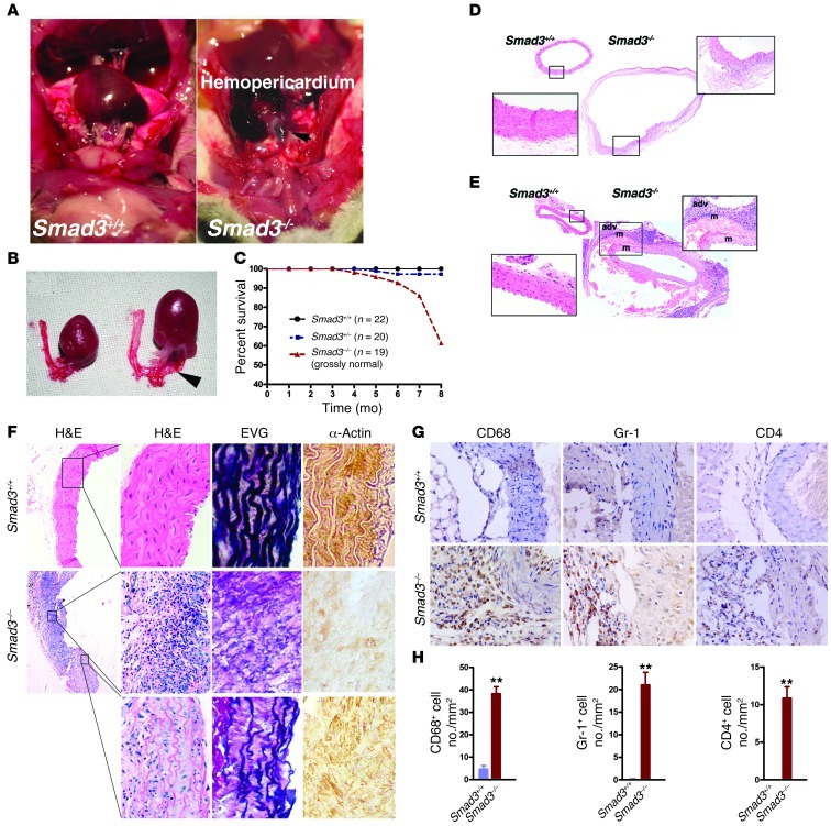
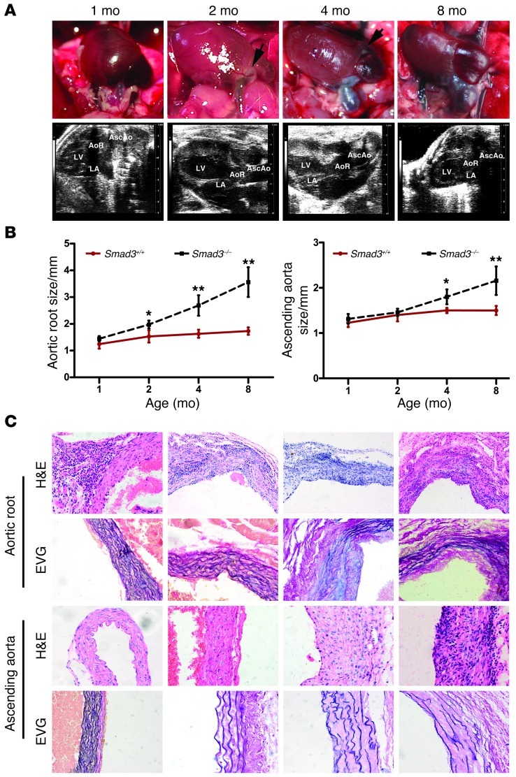
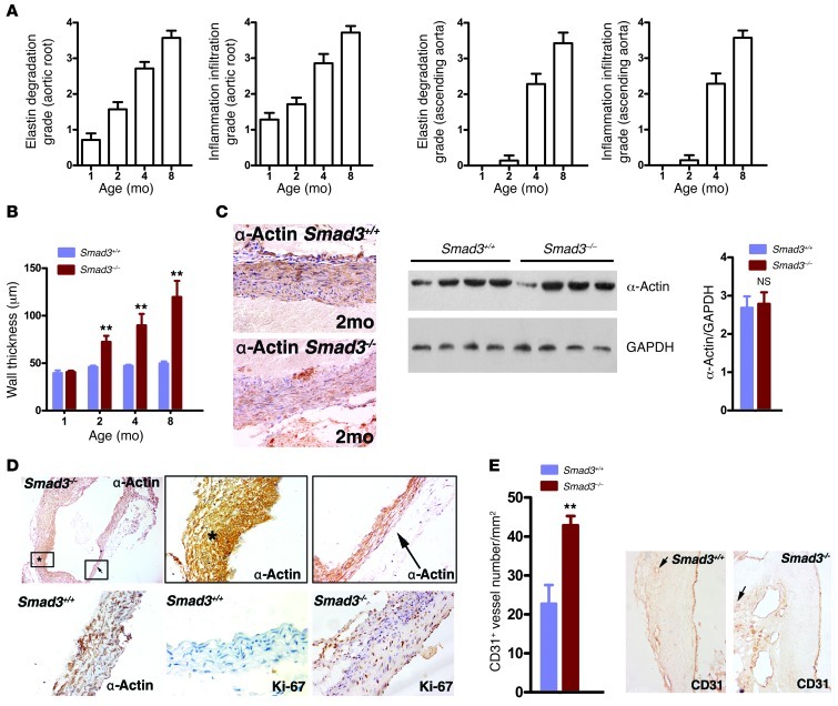
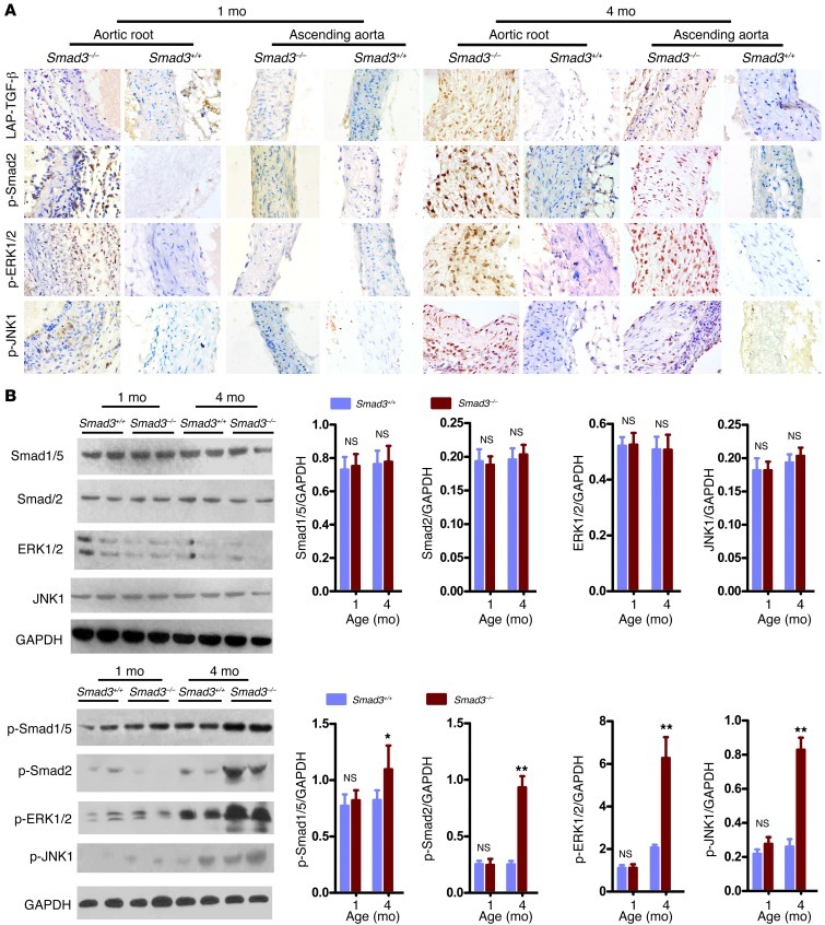
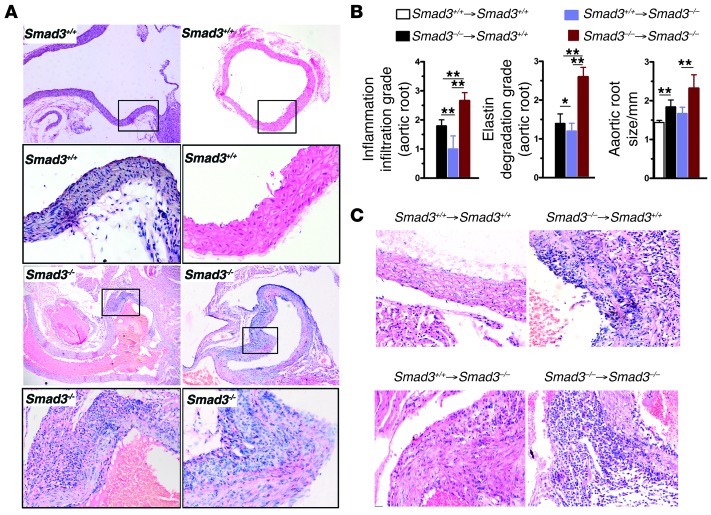
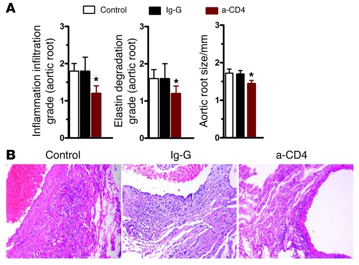
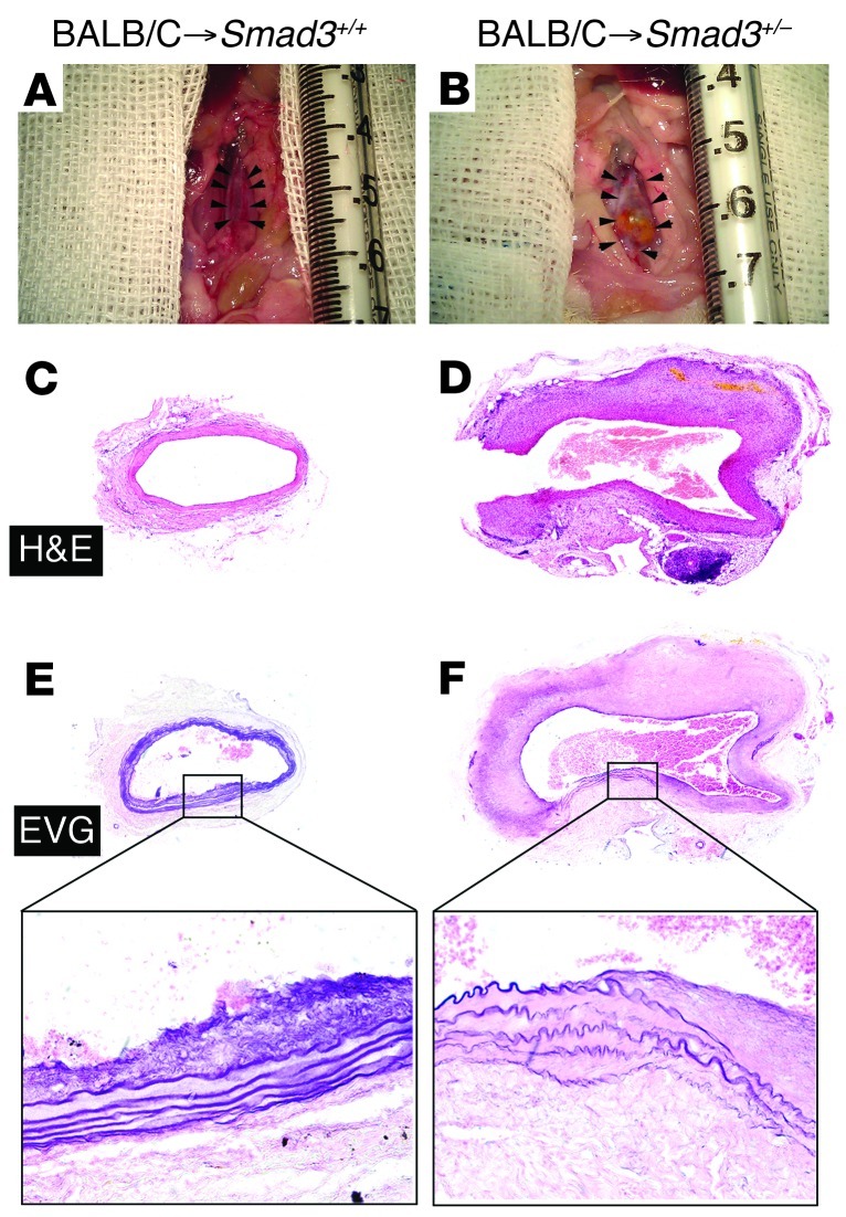
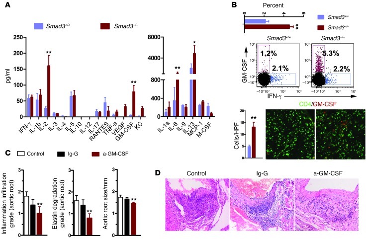
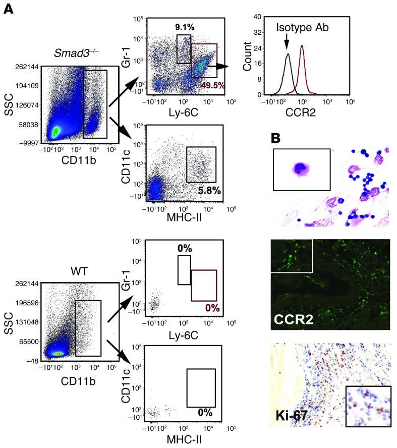
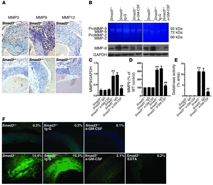
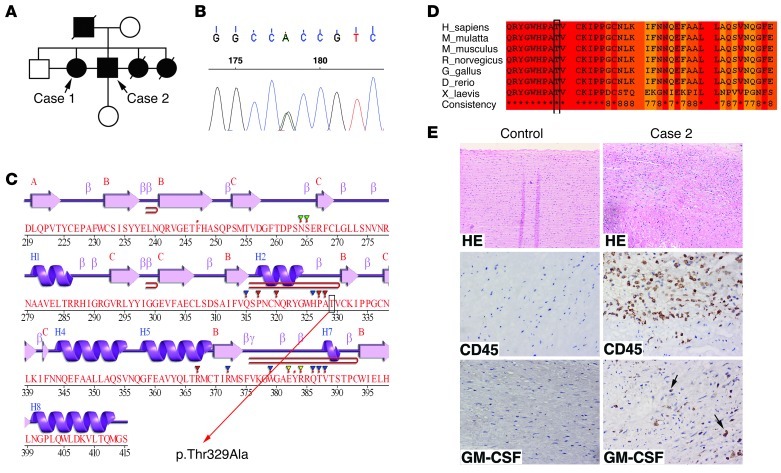
Heterozygous loss-of-function SMAD3 (Mothers against decapentaplegic homolog 3) mutations lead to aneurysm-osteoarthritis syndrome (AOS). In the present study, we found that mice lacking Smad3 had a vascular phenotype similar to AOS, marked by the progressive development of aneurysms. These aneurysms were associated with various pathological changes in transmural inflammatory cell infiltration. Bone marrow transplants from Smad3-/- mice induced aortitis and aortic root dilation in irradiated WT recipient mice. Transplantation of CD4+ T cells from Smad3-/- mice also induced aortitis in Smad3+/+ recipient mice, while depletion of CD4+ T cells in Smad3-/- mice reduced the infiltration of inflammatory cells in the aortic root. Furthermore, IFN-γ deficiency increased, while IL-17 deficiency decreased, disease severity in Smad3+/- mice. Cytokine secretion was measured using a cytokine quantibody array, and Smad3-/- CD4+ T cells secreted more GM-CSF than Smad3+/+ CD4+ T cells. GM-CSF induced CD11b+Gr-1+Ly-6Chi inflammatory monocyte accumulation in the aortic root, but administration of anti-GM-CSF mAb to Smad3-/- mice resulted in significantly less inflammation and dilation in the aortic root. We also identified a missense mutation (c.985A>G) in a family of thoracic aortic aneurysms. Intense inflammatory infiltration and GM-CSF expression was observed in aortas specimens of these patients, suggesting that GM-CSF is potentially involved in the development of AOS.


Figures

Figure 1
Figure 1. Smad3 deficiency induced death due to aneurysm rupture and aortic dissection in mice.
(A) Aneurysm rupture in a Smad3–/– mouse and a Smad3+/+ mouse at 103 days of age. Arrow indicates location of laceration. (B) Comparison of thoracic aorta with that of age- and sex-matched Smad3+/+ mice. Arrow indicates location of laceration. (C) Survival curves of WT (n = 22), Smad3+/– (n = 20), and Smad3–/– (n = 19) mice, which were grossly normal. (D) Representative images of H&E staining of transverse sections of the proximal ascending aorta from Smad3–/– mice dead from aneurysm rupture and normal age- and sex-matched Smad3+/+ mice. (E) H&E staining of transverse sections of the ascending aorta near the aortic arch from WT mice showing normal vascular structures and aneurysm dissection in the ascending aorta from Smad3–/– mice. adv, adventitia; m, media. Original magnification, ×40; ×400 (left insets); ×200 (right insets). (F) H&E staining, EVG staining, and IHC staining for α-SMA of proximal ascending aorta from Smad3–/– mice showed focal inflammatory cell infiltration and some regions of aorta tissue were relatively normal. Original magnification, ×40; ×400 (magnified panels). (G) Sections were serially immunostained for CD68, Gr-1, and CD4 to visualize monocytes/macrophages, neutrophils, and CD4+ T cells in the aortic tissues from age- and sex-matched Smad3–/– and Smad3+/+ mice. Original magnification, ×400. (H) Data are presented as mean ± SEM. **P < 0.001 Smad3–/– versus Smad3+/+ (Mann-Whitney test).
Figure 2
Figure 2. Smad3–/– mice undergo progressive aortic root and ascending aorta dilation.
(A) Representative photographs and ultrasound imaging of the aortic root and ascending aorta in Smad3–/– mice at different ages. Arrows in the photographs of 2-month-old mice identify areas of neovascularization. (B) Aortic root and ascending aortic diameter, measured by echocardiography, at different ages in Smad3+/+ (n = 9/time points) and Smad3–/– (n = 7/time points) mice. *P < 0.01; **P < 0.001, Smad3–/– versus Smad3+/+ at the same age. (C) H&E staining showed inflammatory cell infiltration in the aortic roots and ascending aortas of Smad3–/– mice (n = 12/time points) at different ages. EVG staining showed medial elastin degradation in the aortic roots and ascending aortas of Smad3–/– mice (n = 11/time points) at different ages. Original magnification, ×200 (C).
Figure 3
Figure 3. Various pathological changes associated with aneurysms in Smad3–/– mice.
(A) Inflammatory cell infiltration and elastin degradation were analyzed according to grade indicated in Supplemental Figure 3. (B) Progression of thickening of the aortic media in Smad3–/– mice. *P < 0.01; **P < 0.001, Smad3–/– versus Smad3+/+ at the same age. (C) Sections from the aortic tissue of Smad3–/–mice (2 months) and Smad3+/+ mice were stained for α-SMA to show SMCs. Western blot analysis showed no difference expression of α-SMA at the early stage (2 months). P = 0.16, Smad3–/– versus Smad3+/+. (D) Unbalanced SMC hyperplasia and aortic cross-sectional area reduction appeared in sections from aortic tissue of 4-month-old Smad3–/– mice. (E) CD31+ microvessel contents in aortas from Smad3–/– and WT mice at 2 months. **P < 0.001, Smad3–/– versus Smad3+/+. Representative CD31 staining of aortas from the 2-month time point are shown in the right panels. Original magnification, ×200 (C, E); ×40 (D, upper left panel); ×400 (D, remaining panels).
Figure 4
Figure 4. Evidence for excessive TGF-β signaling in Smad3–/– mouse aortas.
(A) Immunostaining for LAP–TGF-β, p-Smad2, p-ERK1/2 and p-JNK1 on slides of aortic root and ascending aorta from Smad3+/+ and Smad3–/– mice aged 1 or 4 months. IHC analysis reveals that the positive staining of LAP–TGF-β, p-Smad2, p-Erk1/2, and p-JNK1 are predominantly located in the infiltrating inflammatory cells in the aortic root of 1-month-old Smad3–/– mice, while there are increased expression and nuclear translocation of p-Smad2, p-ERK1/2, and p-JNK1 in the aortic media of 4-month-old Smad3–/– mice. Original magnification, ×400. (B) Representative Western blot showing Smad1/5, Smad2, ERK1/2, JNK1, p-Smad1/5, p-Smad2, p-ERK1/2, and p-JNK1 levels in proximal ascending aortas from Smad3+/+ and Smad3–/– mice (1 and 4 months of age). The ratio between Smad1/5, Smad2, ERK1/2, JNK1, p-Smad1/5, p-Smad2, p-ERK1/2, and p-JNK1 to GAPDH levels is shown. *P < 0.01; **P < 0.001.
Figure 5
Figure 5. Aneurysms occur in the context of chronic inflammation.
(A) Representative H&E staining of longitudinal and transverse sections of the ascending aorta from 1-month-old Smad3+/+ and Smad3–/– mouse showing inflammatory cell infiltration in the Smad3–/– aortic root and milder infiltration in that of Smad3+/+ mice. Original magnification, ×100; ×400 (magnified insets). (B) Severity of inflammatory cell infiltration, elastin degradation, and aortic root size in the chimeric mice. The aortic root of Smad3+/+ recipient mouse receiving BM cells from Smad3–/– mouse showed severe inflammatory cell infiltration, elastin degradation, and enlargement of aortic root compared with that of Smad3+/+ recipient mouse receiving BM cells from Smad3+/+ mouse. The aortic root of Smad3–/– mice showed significantly ameliorated pathological changes after receiving Smad3+/+ BM cells. *P < 0.01; **P < 0.001. (C) Representative H&E staining of transverse sections of the aortic root from chimeric mice. Of note, WT recipient mouse receiving BM cells from Smad3–/– mouse revealed a substantially increased accumulation of inflammatory cells within the adventitia and infiltration of inflammatory cells into the media. Original magnification, ×400.
Figure 6
Figure 6. CD4+ T cell depletion inhibits inflammatory aneurysms in Smad3–/– mice.
(A) Two-week-old Smad3–/– mice were given CD4-depleting mAb (GK1.5), control IgG, or nothing. Four weeks later, changes in the aortic root were examined. *P < 0.01 versus control. (B) Representative images of H&E staining of transverse sections of aortic roots from mice from these 3 groups. Original magnification, ×200.
Figure 7
Figure 7. Aortic allografts from Smad3+/– recipients showing aneurysm formation.
(A and B) Gross appearance of aortas from Smad3+/+ recipients and Smad3+/– littermate recipients 12 weeks after transplantation showing aneurysm formation in the latter. (C and D) H&E staining of transverse sections of aortic allograft from Smad3+/+ and Smad3+/– littermate recipients showing greater diameter in the latter than the former. (E and F) EVG staining showing markedly fragmented elastic lamina in the aortic media of transplant from Smad3+/–recipients, which was not observed in the allografts from Smad3+/+ littermate recipient mice. Original magnification, ×40; ×400 (magnified insets).
Figure 8
Figure 8. Smad3–/– CD4+ T cells secrete GM-CSF, which is an important cytokine in AOS.
(A) Analysis of cytokine secretion by CD4+ T cells from Smad3–/– and Smad3+/+ mice (n = 5) using a mouse cytokine antibody array. *P < 0.05; **P < 0.01. (B) Smad3–/– CD4+ T cells produced more GM-CSF than Smad3+/+ CD4+ T cells, which was confirmed by flow cytometry and immunofluorescence staining. This difference was analyzed by comparing the percentage of CD4+GM-CSF+ T cells or counting their number in a high-power field. **P < 0.01. HPF, high-power field. Original magnification, ×400. (C) One-month-old Smad3–/– mice were given anti–GM-CSF mAb, control IgG, or nothing. Four weeks later, changes in the aortic root were examined. **P < 0.01 versus control. (D) Representative images of H&E staining of transverse sections of the aortic roots from mice from these 3 groups. Original magnification, ×200.
Figure 9
Figure 9. The infiltrated cells were predominantly CD11b+Gr-1+ cells, which were regulated by GM-CSF.
(A) Dissociated aortic cells were gated on CD11b; CD11b+ cells were gated on Gr-1 and Ly-6C, which contained CD11b+Gr-1+Ly-6Chi inflammatory monocytes (55.2% ± 4.9%, n = 5) and CD11b+Gr-1+Ly-6Cmid inflammatory granulocytes (7.1% ± 2.3%, n = 5). Histograms showed that the CD11b+Gr-1+Ly-6Chi cells expressed CCR2, which was confirmed by comparison to an isotype control. (B) Wright-Giemsa staining of separated mononuclear cells from aortic tissue revealed the typical inflammatory monocyte shape of these cells. Immunofluorescence staining for CCR2 confirmed the infiltrated cells were inflammatory monocytes. Ki-67 immunostaining of the aortic wall (1-month-old Smad3–/– mice). Approximately 80% of the inflammatory cells stained positive for Ki-67. Original magnification: ×400 (top), ×1000 (top, magnified inset); ×200 (middle), ×400 (middle, magnified inset); ×200 (bottom), ×400 (bottom, magnified inset).
Figure 10
Figure 10. Smad3–/– mouse aorta showed GM-CSF–dependent increase in MMP9 expression and gelatinase.
(A) MMP2, MMP9, and MMP12 immunostaining of the aortic wall (2-month-old Smad3–/– and Smad3+/+ mice received GM-CSF antibody or Ig-G). Original magnification, ×400. (B) Representative sample of 5 separate experiments showed gelatinolytic MMP2 and MMP9 activities in aortas from Smad3+/+ and Smad3–/– mice that received GM-CSF antibody or Ig-G (top). Representative Western blot showing MMP9 levels in aortas from Smad3+/+ and Smad3–/– mice received GM-CSF antibody or Ig-G (bottom). (CE) Semiquantitative analysis of gelatinase activity (MMP9 or all) and MMP9 expression. **P < 0.01 versus Smad3+/+; ##P < 0.01 versus Smad3–/– mice that received Ig-G. (F) Representative samples of all gelatinase activity (green) in aortas of Smad3+/+ mice; Smad3–/– mice received GM-CSF antibody or Ig-G. Original magnification, ×400.
Figure 11
Figure 11. SMAD3 mutations in a family with AOS and the aorta tissue from 1 case showed inflammatory infiltration and increased GM-CSF expression.
(A) Pedigrees with SMAD3 mutations. Squares, males; circles, females. Filled symbols indicate individuals with thoracic aortic aneurysm and/or dissection. Symbols represent individuals with a normal or unknown phenotype. (B) The sequence chromatogram of SMAD3 shows a heterozygous mutation. (C) Secondary structures of SMAD3, which are described by Jpred3 ( http://www.compbio.dundee.ac.uk/www-jpred/). The mutation (p.Thr329Ala) is located in β-folding near H2, which is in the MHI domain of SMAD3. (D) Cross-species protein conservation of SMAD3 around the mutation (p.Thr329Ala) which is described by the IBIVU Server ( http://www.ibi.vu.nl/programs/pralinewww/). (E) H&E staining and immunohistochemistry staining of transverse sections of aorta from a control (donor) and a patient with SMAD3 mutation. It showed inflammatory infiltration and increased GM-CSF expression in the aorta tissues from case 2. Original magnification, ×100 (top panels); ×400 (middle and bottom panels).

References

    1. Lindsay ME, Dietz HC. Lessons on the pathogenesis of aneurysm from heritable conditions. Nature. 2011;473(7347):308–316. doi: 10.1038/nature10145. - DOI - PMC - PubMed
    1. Dietz HC, et al. Marfan syndrome caused by a recurrent de novo missense mutation in the fibrillin gene. Nature. 1991;352(6333):337–339. doi: 10.1038/352337a0. - DOI - PubMed
    1. Loeys BL, et al. A syndrome of altered cardiovascular, craniofacial, neurocognitive and skeletal development caused by mutations in TGFBR1 or TGFBR2. Nat Genet. 2005;37(3):275–281. doi: 10.1038/ng1511. - DOI - PubMed
    1. Dasouki M, et al. Compound heterozygous mutations in fibulin-4 causing neonatal lethal pulmonary artery occlusion, aortic aneurysm, arachnodactyly, and mild cutis laxa. Am J Med Genet A. 2007;143A(22):2635–2641. - PubMed
    1. Dietz HC. TGF-beta in the pathogenesis and prevention of disease: a matter of aneurysmic proportions. J Clin Invest. 2010;120(2):403–407. doi: 10.1172/JCI42014. - DOI - PMC - PubMed

Publication types

MeSH terms