

Defining the Escherichia coli SecA dimer interface residues through in vivo site-specific photo-cross-linking
- PMID: 23585536
- PMCID: PMC3697251
- DOI: 10.1128/JB.02269-12
Defining the Escherichia coli SecA dimer interface residues through in vivo site-specific photo-cross-linking
Abstract
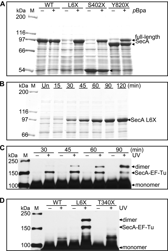
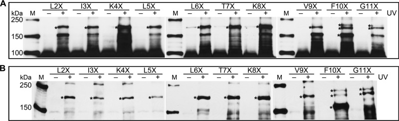
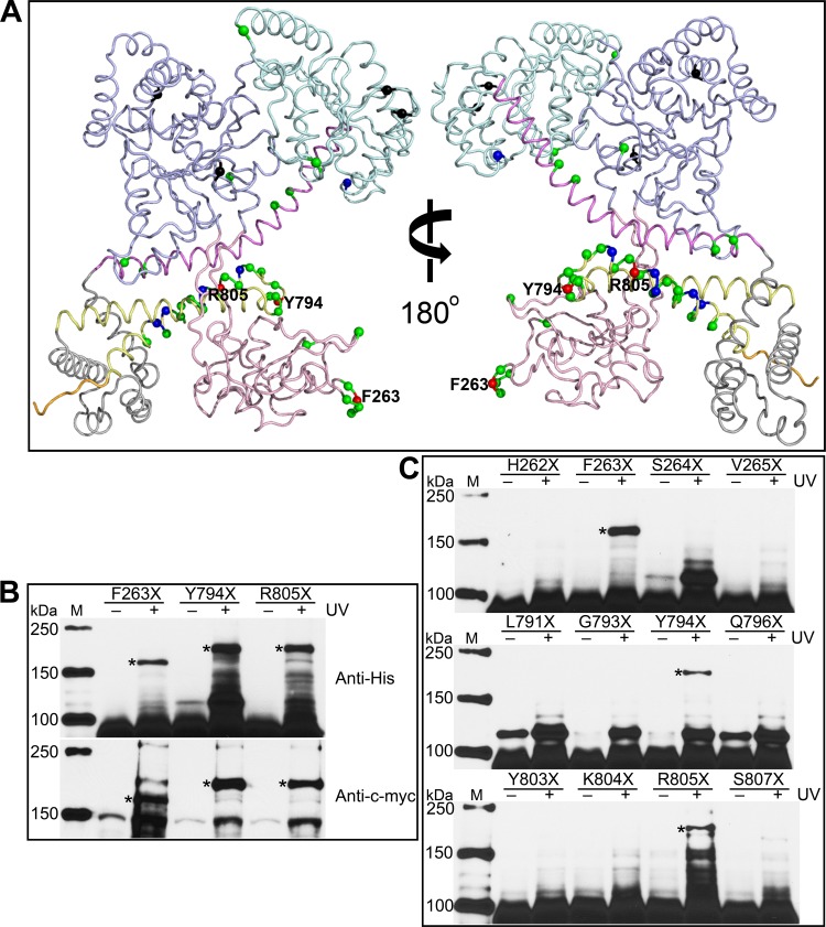
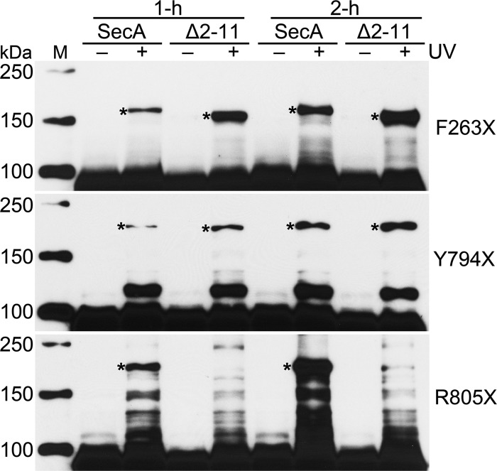
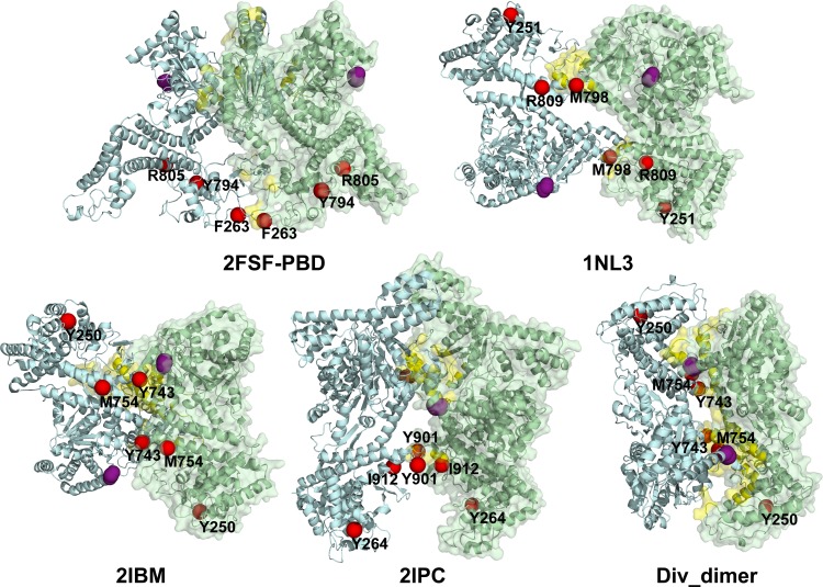
The motor protein SecA is a core component of the bacterial general secretory (Sec) pathway and is essential for cell viability. Despite evidence showing that SecA exists in a dynamic monomer-dimer equilibrium favoring the dimeric form in solution and in the cytoplasm, there is considerable debate as to the quaternary structural organization of the SecA dimer. Here, a site-directed photo-cross-linking technique was utilized to identify residues on the Escherichia coli SecA (ecSecA) dimer interface in the cytosol of intact cells. The feasibility of this method was demonstrated with residue Leu6, which is essential for ecSecA dimerization based on our analytical ultracentrifugation studies of SecA L6A and shown to form the cross-linked SecA dimer in vivo with p-benzoyl-phenylalanine (pBpa) substituted at position 6. Subsequently, the amino terminus (residues 2 to 11) in the nucleotide binding domain (NBD), Phe263 in the preprotein binding domain (PBD), and Tyr794 and Arg805 in the intramolecular regulator of the ATPase 1 domain (IRA1) were identified to be involved in ecSecA dimerization. Furthermore, the incorporation of pBpa at position 805 did not form a cross-linked dimer in the SecA Δ2-11 context, indicating the possibility that the amino terminus may directly contact Arg805 or that the deletion of residues 2 to 11 alters the topology of the naturally occurring ecSecA dimer.
Figures

Similar articles
-
Defining the solution state dimer structure of Escherichia coli SecA using Förster resonance energy transfer.Biochemistry. 2013 Apr 9;52(14):2388-401. doi: 10.1021/bi301217t. Epub 2013 Mar 29. Biochemistry. 2013. PMID: 23484952 Free PMC article.
-
Analysis of SecA dimerization in solution.Biochemistry. 2014 May 20;53(19):3248-60. doi: 10.1021/bi500348p. Epub 2014 May 9. Biochemistry. 2014. PMID: 24786965 Free PMC article.
-
Escherichia coli SecA truncated at its termini is functional and dimeric.FEBS Lett. 2005 Feb 14;579(5):1267-71. doi: 10.1016/j.febslet.2005.01.025. Epub 2005 Jan 26. FEBS Lett. 2005. PMID: 15710424
-
Oligomeric states of the SecA and SecYEG core components of the bacterial Sec translocon.Biochim Biophys Acta. 2007 Jan;1768(1):5-12. doi: 10.1016/j.bbamem.2006.08.013. Epub 2006 Aug 30. Biochim Biophys Acta. 2007. PMID: 17011510 Free PMC article. Review.
-
SecA: a tale of two protomers.Mol Microbiol. 2010 Jun 1;76(5):1070-81. doi: 10.1111/j.1365-2958.2010.07176.x. Epub 2010 Apr 23. Mol Microbiol. 2010. PMID: 20444093 Review.
Cited by
-
Electron-deficient p-benzoyl-l-phenylalanine derivatives increase covalent chemical capture yields for protein-protein interactions.Protein Sci. 2019 Jun;28(6):1163-1170. doi: 10.1002/pro.3621. Epub 2019 Apr 29. Protein Sci. 2019. PMID: 30977234 Free PMC article.
-
Illumination of growth, division and secretion by metabolic labeling of the bacterial cell surface.FEMS Microbiol Rev. 2015 Mar;39(2):184-202. doi: 10.1093/femsre/fuu012. Epub 2015 Jan 23. FEMS Microbiol Rev. 2015. PMID: 25725012 Free PMC article. Review.
-
Outer membrane lipoprotein RlpA is a novel periplasmic interaction partner of the cell division protein FtsK in Escherichia coli.Sci Rep. 2018 Aug 28;8(1):12933. doi: 10.1038/s41598-018-30979-5. Sci Rep. 2018. PMID: 30154462 Free PMC article.
-
An alternate mode of oligomerization for E. coli SecA.Sci Rep. 2017 Sep 18;7(1):11747. doi: 10.1038/s41598-017-11648-5. Sci Rep. 2017. PMID: 28924213 Free PMC article.
-
The Sec System: Protein Export in Escherichia coli.EcoSal Plus. 2017 Nov;7(2):10.1128/ecosalplus.ESP-0002-2017. doi: 10.1128/ecosalplus.ESP-0002-2017. EcoSal Plus. 2017. PMID: 29165233 Free PMC article. Review.
References
-
- Papanikou E, Karamanou S, Economou A. 2007. Bacterial protein secretion through the translocase nanomachine. Nat. Rev. Microbiol. 5:839–851 - PubMed
-
- Fekkes P, de Wit JG, van der Wolk JP, Kimsey HH, Kumamoto CA, Driessen AJ. 1998. Preprotein transfer to the Escherichia coli translocase requires the co-operative binding of SecB and the signal sequence to SecA. Mol. Microbiol. 29:1179–1190 - PubMed
-
- Economou A, Wickner W. 1994. SecA promotes preprotein translocation by undergoing ATP-driven cycles of membrane insertion and deinsertion. Cell 78:835–843 - PubMed
-
- Papanikolau Y, Papadovasilaki M, Ravelli RBG, McCarthy AA, Cusack S, Economou A, Petratos K. 2007. Structure of dimeric SecA, the Escherichia coli preprotein translocase motor. J. Mol. Biol. 366:1545–1557 - PubMed
-
- Cabelli RJ, Dolan KM, Qian LP, Oliver DB. 1991. Characterization of membrane-associated and soluble states of SecA protein from wild-type and SecA51(TS) mutant strains of Escherichia coli. J. Biol. Chem. 266:24420–24427 - PubMed
Publication types
MeSH terms
Substances
Grants and funding
LinkOut - more resources
Full Text Sources
Other Literature Sources
Molecular Biology Databases
