. 2013 Jun;195(12):2852-63.
doi: 10.1128/JB.00244-13. Epub 2013 Apr 12.

Analysis of novel iron-regulated, surface-anchored hemin-binding proteins in Corynebacterium diphtheriae

Affiliations

Analysis of novel iron-regulated, surface-anchored hemin-binding proteins in Corynebacterium diphtheriae

Courtni E Allen et al. J Bacteriol. 2013 Jun.

Abstract

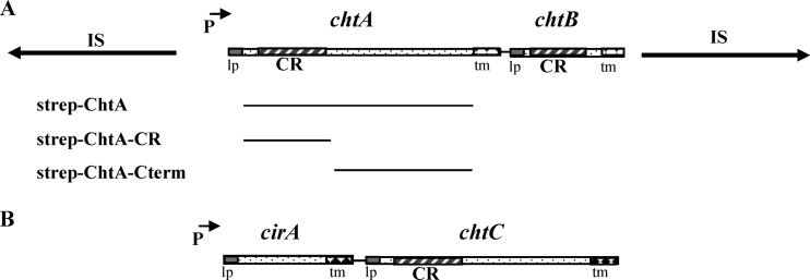
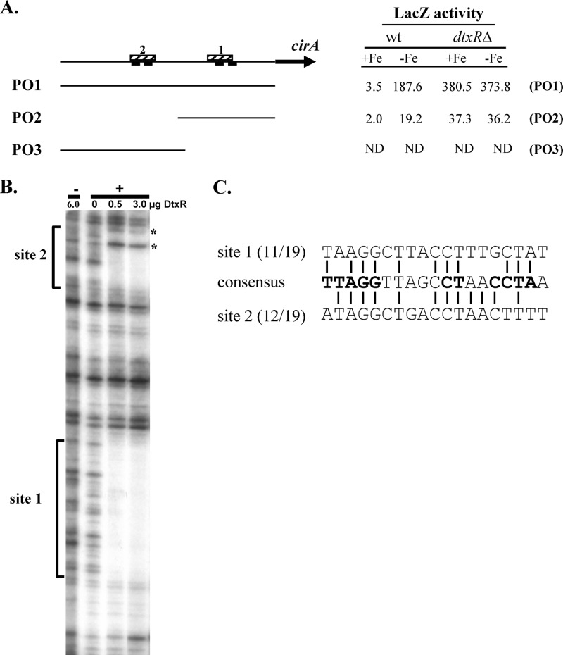
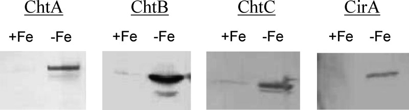
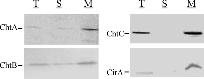
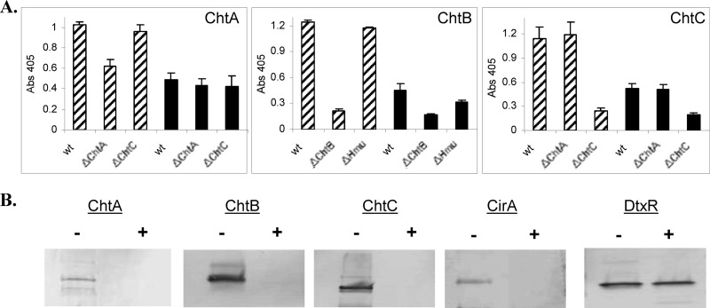
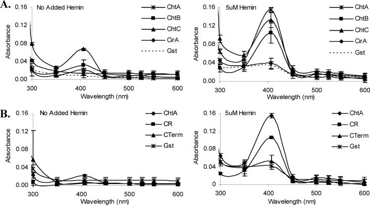
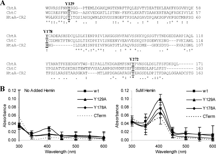
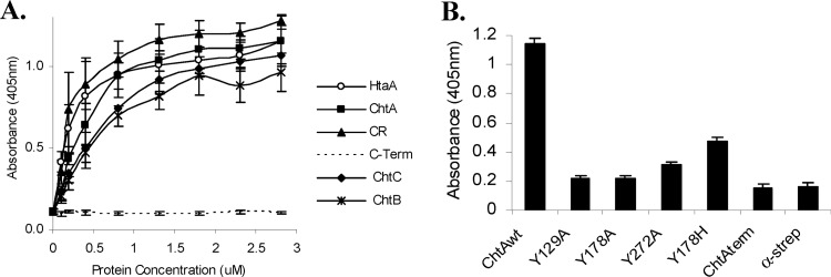
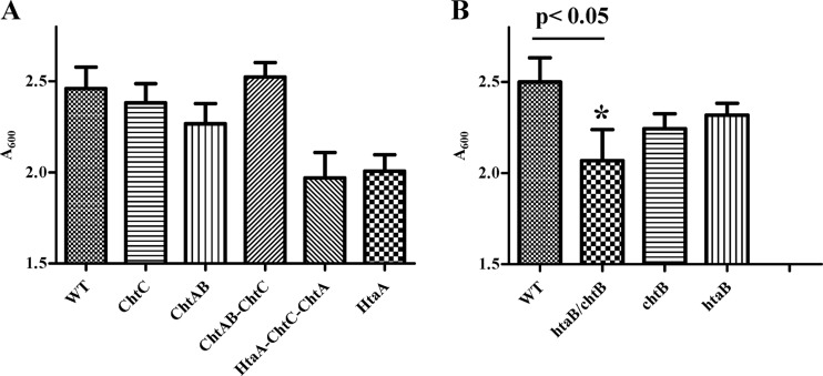
Corynebacterium diphtheriae utilizes hemin and hemoglobin (Hb) as iron sources during growth in iron-depleted environments, and recent studies have shown that the surface-exposed HtaA protein binds both hemin and Hb and also contributes to the utilization of hemin iron. Conserved (CR) domains within HtaA and in the associated hemin-binding protein, HtaB, are required for the ability to bind hemin and Hb. In this study, we identified and characterized two novel genetic loci in C. diphtheriae that encode factors that bind hemin and Hb. Both genetic systems contain two-gene operons that are transcriptionally regulated by DtxR and iron. The gene products of these operons are ChtA-ChtB and ChtC-CirA (previously DIP0522-DIP0523). The chtA and chtB genes are carried on a putative composite transposon associated with C. diphtheriae isolates that dominated the diphtheria outbreak in the former Soviet Union in the 1990s. ChtA and ChtC each contain a single N-terminal CR domain and exhibit significant sequence similarity to each other but only limited similarity with HtaA. The chtB and htaB gene products exhibited a high level of sequence similarity throughout their sequences, and both proteins contain a single CR domain. Whole-cell binding studies as well as protease analysis indicated that all four of the proteins encoded by these two operons are surface exposed, which is consistent with the presence of a transmembrane segment in their C-terminal regions. ChtA, ChtB, and ChtC are able to bind hemin and Hb, with ChtA showing the highest affinity. Site-directed mutagenesis showed that specific tyrosine residues within the ChtA CR domain were critical for hemin and Hb binding. Hemin iron utilization assays using various C. diphtheriae mutants indicate that deletion of the chtA-chtB region and the chtC gene has no affect on the ability of C. diphtheriae to use hemin or Hb as iron sources; however, a chtB htaB double mutant exhibits a significant decrease in hemin iron use, indicating a role in hemin transport for HtaB and ChtB.


Figures

Fig 1
Fig 1
(A) Genetic map of the chtA-chtB region. The chtA-chtB genes in NCTC13129 are present on a putative transposon that is composed of two identical and inverted insertion sequences (IS elements, indicated by lines with arrows). CR, conserved region; lp, leader peptide; tm, transmembrane region; P with arrow, DtxR and iron-regulated promoter. Below the genetic map are shown the regions of ChtA that were used to construct various Strep-tagged fusion proteins. (B) Genetic map of the cirA-chtC region in NCTC13129.
Fig 2
Fig 2
The cirA promoter is regulated by iron and DtxR. (A) Map of the region upstream of the cirA gene, showing putative −10 elements (solid bars below line) and DtxR-binding sites 1 and 2 (hatched bars above line). DNA sequences carried by the various promoter fusions PO1, PO2, and PO3 are shown. LacZ activities from the various promoter fusions in wt C. diphtheriae C7 and a dtxRΔ mutant from strains grown in high (+Fe)- or low (−Fe)-iron medium are shown. ND, none detected. (B) DNase I protection at the cirA promoter region. DtxR protects two distinct regions upstream of cirA, indicated by brackets as site 1 and site 2. Asterisks indicate hypercleavable sites. +, presence of divalent metal Co2+; −, absence of divalent metal Co2+. (C) Alignment of the two DtxR-binding sites upstream of cirA with the 19-bp consensus DtxR-binding site. The most highly conserved bases in the consensus sequence are in bold.
Fig 3
Fig 3
Expression of ChtA, ChtB, ChtC, and CirA is repressed by iron in the C. diphtheriae wild-type strain 1737. Bacteria were grown in high- or low-iron HIBTW medium. Proteins from either whole cells (ChtC and CirA) or supernatant fractions (ChtA and ChtB were located predominately in the supernatant in HIBTW medium) were detected by Western blotting using polyclonal antiserum specific to the proteins indicated. Samples were loaded using equivalent protein levels. +Fe, high-iron medium; −Fe, iron-depleted medium.
Fig 4
Fig 4
Protein localization studies show that ChtA, ChtB, ChtC, and CirA are associated predominately with the membrane fraction in C. diphtheriae 1737. Cultures were grown in mPGT medium under low-Fe (0.5 μM) conditions and bacteria were fractionated as described in Materials and Methods. Polyclonal antiserum specific to the proteins indicated was used for detection of total cellular proteins (T), soluble cytosolic proteins (S), and membrane-associated proteins (M). Samples were loaded using equivalent protein levels.
Fig 5
Fig 5
ChtA, ChtB, ChtC and CirA are surface exposed in C. diphtheriae. (A) A whole-cell ELISA was used to detect surface proteins in strain 1737. C. diphtheriae strains were grown in mPGT medium under either high-iron (solid bars) or low-iron (hatched bars) conditions. Equivalent amounts of cells were fixed to the plates, and antiserum specific to the protein of interest was used to detect surface proteins. Values represent the means from three independent experiments (± standard deviations [SD]). (B) Proteinase K was used to detect the presence of surface proteins in C. diphtheriae. See Materials and Methods for experimental details. Bacteria were grown in low-iron mPGT medium, and intact bacteria were incubated in the presence (+) or absence (−) of proteinase K. Proteins from whole cells were examined by Western blotting using the relevant antiserum.
Fig 6
Fig 6
(A) ChtA, ChtB, and ChtC are hemin-binding proteins. UV-visible spectroscopy was used to examine the hemin-binding properties of purified GST-CirA, Strep-ChtA, Strep-ChtB, and Strep-ChtC. Proteins at 2 μM were incubated for 15 min in the presence of 5 μM hemin or with no added hemin prior to spectral analysis. Similar results were observed for GST-tagged ChtA, ChtB, and ChtC (data not shown). GST was included as a negative control. Values are the means from three independent experiments (± SD). (B) The CR domain of ChtA binds hemin. UV-visible spectroscopy was used to examine the hemin-binding properties of the CR domain (CR) of ChtA and the C-terminal region (CTerm) of ChtA using Strep-tagged fusion proteins harboring these specific regions. The same procedures described for panel A above were used for these studies.
Fig 7
Fig 7
(A) Sequence alignment of CR domains from the C. diphtheriae ChtA, ChtC, and HtaA proteins. Conserved tyrosine residues are indicated in bold and above the sequences. Asterisks indicate identity; colons and periods indicate sequence similarity. (B) Conserved tyrosine residues in the CR domain of ChtA are critical for optimal hemin binding. UV-visible spectroscopy was used to assess the hemin binding capability of proteins with various tyrosine-to-alanine substitutions in the CR domain of Strep-tagged ChtA. Proteins at 2 μM were incubated for 15 min in the presence of 5 μM hemin or with no added hemin prior to analysis. Values are the means from three independent experiments (± SD). The difference in peak absorbance (406 nm) between the wt and the Y129A mutant is significant at a P value of <0.01, and the difference in peak absorbance between wt and the Y178A, Y178H, and Y272A mutants is significant at a P value of <0.05.
Fig 8
Fig 8
ChtA, ChtB, and ChtC are hemoglobin-binding proteins. (A) Hb binding by Strep-tagged ChtA, ChtA-CR (CR), ChtA-CTerm (C-Term), ChtC, and ChtB was assessed at various protein concentrations as indicated using an ELISA. The Hb-binding protein HtaA was used as a positive control. Values represent the means from three independent experiments (±SD). Binding was detected using anti-Strep antibodies. (B) The effect of amino acid substitutions in ChtA on Hb binding. Hb binding by the various Strep constructs (200 nM) was assessed as for panel A. Values represent the means from three independent experiments (±SD). Binding was detected using anti-Strep antibodies. α-strep, negative-control wells coated only with Hb.
Fig 9
Fig 9
Hb iron utilization assays. Nonpolar deletion mutants were constructed in C. diphtheriae 1737 and assessed for their ability to use Hb as the sole iron source for growth in low-iron mPGT medium. Cultures were grown overnight at 37°C, and cell density was measured by A600. Results are the means from three independent experiments ± standard deviations. The growth difference between the wt and the chtB-htaB double mutant (*) was significant at a P value of <0.05.

References

    1. Collier RJ. 2001. Understanding the mode of action of diphtheria toxin: a perspective on progress during the 20th century. Toxicon 39:1793–1803 - PubMed
    1. Barksdale L. 1970. Corynebacterium diphtheriae and its relatives. Bacteriol. Rev. 34:378–422 - PMC - PubMed
    1. Cornelis P, Andrews SC. 2010. Iron uptake and homeostasis in microorganisms. Caister Academic Press, Norfolk, United Kingdom
    1. Crosa JH, Mey AR, Payne SM. 2004. Iron transport in bacteria. ASM Press, Washington DC
    1. Boyd J, Oza MN, Murphy JR. 1990. Molecular cloning and DNA sequence analysis of a diphtheria tox iron-dependent regulatory element (dtxR) from Corynebacterium diphtheriae. Proc. Natl. Acad. Sci. U. S. A. 87:5968–5972 - PMC - PubMed

Publication types

MeSH terms

LinkOut - more resources