. 2013 Jul;28(7):1874-89.
doi: 10.1093/humrep/det064. Epub 2013 Apr 12.

Intracellular translocation and differential accumulation of cell-penetrating peptides in bovine spermatozoa: evaluation of efficient delivery vectors that do not compromise human sperm motility

Affiliations

Intracellular translocation and differential accumulation of cell-penetrating peptides in bovine spermatozoa: evaluation of efficient delivery vectors that do not compromise human sperm motility

Sarah Jones et al. Hum Reprod. 2013 Jul.

Abstract

Study question: Do cell penetrating peptides (CPPs) translocate into spermatozoa and, if so, could they be utilized to deliver a much larger protein cargo?

Summary answer: Chemically diverse polycationic CPPs rapidly and efficiently translocate into spermatozoa. They exhibit differential accumulation within intracellular compartments without detrimental influences upon cellular viability or motility but they are relatively ineffective in transporting larger proteins.

What is already known: Endocytosis, the prevalent route of protein internalization into eukaryotic cells, is severely compromised in mature spermatozoa. Thus, the translocation of many bioactive agents into sperm is relatively inefficient. However, the delivery of bioactive moieties into mature spermatozoa could be significantly improved by the identification and utility of an efficient and inert vectorial delivery technology.

Study design: CPP translocation efficacies, their subsequent differential intracellular distribution and the influence of peptides upon viability were determined in bovine spermatozoa. Temporal analyses of sperm motility in the presence of exogenously CPPs utilized normozoospermic human donor samples.

Materials and methods: CPPs were prepared by manual, automated and microwave-enhanced solid phase synthesis. Confocal fluorescence microscopy determined the intracellular distribution of rhodamine-conjugated CPPs in spermatozoa. Quantitative uptake and kinetic analyses compared the translocation efficacies of chemically diverse CPPs and conjugates of biotinylated CPPs and avidin. 3-(4,5-dimethylthiazol-2-yl)-5-(3-carboxymethoxyphenyl)-2-(4-sulfophenyl)-2H-tetrazolium, inner salt (MTS) conversion assays were employed to analyse the influence of CPPs upon sperm cell viability and sperm class assays determined the impact of CPPs on motility in capacitated and non-capacitated human samples.

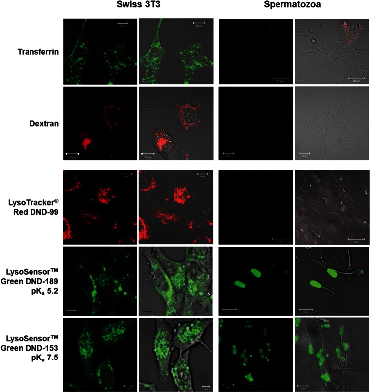
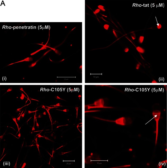
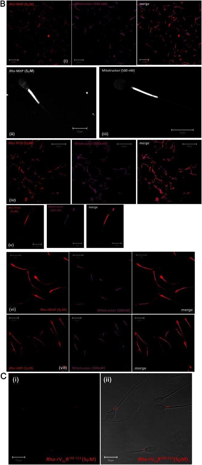
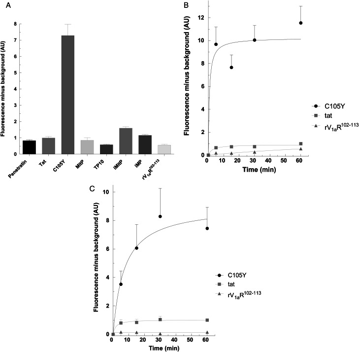
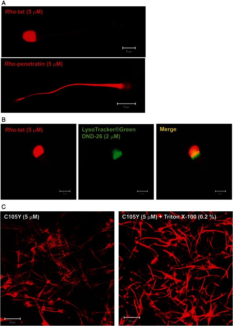
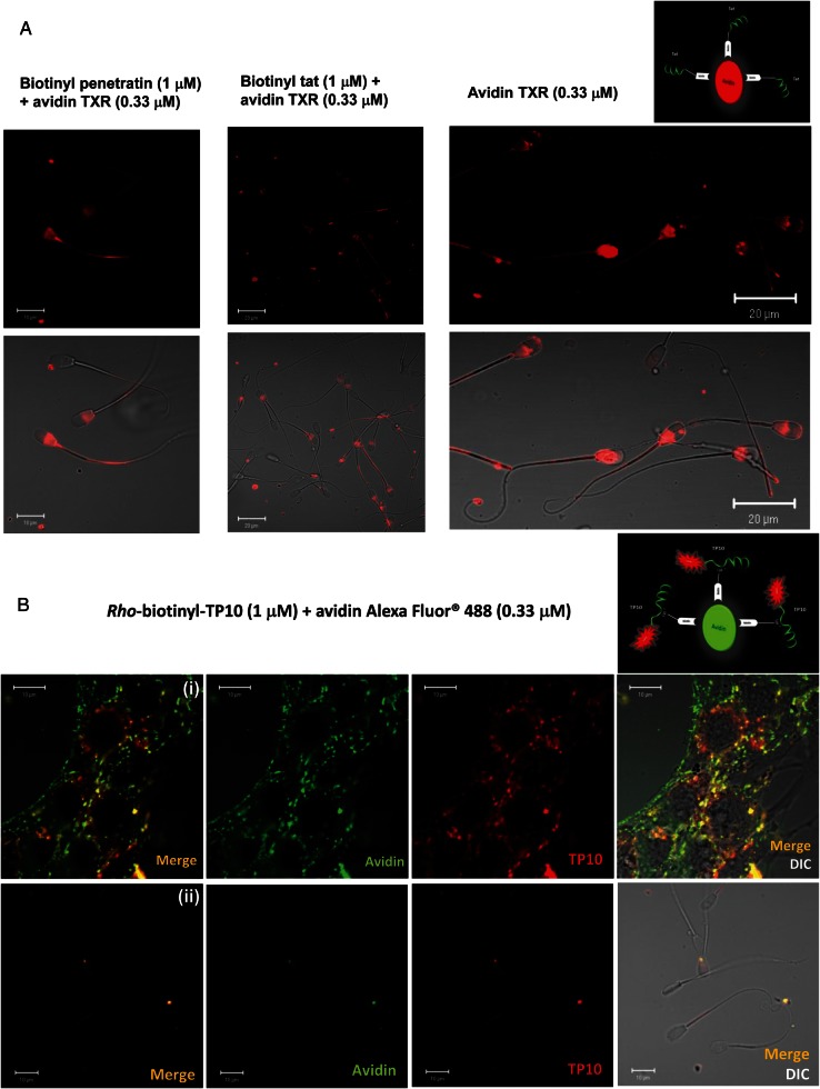
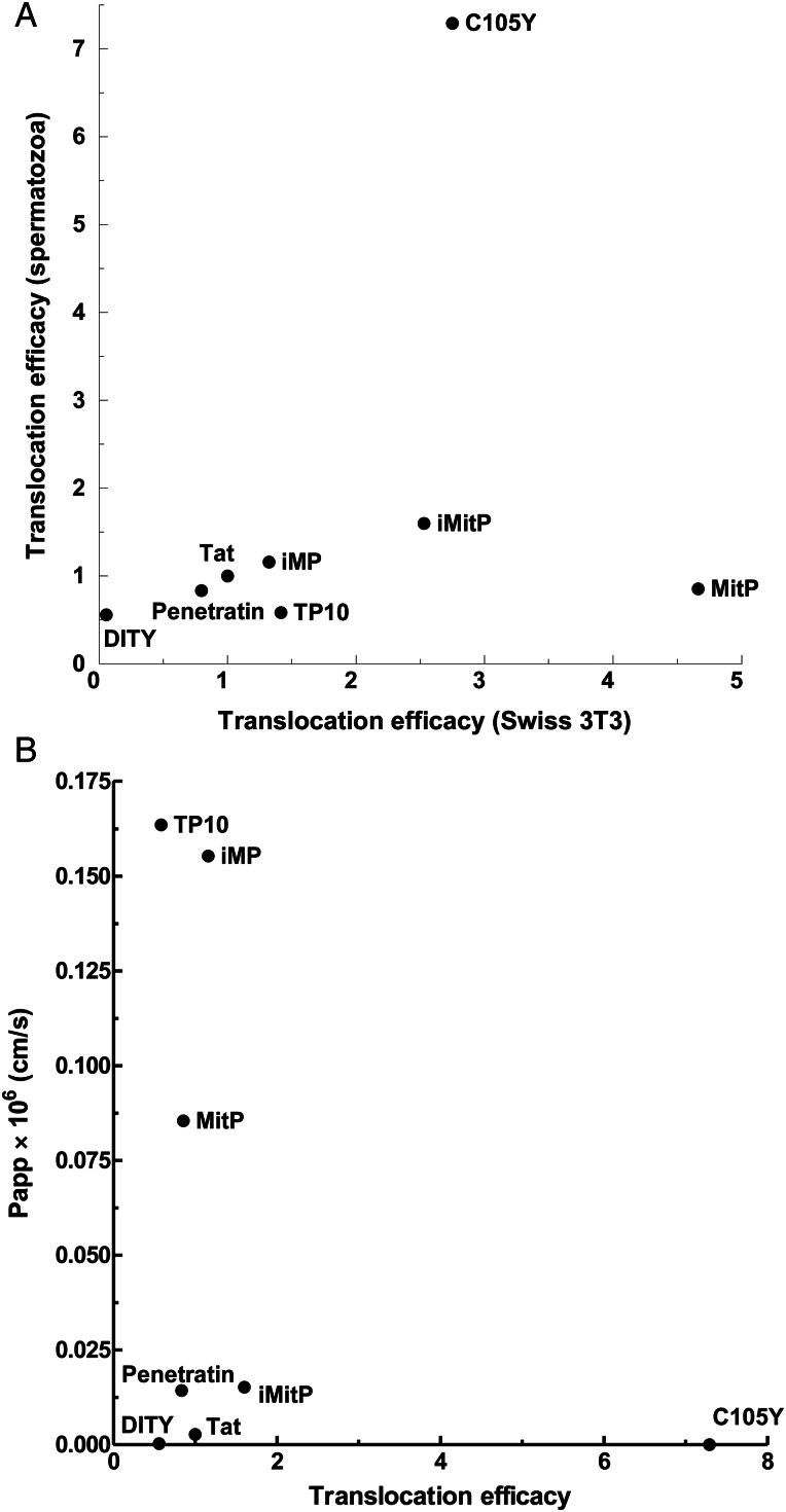
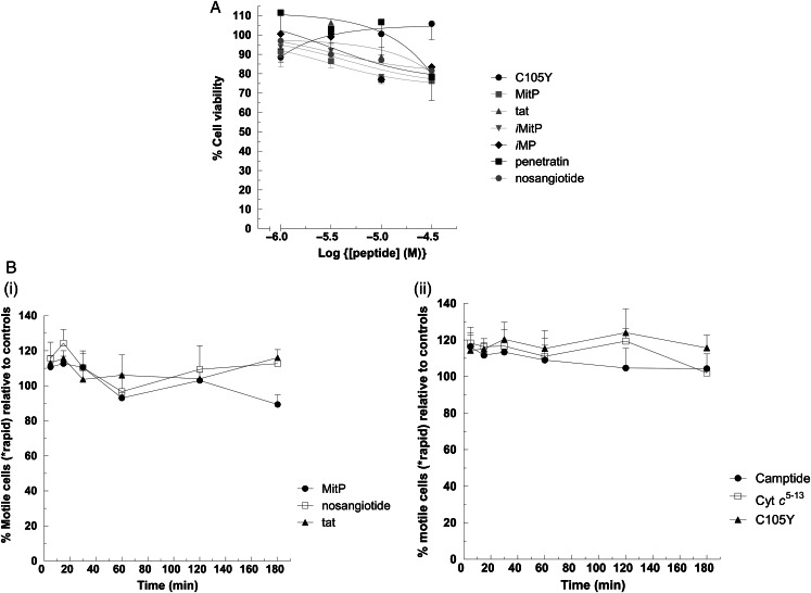
Main results: Chemically heterogeneous CPPs readily translocated into sperm to accumulate within discrete intracellular compartments. Mitoparan (INLKKLAKL(Aib)KKIL), for example, specifically accumulated within the mitochondria located in the sperm midpiece. The unique plasma membrane composition of sperm is a critical factor that directly influences the uptake efficacy of structurally diverse CPPs. No correlations in efficacies were observed when comparing CPP uptake into sperm with either uptake into fibroblasts or direct translocation across a phosphatidylcholine membrane. These comparative investigations identified C105Y (CSIPPEVKFNKPFVYLI) as a most efficient pharmacokinetic modifier for general applications in sperm biology. Significantly, CPP uptake induced no detrimental influence upon either bovine sperm viability or the motility of human sperm. As a consequence of the lack of endocytotic machinery, the CPP-mediated delivery of much larger protein complexes into sperm is relatively inefficient when compared with the similar process in fibroblasts.

Limitations, reasons for caution: It is possible that some CPPs could directly influence aspects of sperm biology and physiology that were not analysed in this study.

Wider implications of the findings: CPP technologies have significant potential to deliver selected bioactive moieties and so could modulate the biology and physiology of human sperm biology both prior- and post-fertilization.

Keywords: cell-penetrating peptide; cytotoxicity; membrane translocation; mitochondrion; motility; spermatozoa.


Figures

Figure 1
Figure 1
Endocytosis incompetent bovine spermatozoa. Swiss 3T3 cells and bovine spermatozoa were treated with fluorescently labelled markers of endocytosis and viewed by live confocal cell imaging analysis at 37°C. As a marker of clathrin-mediated endocytosis, Swiss 3T3 cells and bovine spermatozoa were treated with Transferrin Alexa Fluor®488 (50 μg/ml) and Transferrin Texas Red® (50 μg/ml), respectively. As a marker of macropinocytosis, both cell types were treated with Dextran Texas Red® (10 μM). To visualize lysosomal formation, Swiss 3T3 cells and bovine spermatozoa were treated with LysoTracker® Red DND-99 (75 nM). Though LysoTracker stains have been utilized at much higher concentrations (≥333 nM; Thomas et al., 1998) as acrosomal markers, a maximum application concentration of 75 nM, as recommended by the manufacturers, gives no discernible intracellular labelling of bovine spermatozoa. In contrast, application of 75 nM LysoTracker® Red DND-99 strongly labels lysosomal vesicles in Swiss 3T3 cells. To refine these results, both cell types were treated with two LysoSensorTM probes, LysoSensorTM Green DND-189 pKa 5.2 (1 μM) and LysoSensorTM Green DND-153 pKa 7.5 (1 μM), which exhibit a pH-dependent increase in fluorescence intensity. Most significantly and in accordance with an absence of endocytotic machinery, there is no evidence of vesicularized ultrastructures in LysoSensor-treated spermatozoa as is observed in Swiss 3T3 cells. Both LysoSensors do demonstrate diffuse labelling of the head regions of bovine spermatozoa and signal intensity is enhanced with LysoSensorTM Green DND-189 pKa 5.2. However, this acidotropic probe does not specifically label acrosomal regions (Castro-González et al., 2010). Fluorescent distributions of endocytotic markers (first and third columns) are also presented merged with images taken under differential interference contrast (second and fourth columns) so as to assist with the visualization of subcellular distribution. Scale bars: for Swiss 3T3 cells incubated with transferrin, dextran and LysoTracker® Red DND-99, bars = 10 μm. For Swiss 3T3 cells incubated with LysoSensor™ Green DND-189 and LysoSensor™ Green DND-153, bars = 5 μm. For spermatozoa incubated with transferrin and dextran, bars = 20 μm. For spermatozoa incubated with LysoTracker® Red DND-99, bars = 50 μm and for spermatozoa incubated with with LysoSensor™ Green DND-189 and LysoSensor™ Green DND-153, bars = 10 μm.
Figure 2
Figure 2
Intracellular accumulation and differential subcellular localization of CPPs in bovine spermatozoa. (A) Differential subcellular distributions of the CPPs Rho-penetratin, Rho-tat and Rho-C105Y. Isolated bovine spermatozoa were incubated for 1 h with 5 μM TAMRA-labelled peptides and viewed by live confocal cell imaging. Whilst Rho-penetratin demonstrates a more diffuse and generalized distribution (i), Rho-tat is confined to the spermatozoa head, with a predominant fluorescent labelling of the acrosomal region (ii). Rho-C105Y accumulates predominantly and strongly within the post-equatorial and equatorial subdomains of the head, the midpiece and posterior ring and neck (iii, iv) and panel (iv) clearly demonstrates an absence of rhodamine-labelled peptide within the interior head and acrosome. (B) Subcellular distributions of mitoparans and related analogues. Isolated bovine spermatozoa were treated for 45 min with both 5 μM TAMRA-labelled peptides and 500 nM MitoTracker® Deep Red 633 prior to live confocal cell imaging analysis (i, iv, v, vi, vii). Rho-MitP (i, ii) co-localizes strongly with the mitochondrial midpiece. Panel (ii) represents a bovine spermatozoon solely labelled with Rho-MitP and compares closely to a spermatozoon solely labelled with MitoTracker® Deep Red 633 (iii). Mitochondrial midpiece labelling by Rho-MitP is purposefully shown here in the absence of MitoTracker staining so as to eliminate any bleed-through artefacts. Rho-TP10 (iv, v) demonstrates a similar subcellular distribution to that of Rho-MitP, whereas the subcellular distributions of Rho-iMP and Rho-iMitP are more generalized (vi, vii). All treatments were performed at 37°C including live cell imaging analysis. (C) The TAMRA-labelled non-penetrant peptide, rV1aR102–113 was used as a negative control and bovine spermatozoa were treated as above. The confocal image (i) is also presented here merged with an image taken under differential interference contrast (DIC) so as to better visualize sperm morphology (ii). Scale bars: Panel A (i) and (iii) = 20 μm, (ii) and (iv) = 10 μm. Panel B (i) = 20 μm, (ii) and (iii) = 10 μm, (iv) = 100 μm, (v) = 10 μm, (vi) = 20 μm and (vii) = 20 μm. Panel C (i) and (ii) = 10 μm.
Figure 2
Figure 2
Continued
Figure 3
Figure 3
Quantitative analysis of peptide translocation. (A) Comparative analysis of CPP translocation efficacies into bovine spermatozoa. Bovine spermatozoa were incubated with TAMRA-labelled CPP (5 μM) for 1 h at 37°C. (B and C) Temporal-dependent intracellular uptake of C105Y into bovine spermatozoa and Swiss 3T3 cells. Bovine spermatozoa (B) and Swiss 3T3 fibroblasts (C) were incubated at 37°C with TAMRA-labelled peptides (5 μM) for the times indicated. Data are the mean ± s.e.m. of three experiments performed in triplicate and are expressed as mean fluorescence (minus background) ± s.e.m. from three experiments normalized so that the mean value for tat is equal to 1.0 (see the section Materials and methods).
Figure 4
Figure 4
Verification of CPPs accretion in intracellular structures of bovine spermatozoa. (A) Degradation of possible surface-associated CPP. To establish that detected fluorescence was not attributable to surface-associated CPP, isolated bovine spermatozoa were incubated for 1 h with 5 μM TAMRA-labelled CPP and subsequently treated with 1% (wt/vol) trypsin at 37°C prior to visualization using live confocal cell imaging. Intracellular accumulation of TAMRA-labelled CPPs, presented here, demonstrates no discernible difference compared with bovine spermatozoa treated with TAMRA-labelled CPP in the absence of trypsin. (B) To further establish that our fluorescently labelled CPPs were not merely surface associated we extended our investigations of polycationic CPP to include any observed co-localizations with established intracellular probes. Isolated bovine spermatozoa were incubated for 1 h with Rho-tat (5 μM) and LysoTracker®Green DND-26 (2 μM) and subsequently treated with 1% (wt/vol) trypsin at 37°C prior to visualization using live confocal cell imaging. The merged panel demonstrates a clear area of intracellular co-localization (yellow) between LysoTracker®Green DND-26 (2 μM) and Rho-tat (5 μM). (C) Demembranation of spermatozoa does not decrease CPP accretion in intracellular structures. Rho-C105Y (5 μM)-treated spermatozoa were treated with 1% (wt/vol) trypsin and subsequently incubated in the presence and absence of 0.2% Triton X-100 for 10 min at 37°C. Under both conditions, spermatozoa show a clear accumulation of Rho-C105Y in intracellular structures, whilst an enhancement in fluorescence intensity is evident in those spermatozoa treated with Triton-X100. Scale bars: Panel A, rho-tat = 5 μm and rho-penetratin = 10 μm. Panel B = 5 μm and Panel C = 20 μm.
Figure 5
Figure 5
CPP-mediated protein delivery into bovine spermatozoa. (A) Biotinyl-CPP constructs of tat and penetratin were synthesized and complexed with avidin Terxas Red (TXR) at a 3:1 molar ratio to assess the utility of CPP for the intracellular delivery of large protein cargoes such as avidin (see caption). Following 1 h incubation with bovine spermatozoa, live confocal cell imaging analysis demonstrated a distinct absence of CPP-mediated intracellular uptake of TXR-labelled avidin. Moreover, avidin TXR alone clearly demonstrated a degree of non-specific binding to equatorial and post-equatorial ridge subdomains of live spermatozoa. Corresponding lower panels are presented here merged with images taken under differential interference contrast (DIC) so as to assist with visualization of subcellular distribution. (B) A TAMRA-labelled biotinylated construct of TP10 (rho-biotinyl-TP10) was synthesized and complexed with avidin Alexa Fluor® 488 at a 3:1 molar ratio to yield a dual-labelled biosensor of both CPP and protein uptake (see caption). Swiss 3T3 (upper panel) and bovine spermatozoa (lower panel) were treated for 1 h with the dual-labelled biosensor and viewed by confocal live cell imaging. Both rho-biotinyl-TP10 and avidin Alexa Fluor® 488 assume intracellular vesicular distributions within Swiss 3T3 cells, including liberated cargo (green fluorescence), liberated TP10 (red fluorescence) and colocalized moieties (yellow). In contrast, a distinct absence of both rho-biotinyl-TP10 and avidin Alexa Fluor® 488 is evident in bovine spermatozoa. Scale bars: Panel A, for biotinyl penetratin + avidin TXR, bars = 10 μm, for biotinyl tat + avidin TXR, bars = 20 μm and for avidin TXR bars = 20 μm. Panel B, all bars = 10 μm.
Figure 6
Figure 6
(A) Correlation analysis between CPPs translocation efficacies in bovine spermatozoa (ordinate) and mammalian Swiss 3T3 cells (abscissa). (B) Correlation analysis between passive permeability across a phosphatidylcholine membrane (ordinate) and CPP translocation efficacy in bovine spermatozoa (abscissa).
Figure 7
Figure 7
CPP import is compatible with sperm viability and motility. (A) Isolated bovine spermatozoa were treated with CPP for 1 h at the concentrations indicated. Cell viability was measured by MTS conversion and expressed as a percentage of those spermatozoa treated with vehicle alone (sEBSS). Data points are means ± s.e.m. from three independent experiments performed on each of two sperm samples (n = 6). (B) Motility data were collected from human sperm cells treated with six different CPPs. Each peptide was tested on samples from three individual donors. Data are shown as % rapid cells from treated samples relative to that of controls at six time points over 3 h (5, 15, 30, 60, 120 and 180 min) and expressed as the mean ± s.e.m. MitP, nosangiotide and tat peptides were tested in non-capacitating buffer (i) and camptide, Cyt c5–13 and C105Y were tested in capacitating media (ii). *rapid cells = velocity (average path) ≥25 µms−1 and straightness ≥80%.

Similar articles

Cited by

References

    1. Abou-haila A, Tulsiani DRP. Signal transduction pathways that regulate sperm capacitation and the acrosome reaction. Arch Biochem Biophys. 2009;485:72–81. - PubMed
    1. Bedu-Addo K, Lefièvre L, Mosely FLC, Barratt CLR, Publicover SJ. Bicarbonate and bovine serum albumin reversibly ‘switch’ capacitation-induced events in human spermatozoa. Mol Hum Reprod. 2005;11:683–691. - PubMed
    1. Boerke A, Dielman SJ, Gadella BM. A possible role for sperm RNA in early embryo development. Theriogenology. 2007;68S:S147–S155. - PubMed
    1. Castro-González D, Alvarez M, Muro J, Esteso MC, de Paz P, Anel L, Martínez-Pastor F. The acidic probe LysoSensor is not useful for acrosome evaluation of cryopreserved ram spermatozoa. Reprod Domest Anim. 2010;45:363–367. - PubMed
    1. Costello S, Michelangeli F, Nash K, Lefievre L, Morris J, Machado-Oliveira G, Barratt C, Kirman-Brown J, Publicover S. Ca2+-stores in sperm: their identities and functions. Reproduction. 2009;138:425–437. - PMC - PubMed

Publication types

Substances