. 2013 Jun;162(2):567-80.
doi: 10.1104/pp.112.210450. Epub 2013 Apr 12.

Arabidopsis 3-ketoacyl-coenzyme a synthase9 is involved in the synthesis of tetracosanoic acids as precursors of cuticular waxes, suberins, sphingolipids, and phospholipids

Affiliations

Arabidopsis 3-ketoacyl-coenzyme a synthase9 is involved in the synthesis of tetracosanoic acids as precursors of cuticular waxes, suberins, sphingolipids, and phospholipids

Juyoung Kim et al. Plant Physiol. 2013 Jun.

Abstract

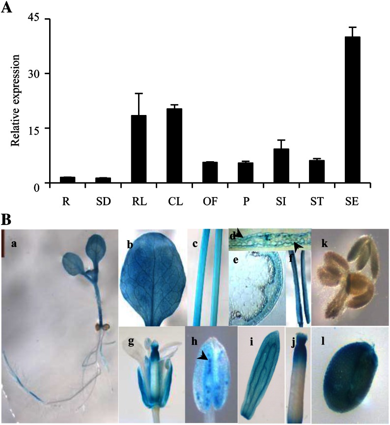
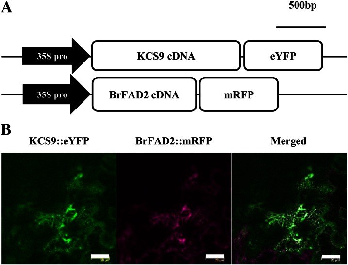
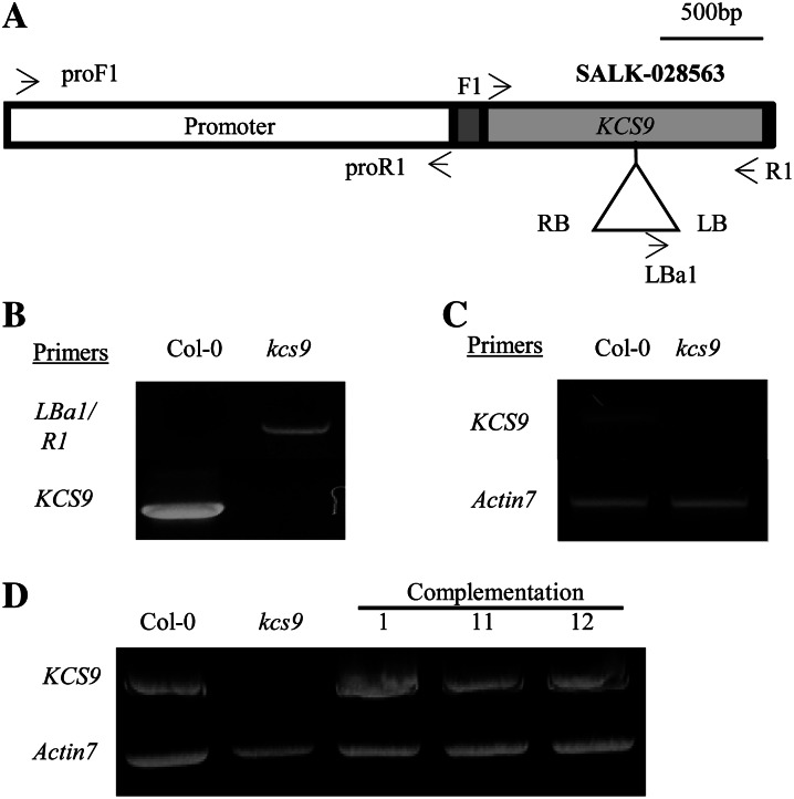
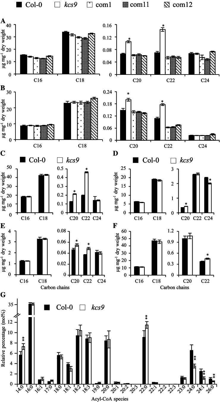
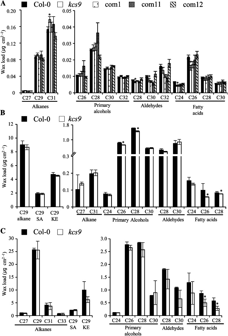
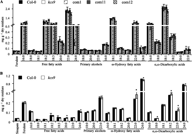
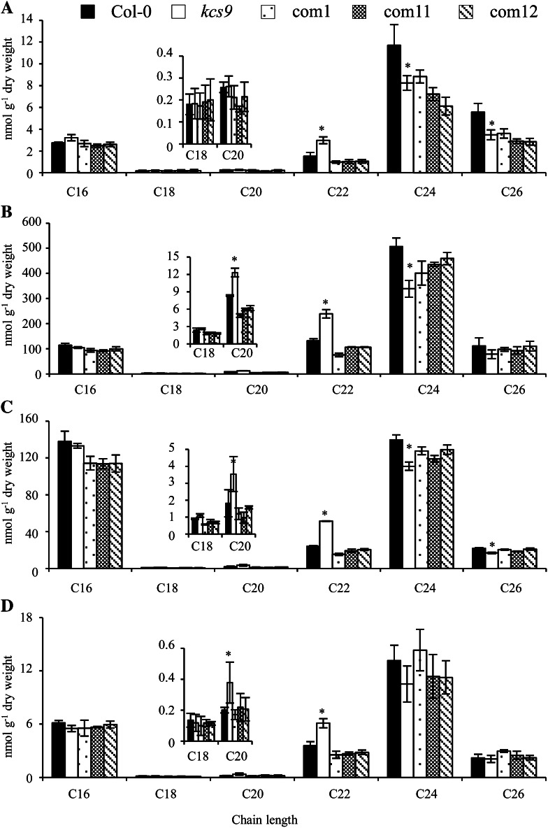
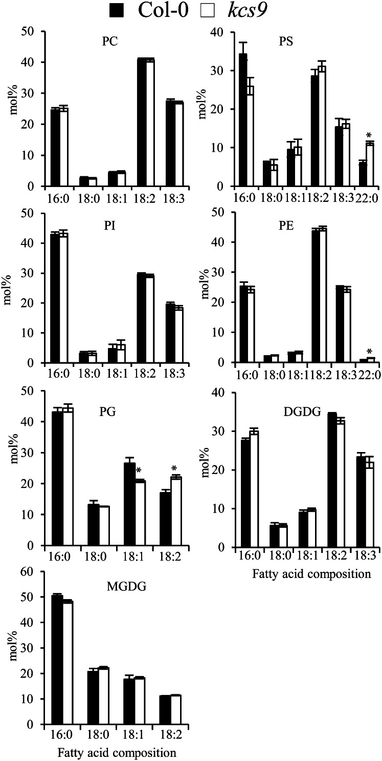
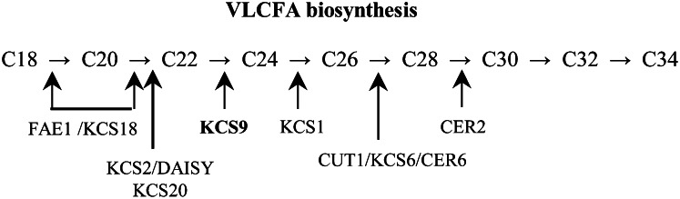
Very-long-chain fatty acids (VLCFAs) with chain lengths from 20 to 34 carbons are involved in diverse biological functions such as membrane constituents, a surface barrier, and seed storage compounds. The first step in VLCFA biosynthesis is the condensation of two carbons to an acyl-coenzyme A, which is catalyzed by 3-ketoacyl-coenzyme A synthase (KCS). In this study, amino acid sequence homology and the messenger RNA expression patterns of 21 Arabidopsis (Arabidopsis thaliana) KCSs were compared. The in planta role of the KCS9 gene, showing higher expression in stem epidermal peels than in stems, was further investigated. The KCS9 gene was ubiquitously expressed in various organs and tissues, including roots, leaves, and stems, including epidermis, silique walls, sepals, the upper portion of the styles, and seed coats, but not in developing embryos. The fluorescent signals of the KCS9::enhanced yellow fluorescent protein construct were merged with those of BrFAD2::monomeric red fluorescent protein, which is an endoplasmic reticulum marker in tobacco (Nicotiana benthamiana) epidermal cells. The kcs9 knockout mutants exhibited a significant reduction in C24 VLCFAs but an accumulation of C20 and C22 VLCFAs in the analysis of membrane and surface lipids. The mutant phenotypes were rescued by the expression of KCS9 under the control of the cauliflower mosaic virus 35S promoter. Taken together, these data demonstrate that KCS9 is involved in the elongation of C22 to C24 fatty acids, which are essential precursors for the biosynthesis of cuticular waxes, aliphatic suberins, and membrane lipids, including sphingolipids and phospholipids. Finally, possible roles of unidentified KCSs are discussed by combining genetic study results and gene expression data from multiple Arabidopsis KCSs.


Figures

Figure 1.
Figure 1.
A, qRT-PCR analysis of KCS9 in various Arabidopsis organs including stem epidermis. Total RNAs were isolated from 10-d-old seedlings and various organs of 6-week-old Arabidopsis and subjected to qRT-PCR analysis. EIF4-a was used as an internal standard to verify RNA quantity and quality (Gutierrez et al., 2008). R, Roots; SD, seedlings; RL, rosette leaves; CL, cauline leaves; OF, open flowers; P, pollen grains; SI, siliques harvested at 7 d after flowering; ST, stems; SE, stem epidermis. B, GUS expression under the control of the KCS9 promoter in transgenic Arabidopsis. Panels are as follows: a, young seedling; b, mature leaf; c, stem; d, cross section of leaf; e, cross section of stem; f, silique walls; g, flower; h, anther (arrowhead indicates pollen grain); i, petal; j, style; k, developing embryos; l, developing seed.
Figure 2.
Figure 2.
Subcellular localization of Arabidopsis KCS9 in tobacco epidermis. A, Schematic diagrams of pKCS9::eYFP and pBrFAD2::mRFP constructs. 35S pro, Promoter of cauliflower mosaic virus 35S RNA. B, Fluorescent signals of KCS9::eYFP and BrFAD2::mRFP in tobacco epidermal cells. Genes encoding fluorescent proteins were translationally fused to KCS9 and BrFAD2 under the control of the CaMV 35S promoter. The constructed vectors were coinfiltrated into tobacco epidermis via A. tumefaciens-mediated transformation, and the fluorescent signals were observed using a laser confocal scanning microscope 48 h after infiltration. BrFAD2::mRFP was used as an ER marker (Jung et al., 2011). Bars = 10 µm.
Figure 3.
Figure 3.
Isolation of T-DNA-inserted kcs9 mutant (A–C) and generation of complementation lines of the kcs9 mutant (D). A, Genomic organization of the T-DNA-tagged KCS9 gene. T-DNA-inserted kcs9 seeds were obtained from SALK (SALK 028563). The promoter region, 5′ and 3′ untranslated regions, and coding region of the KCS9 gene are shown in the white box, gray boxes, and black box, respectively. LB, Left border; RB, right border. B, Genomic DNA was isolated from the wild type (Col-0) and the kcs9 mutant, and T-DNA insertion was confirmed by genomic DNA PCR using the LBa1 and R1 primers shown in Supplemental Table S1. C, The levels of KCS9 transcripts in 10-d-old seedlings of the wild type (Col-0) and the kcs9 mutant were analyzed by RT-PCR using the F1 and R1 primers shown in Supplemental Table S1. Actin7 was used to determine the quantity and quality of the cDNAs. D, The binary vector harboring KCS9::eYFP under the control of the CaMV 35S promoter was transformed into Arabidopsis for complementation of the kcs9 mutant. The transgenic seedlings were selected on 1/2 MS medium with kanamycin, and leaves of 3-week-old plants were used for RNA isolation to analyze the expression of KCS9 by RT-PCR analysis.
Figure 4.
Figure 4.
Fatty acid analysis (A–F) and fatty acyl-CoA profiling (G). A to F, Fatty acids were extracted from lyophilized leaves (A), stems (B), aerial parts (C) and roots (D) of young seedlings, flowers (E), and silique walls (F) of the wild type (Col-0), kcs9, and complementation lines (com1, com11, and com12) and analyzed using GC. G, Fatty acyl-CoA profiling was performed on lyophilized roots of wild-type and kcs9 mutant plants. The x axis represents the carbon chain length of the fatty acids. Values shown are means of four experiments ± sd. Asterisks denote statistical differences with respect to the wild type: *P < 0.05, **P < 0.01.
Figure 5.
Figure 5.
Cuticular wax composition and amount in leaves (A), stems (B), and silique walls (C) of the wild type (Col-0), kcs9, and complementation lines (com1, com11, and com12). Cuticular waxes were extracted from 5-week-old plants with chloroform and analyzed using GC. The x axis represents the carbon chain length of VLCFAs and their derivatives. Values shown are means of four experiments ± sd. Asterisks indicate statistical differences with respect to the wild type: *P < 0.05. KE, Ketone; SA, secondary alcohol.
Figure 6.
Figure 6.
Aliphatic suberin composition and amount in roots (A) and seed coats (B) of the wild type (Col-0), kcs9, and complementation lines (com1, com11, and com12). Two-week-old roots and seed coats were lyophilized, delipidated, and hydrolyzed, and then lipid-soluble extracts were analyzed using GC and GC-mass spectrometry. Values shown are means of three experiments ± sd. Asterisks indicate statistical differences with respect to the wild type: *P < 0.05.
Figure 7.
Figure 7.
Sphingolipid composition and amount in young seedling of the wild type, kcs9, and complementation lines (com1, com11, and com12). A, Ceramide. B, Glycosyl inositolphosphoceramide. C, Glucosylceramide. D, Hydroxyceramide. The amounts of all components with equal carbon chain lengths in each class of sphingolipids were combined. Values shown are means of three experiments ± sd. Asterisks indicate statistical differences with respect to the wild type: *P < 0.05.
Figure 8.
Figure 8.
Glycerolipid analysis in roots of the wild type and the kcs9 mutant. Glycerolipids were extracted from 2-week-old roots and separated by thin-layer chromatography. Fatty acid composition from individual lipids was analyzed by GC. Values shown are means of three experiments ± sd. Asterisks indicate statistical differences with respect to the wild type: *P < 0.05. DGDG, Digalactosyl diacylglycerol; MGDG, monogalactosyl diacylglycerol; PC, phosphatidylcholine; PG, phosphatidylglycerol; PI, phosphatidylinositol.
Figure 9.
Figure 9.
Substrate specificity of KCSs and CER2, which are involved in the production of VLCFAs in Arabidopsis. Numbers correspond to the number of carbon chains of VLCFAs. The elongation steps that are catalyzed by KCSs and CER2 are indicated by arrows.

References

    1. Aarts MG, Hodge R, Kalantidis K, Florack D, Wilson ZA, Mulligan BJ, Stiekema WJ, Scott R, Pereira A. (1997) The Arabidopsis MALE STERILITY 2 protein shares similarity with reductases in elongation/condensation complexes. Plant J 12: 615–623 - PubMed
    1. An G. (1987) Binary Ti vectors for plant transformation and promoter analysis. Methods Enzymol 153: 292–305
    1. Bach L, Michaelson LV, Haslam R, Bellec Y, Gissot L, Marion J, Da Costa M, Boutin JP, Miquel M, Tellier F, et al. (2008) The very-long-chain hydroxy fatty acyl-CoA dehydratase PASTICCINO2 is essential and limiting for plant development. Proc Natl Acad Sci USA 105: 14727–14731 - PMC - PubMed
    1. Barthlott W, Neinhuis C. (1997) Characterization and distribution of water-repellent, self-cleaning plant surfaces. Ann Bot (Lond) 79: 667–677
    1. Beaudoin F, Wu X, Li F, Haslam RP, Markham JE, Zheng H, Napier JA, Kunst L. (2009) Functional characterization of the Arabidopsis β-ketoacyl-coenzyme A reductase candidates of the fatty acid elongase. Plant Physiol 150: 1174–1191 - PMC - PubMed

Publication types

MeSH terms