. 2013 May 15;190(10):5216-25.
doi: 10.4049/jimmunol.1300097. Epub 2013 Apr 12.

Mouse, but not human STING, binds and signals in response to the vascular disrupting agent 5,6-dimethylxanthenone-4-acetic acid

Affiliations

Mouse, but not human STING, binds and signals in response to the vascular disrupting agent 5,6-dimethylxanthenone-4-acetic acid

Joseph Conlon et al. J Immunol. .

Abstract

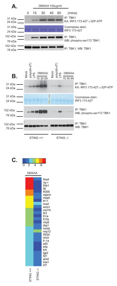
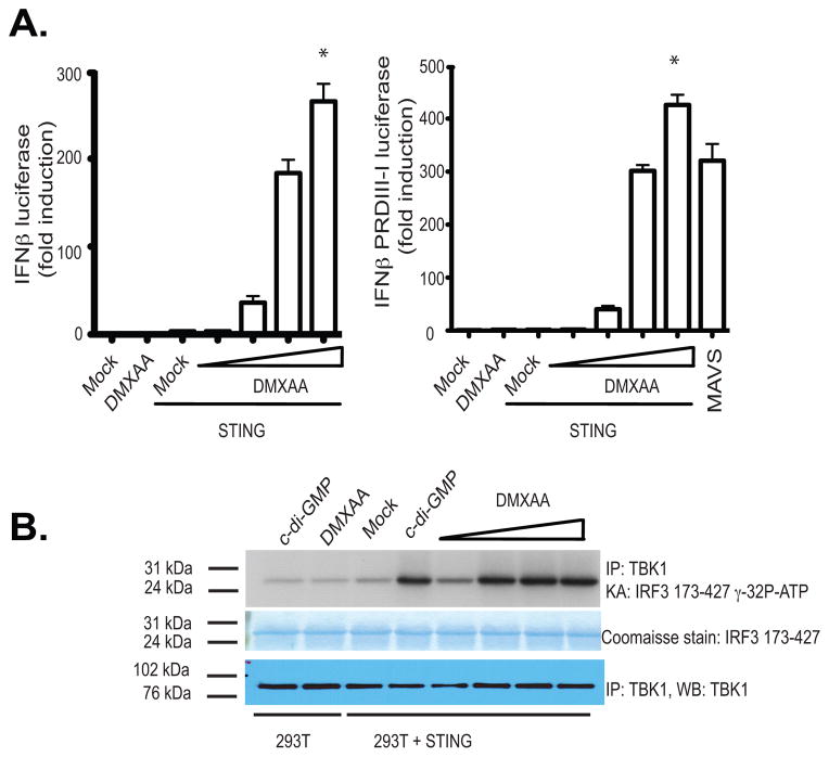
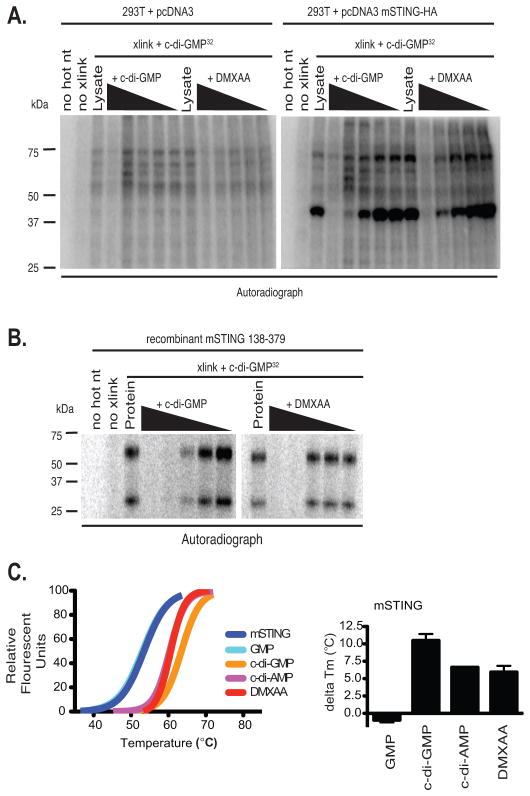
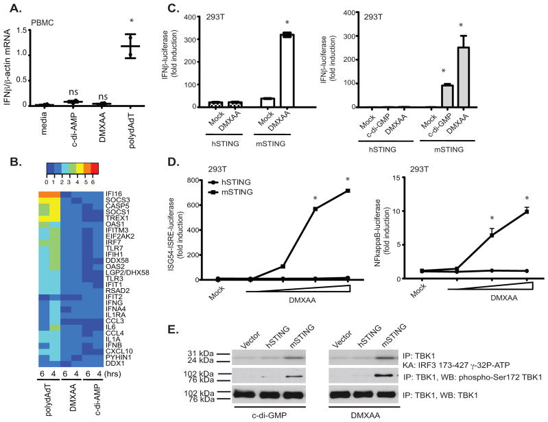
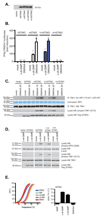
Vascular disrupting agents such as 5,6-dimethylxanthenone-4-acetic acid (DMXAA) represent a novel approach for cancer treatment. DMXAA has potent antitumor activity in mice and, despite significant preclinical promise, failed human clinical trials. The antitumor activity of DMXAA has been linked to its ability to induce type I IFNs in macrophages, although the molecular mechanisms involved are poorly understood. In this study, we identify stimulator of IFN gene (STING) as a direct receptor for DMXAA leading to TANK-binding kinase 1 and IFN regulatory factor 3 signaling. Remarkably, the ability to sense DMXAA was restricted to murine STING. Human STING failed to bind to or signal in response to DMXAA. Human STING also failed to signal in response to cyclic dinucleotides, conserved bacterial second messengers known to bind and activate murine STING signaling. Collectively, these findings detail an unexpected species-specific role for STING as a receptor for an anticancer drug and uncover important insights that may explain the failure of DMXAA in clinical trials for human cancer.


Figures

Figure 1
Figure 1. DMXAA signals via STING in macrophages
(A) Wild-type immortalized bone marrow-derived macrophages were stimulated with DMXAA (100μg/ml) at the indicated time points. Whole cell lysates were prepared and endogenous TBK1 immunoprecipitated (IP) and analyzed by in vitro kinase assay and western blotted (WB) for phospho-TBK1 (Ser172) and total TBK1. (B) Wild-type or STING−/− bone marrow-derived macrophages were stimulated with poly(dA-dT) (3μg/ml), LPS [10ng/ml] or DMXAA (75μg/ml) as indicated and analyzed as in A. (C) BMDM from wild-type and STING−/− mice were stimulated with DMXAA (75μg/ml) for 4 hours and mRNA levels for a selection of innate immune genes were analyzed by Nanostring analysis. Heatmaps representing differentially regulated genes are presented and scaled by log2(X-min(X) + 1). Data are presented from one experiment which is representative of three experiments (A, B) or two experiments (C).
Figure 2
Figure 2. Reconstitution of DMXAA induced IFNβ signaling in STING expressing HEK293 cells
(A) 293T cells were transfected with either empty vector or mSTING in the presence of an IFN-β luciferase reporter gene (left panel) or a multimerized PRDIII-I reporter gene (right panel) and transfected cells stimulated with DMXAA from 10–100 μg/ml for 18hours and luciferase activity was measured. Data are presented as the mean ± s.e.m of one experiment representative of three experiments. * indicates p <0.05 for the comparison of the highest dose of DMXAA relative to vector control. (B) 293T cells transfected as above were stimulated with cyclic-di-GMP (10μg/ml) or with increasing amounts of DMXAA as in A and endogenous TBK1 activity was analyzed by in vitro kinase assay.
Figure 3
Figure 3. STING binds to DMXAA and is a direct sensor for DMXAA
(A) 293T cells were transfected with empty vector or mSTING and cell lysates were UV-crosslinked to c-di-GMP in the presence of cold competing c-di-GMP or DMXAA in 10-fold serial dilutions. (B) One μg His6-mSTING 138–378 was analyzed as described in C. (C) Left panel, thermal shift analysis of mouse STING in the presence of GMP, c-di-GMP, c-di-AMP, and DMXAA. The melting temperature shifts in the presence of the ligands are plotted in the graph to the right of the melting curves. Data are representative of two independent experiments (A–C).
Figure 4
Figure 4. Mouse, but not human, STING signals in response to DMXAA and c-di nucleotides
(A)PBMC from two volunteers were stimulated with DMXAA (10μg/ml), c-di-AMP (5μM) or poly (dA-dT) (4μg/ml) for 6 hours and IFN-β mRNA levels measured by quantitative PCR. * indicates p <0.05 for the comparison of each ligand relative to medium control. (B) RNA isolated from PBMCs stimulated as in A for 4 and 6 hours was subject to Nanostring analysis to monitor expression of 30 innate immune genes. (C–D) 293T cells were transfected with empty vector, pEF-BOS hSTING-Flag-His, or pEF-BOS mSTING-Flag-His, followed by stimulation with either DMXAA or c-di-GMP or with increasing concentrations of DMXAA (D). IFN-β luciferase (C) ISG54-ISRE luciferase, and NF-κB luciferase (D) activities were measured. * indicates p <0.05 for the comparison of DMXAA or c-d-GMP relative to vector control. (E) 293T cells were transfected as above and stimulated with DMXAA and cyclic-di-GMP. Endogenous TBK1 was immunoprecipitated and analyzed as in Figure 1. Data are presented as the mean ± s.e.m of one experiment representative of three experiments (A). (B–E) data is representative of three separate experiments.
Figure 5
Figure 5. DMXAA-induced signaling is dependent on the mouse STING C-terminal domain
293T cells were transfected with human, mouse, or the indicated chimeric STING molecules as well as the IFNβ PRDIII-I luciferase reporter gene. Transfected cells were (A) subjected to immunoblotting with anti-Flag antibody or (B) stimulated with DMXAA or cyclic-di-GMP as indicated and analyzed for IFN-β luciferase reporter gene activation. hu-STING, hu-moSTING, and mo-huSTING were compared to moSTING for each treatment and the statistical significance (p <0.05) was indicated by *. ns=non-significant. (C–D) Cells transfected as above without reporter genes were treated as indicated and analyzed by immunoblotting for phospho-TBK1 (Ser172), phospho-IRF3 (Ser396), total TBK1, β-actin, and Flag (to detect expression of recombinant STING) as indicated. (E) Left panel, thermal shift analysis of human STING CTD in the presence of GMP, c-di-GMP, c-di-AMP, and DMXAA. The melting temperature shifts in the presence of the ligands are plotted in the right panel.

References

    1. Rakoff-Nahoum S, Medzhitov R. Role of toll-like receptors in tissue repair and tumorigenesis. Biochemistry Biokhimiia. 2008;73:555–561. - PubMed
    1. Rakoff-Nahoum S, Medzhitov R. Toll-like receptors and cancer. Nature reviews Cancer. 2009;9:57–63. - PubMed
    1. Baguley BC, Ching LM. Immunomodulatory actions of xanthenone anticancer agents. BioDrugs: clinical immunotherapeutics, biopharmaceuticals and gene therapy. 1997;8:119–127. - PubMed
    1. Wallace A, LaRosa DF, Kapoor V, Sun J, Cheng G, Jassar A, Blouin A, Ching LM, Albelda SM. The vascular disrupting agent, DMXAA, directly activates dendritic cells through a MyD88-independent mechanism and generates antitumor cytotoxic T lymphocytes. Cancer research. 2007;67:7011–7019. - PubMed
    1. Jassar AS, Suzuki E, Kapoor V, Sun J, Silverberg MB, Cheung L, Burdick MD, Strieter RM, Ching LM, Kaiser LR, Albelda SM. Activation of tumor-associated macrophages by the vascular disrupting agent 5,6-dimethylxanthenone-4-acetic acid induces an effective CD8+ T-cell-mediated antitumor immune response in murine models of lung cancer and mesothelioma. Cancer research. 2005;65:11752–11761. - PubMed

Publication types

MeSH terms