. 2013 Apr 9;8(4):e60032.
doi: 10.1371/journal.pone.0060032. Print 2013.

Mutation of the fiber shaft heparan sulphate binding site of a 5/3 chimeric adenovirus reduces liver tropism

Affiliations

Mutation of the fiber shaft heparan sulphate binding site of a 5/3 chimeric adenovirus reduces liver tropism

Anniina Koski et al. PLoS One. .

Abstract

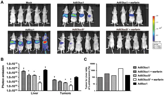
Natural tropism to the liver is a major obstacle in systemic delivery of adenoviruses in cancer gene therapy. Adenovirus binding to soluble coagulation factors and to cellular heparan sulphate proteoglycans via the fiber shaft KKTK domain are suggested to cause liver tropism. Serotype 5 adenovirus constructs with mutated KKTK regions exhibit liver detargeting, but they also transduce tumors less efficiently, possibly due to altered fiber conformation. We constructed Ad5/3lucS*, a 5/3 chimeric adenovirus with a mutated KKTK region. The fiber knob swap was hypothesized to facilitate tumor transduction. This construct was studied with or without additional coagulation factor ablation. Ad5/3lucS* exhibited significantly reduced transduction of human hepatic cells in vitro and mouse livers in vivo. Combination of coagulation factor ablation by warfarinization to Ad5/3lucS* seemed to further enhance liver detargeting. Cancer cell transduction by Ad5/3lucS* was retained in vitro. In vivo, viral particle accumulation in M4A4-LM3 xenograft tumors was comparable to controls, but Ad5/3lucS* transgene expression was nearly abolished. Coagulation factor ablation did not affect tumor transduction. These studies set the stage for further investigations into the effects of the KKTK mutation and coagulation factor ablation in the context of 5/3 serotype chimerism. Of note, the putative disconnect between tumor transduction and transgene expression could prove useful in further understanding of adenovirus biology.


Conflict of interest statement

Competing Interests: A.H. is shareholder and consultant to Oncos Therapeutics, Ltd. This does not alter the authors’ adherence to all the PLOS ONE policies on sharing data and materials.

Figures

Figure 1
Figure 1. Cancer cell transduction in comparison to Ad5 is retained despite KKTK motif mutation of the Ad5/3 shaft.
To study the cell transduction properties of the mutated virus A) M4A4-LM3 human breast ductal carcinoma cells were infected with indicated viruses at 40, 200, and 1000 viral particles (VP) per cell. B) Hey ovarian adenocarcinoma, PC-3 prostate cancer, SKOV3.ip1 ovarian adenocarcinoma cells were infected with 200 VP/cell. Unbound virus was removed after 1 h incubation and luciferase activity was measured from cell lysates after 24 hours of incubation. C) SKOV3.ip1 cells were preincubated with indicated concentrations of free recombinant knob 5 or knob 3 proteins and thereafter infected with 1000 VP/cell of Ad5/3luc1 or Ad5/3lucS* alone or virus preincubated with indicated concentrations of heparin. Luciferase transgene activity was quantified with a luminometer and expressed as relative light units (RLU) per ml of cell lysate. Assays were performed in triplicates, expressed as mean+SD. A–B) *** p<0.001 against Ad5/3luc1, p<0.01 Ad5luc1 vs. Ad5/3lucS*.
Figure 2
Figure 2. KKTK mutated 5/3 virus is detargeted from human hepatocytes but coagulation factors result in enhanced transduction.
A) Monolayers of HepG2 cells were infected with increasing doses of Ad5/3luc1, Ad5/3lucS* or Ad5luc1 and luciferase expression was quantified 24 hours later. * p<0.05, *** p<0.001 vs. Ad5/3luc1, p<0.05 Ad5luc1 vs. Ad5/3lucS*. B–D) HepG2 cells were infected with either virus only or virus preincubated with physiological concentrations of factor X (FX) or factor IX (FIX) and luciferase activity was measured 24h later. * p<0.05, ** p<0.01, *** p<0.001 vs. virus only, p<0.05 vs. virus+FIX. Assays were performed in triplicates, expressed as mean+SD.
Figure 3
Figure 3. Warfarinization enhances liver detargeting of Ad5/3lucS*.
Nude NMRI mice carrying M4A4 -LM3 mammary fat pad tumors were treated with 4×10¹⁰ VP of Ad5/3luc1, Ad5/3lucS* or Ad5luc1 or NaCl only to the tail vein. Indicated groups had been pretreated with warfarin to deplete vitamin K-dependent coagulation factors. A) 24 hours later mice were imaged for transgene activity by injecting D-luciferin i.p. and IVIS luminosity imaging. B) Regions of interest were drawn around liver and tumor areas to quantify photon emission signals and mock was subtracted. C) Ratio of luciferase expression quantified from tumors and livers was calculated. KKTK mutation reduces liver transduction to similar degree as warfarin. Both also improve the ratio of tumor to liver transduction, best result with Ad5/3lucS*+warfarin. Mean+SD, n = 4–5 mice per group. * p<0.05 vs. Ad5/3luc1, p<0.05 vs. Ad5/3lucS*.
Figure 4
Figure 4. Warfarinization and KKTK mutation both result in reduced liver gene expression.
Nude NMRI mice carrying M4A4 -LM3 mammary fat pad tumors were treated with 4×10¹⁰ VP of Ad5/3luc1, Ad5/3lucS* or Ad5luc1 or NaCl only to the tail vein. Indicated groups had been pretreated with warfarin to deplete vitamin K-dependent coagulation factors. A) 48 hours later mice were imaged for transgene activity by injecting D-luciferin i.p. and IVIS luminosity imaging. B) Regions of interest were drawn around liver and tumor areas to quantify photon emission signals and mock was subtracted. C) Ratio of luciferase expression quantified from tumors and livers was calculated. KKTK mutation and warfarinization reduce liver transduction and improve the ratio of tumor to liver transduction, best result with warfarinization. p<0.05 vs. Ad5/3lucS*.
Figure 5
Figure 5. Hepatic luciferase expression is reduced with both KKTK mutation and warfarinization.
A–F) Nude NMRI mice carrying mammary fat pad tumors were treated intravenously with 4×10¹⁰ VP of Ad5/3luc1, Ad5/3lucS* or Ad5luc1. Some groups were pretreated with warfarin to deplete coagulation factors. 48 hours after virus injection mice were sacrificed and luciferase expression in homogenized tissue lysates was quantified and normalized for total protein content of lysates. KKTK mutation reduced hepatic transgene expression, but reduction was also seen in tumors and other tissues. Mean+SD, n = 3–5 mice per group. *p<0.05, **p<0.01, ***p<0.001 vs. Ad5/3luc1.
Figure 6
Figure 6. Distribution of viral particles is not affected by KKTK mutation at lower viral dose or warfarinization.
Mice carrying xenograft mammary fat pad tumors were injected with 4×10¹⁰ VP of Ad5/3luc1, Ad5/3lucS* or Ad5luc1 into the tail vein. Indicated groups had been pretreated with warfarin. Mice were sacrificed A) 30 minutes or B) 3 hours after virus injection and organs, tumors and whole blood was collected. Viral loads in the samples were quantified by adenoviral E4 region qPCR. Mouse β-actin for organs and blood and human β-actin for tumor tissue was used to normalize viral titers to genomic DNA. At 30 minutes there was less liver uptake with Ad5/3luS*+warfarin, this difference disappeared by 3 hours. C) At a higher dose of 5×10¹⁰ VP (analysis at 30min), there was a significant decrease in liver uptake of Ad5/3lucS* virus with or without warfarin compared to Ad5/3luc1, and a trend for higher tumor uptake in the Ad5/3lucS* virus with warfarin group. Mean+SD, n = 3 mice per group. *p<0.05vs.Ad5/3luc1.
Figure 7
Figure 7. Subtle differences are observed in viral antigen expression pattern and DNA copy numbers in the liver.
Mammary fat pad tumor carrying mice were injected intravenously with 4×10¹⁰ VP of the indicated viruses or NaCl only. The indicated groups had been pretreated with warfarin to deplete coagulation factors. Immunohistology for adenovirus hexon antigen expression in the liver A) 30 min after virus administration. Viral antigen (stains brown) is observed within Kupffer cells (arrowheads) and in individual hepatocytes (black arrows) and occasional leukocytes in sinus (white arrow). Inset: Viral antigen can also be seen in neutrophils in the circulating blood in a central vein (arrowheads) and in an adjacent sinus (arrow). Note the generally weak antigen expression and its restriction to Kupffer cells (arrowheads) and some random hepatocytes (arrows) in an Ad5/3lucS*+warfarin treated mouse. Bars = 20 µm for Ad5/3luc1 and Ad5/3lucS* and Ad5/3lucS*+warfarin, Bars = 10 µm for inset and Ad5/3luc1+ warfarin. B)3h after virus administration. Viral antigen expression in Kupffer cells (arrowheads), hepatocytes (arrows), some leukocytes in sinuses (white arrows) and occasional neutrophils within portal veins (white arrowheads). PV = portal vein. Inset: Portal vein. Viral antigen expression in hepatocyte (H), neutrophil (arrow) and endothelial cell (arrowhead). Note the strong antigen expression in an Ad5/3luc1 treated mouse in hepatocytes surrounding portal areas and the weak antigen expression with restriction to Kupffer cells in an Ad5/3lucS*+warfarin treated mouse. Bar = 50 µm for Ad5/3luc1, Bars = 20 µm for Ad5/3luc1+warfarin, Ad5/3lucS* and Ad5/3lucS*+warfarin, Bar = 10 µm for inset. C) Semi-quantitative assessment of virus antigen expression in hepatocytes, n = 2–4 mice/group. D-E) Mice were sacrificed 3 h after virus administration and cells of the liver were freshly isolated and separated into liver parenchymal cells (PC; hepatocytes) and non-parenchymal cells (NPC; include Kupffer cells and endothelial cells). D) DNA was extracted and the amount of virus particles determined by quantitative PCR with primers and probes targeting the adenoviral E4 region. Primers and probes for mouse β-actin were used to normalize viral titers to sample genomic DNA. E) The ratio of virus present in non-parenchymal versus parenchymal cells was calculated. D-E) Mean+SD, n = 3–4 mice per group.
Figure 8
Figure 8. Blood cell counts and liver enzymes after intravenous virus administration.
Mammary fat pad tumor carrying mice were injected intravenously with 4×10¹⁰ VP of the indicated viruses or NaCl only for mock, through the tail vein. Indicated groups had been pretreated with warfarin to deplete coagulation factors. Blood samples were collected 48 hours after injection. A) White blood cell (WBC) and red blood cell (RBC) and B) platelet counts were measured. C) Aspartate amino transferase (AST) and alanine amino transferase (AST) levels determined from plasma samples. D) Serum samples were collected 6 hours after treatment and interleukin (IL)-6, MCP-1 and TNF-alpha levels determined by FACSArray. Mean+SD, n = 2–5. Neither warfarinization nor KKTK mutation affected circulatory amounts of blood cells and the given virus treatments did not provoke liver enzyme elevations, but KKTK mutation resulted in milder cytokine responses. Mean+SD, n = 2–5 mice per group, * p<0.05, **p<0.01 vs. Ad5/3luc1, p<0.05 vs. mock.

References

    1. Fukuhara H, Homma Y, Todo T (2009) Oncolytic virus therapy for prostate cancer. Int J Urol 17: 20–30. - PubMed
    1. Liang M (2011) Clinical Development of Oncolytic viruses in China. Curr Pharm Biotechnol. - PubMed
    1. Toth K, Wold WS (2010) Increasing the efficacy of oncolytic adenovirus vectors. Viruses 2: 1844–1866. - PMC - PubMed
    1. Nemunaitis J, Senzer N, Sarmiento S, Zhang YA, Arzaga R, et al. (2007) A phase I trial of intravenous infusion of ONYX-015 and enbrel in solid tumor patients. Cancer Gene Ther 14: 885–893. - PubMed
    1. Small EJ, Carducci MA, Burke JM, Rodriguez R, Fong L, et al. (2006) A phase I trial of intravenous CG7870, a replication-selective, prostate-specific antigen-targeted oncolytic adenovirus, for the treatment of hormone-refractory, metastatic prostate cancer. Mol Ther 14: 107–117. - PubMed

Publication types

MeSH terms