. 2013 Apr 9;8(4):e61064.
doi: 10.1371/journal.pone.0061064. Print 2013.

Oncomir miR-125b suppresses p14(ARF) to modulate p53-dependent and p53-independent apoptosis in prostate cancer

Affiliations

Oncomir miR-125b suppresses p14(ARF) to modulate p53-dependent and p53-independent apoptosis in prostate cancer

Sumaira Amir et al. PLoS One. .

Abstract

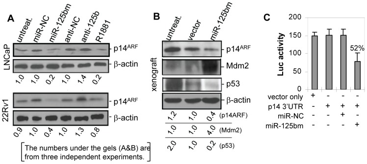
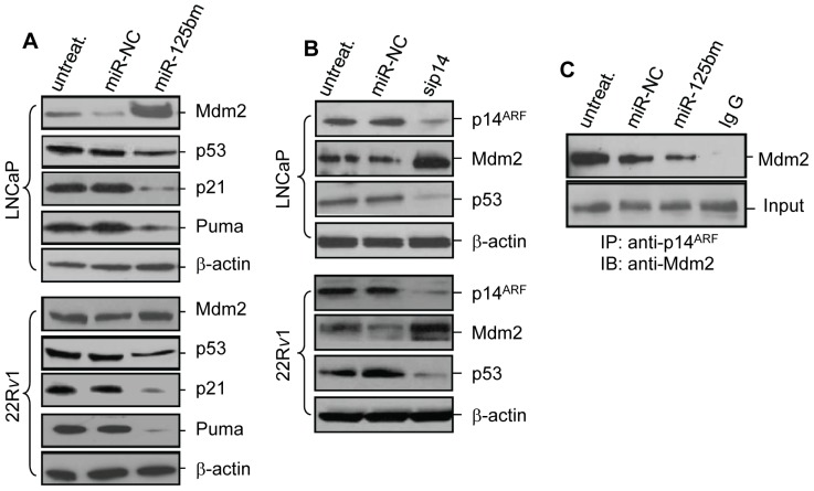
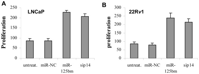
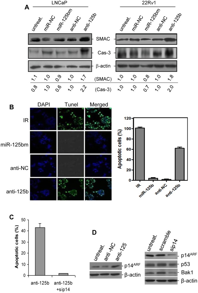
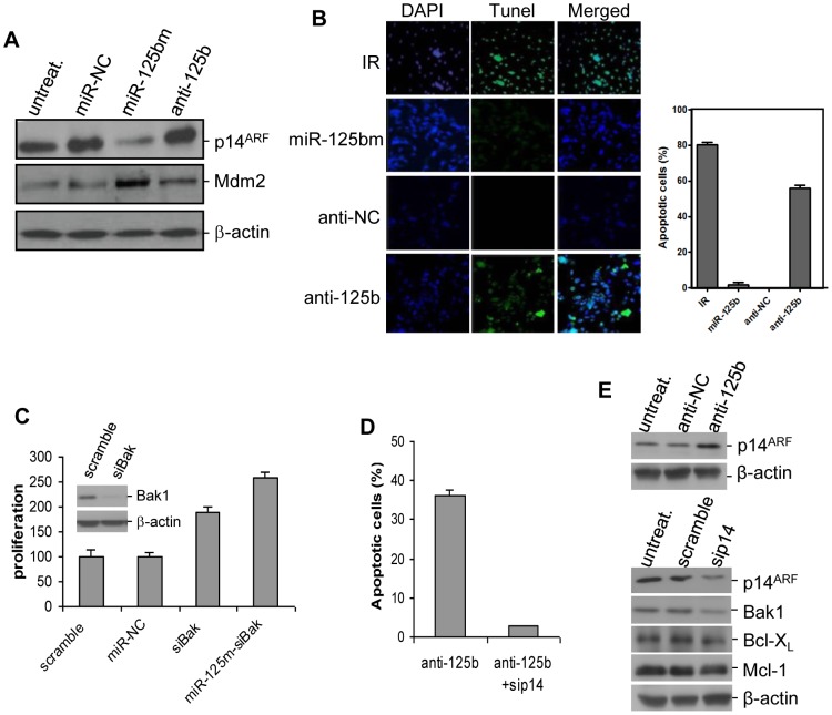
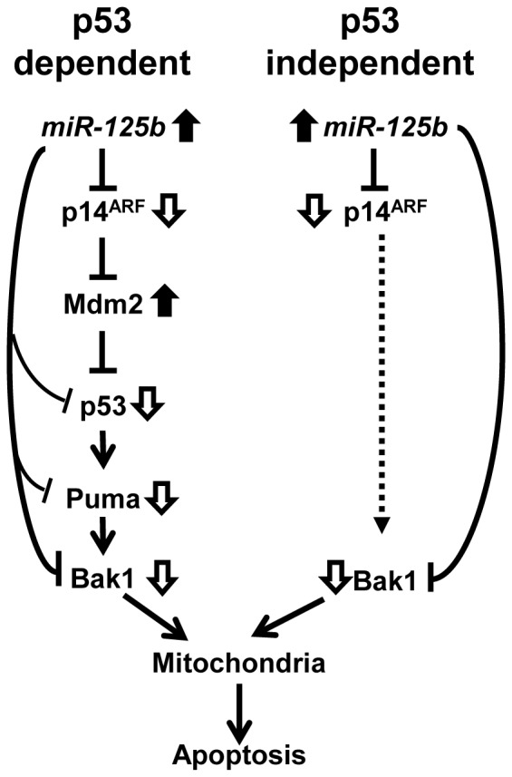
MicroRNAs are a class of naturally occurring small non-coding RNAs that target protein-coding mRNAs at the post-transcriptional level and regulate complex patterns of gene expression. Our previous studies demonstrated that in human prostate cancer the miRNA miR-125b is highly expressed, leading to a negative regulation of some tumor suppressor genes. In this study, we further extend our studies by showing that miR-125b represses the protein product of the ink4a/ARF locus, p14(ARF), in two prostate cancer cell lines, LNCaP (wild type-p53) and 22Rv1 (both wild type and mutant p53), as well as in the PC-346C prostate cancer xenograft model that lentivirally overexpressed miR-125b. Our results highlight that miR-125b modulates the p53 network by hindering the down-regulation of Mdm2, thereby affecting p53 and its target genes p21 and Puma to a degree sufficient to inhibit apoptosis. Conversely, treatment of prostate cancer cells with an inhibitor of miR-125b (anti-miR-125b) resulted in increased expression of p14(ARF), decreased level of Mdm2, and induction of apoptosis. In addition, overexpression of miR-125b in p53-deficient PC3 cells induced down-regulation of p14(ARF), which leads to increased cell proliferation through a p53-independent manner. Thus, we conclude that miR-125b acts as an oncogene which regulates p14(ARF)/Mdm2 signaling, stimulating proliferation of prostate cancer cells through a p53-dependent or p53-independent function. This reinforces our belief that miR-125b has potential as a therapeutic target for the management of patients with metastatic prostate cancer.


Conflict of interest statement

Competing Interests: The authors have declared that no competing interests exist.

Figures

Figure 1
Figure 1. MiR-125b down-regulates p14ARF in CaP cells.
A) Western blot analysis of expression levels of p14ARF in LNCaP (top) and 22Rv1 cells (bottom). Cells grown in 10% FBS media were transfected with 50 nM of miR-125bm or anti-miR-125b (anti-125b) for 72 hrs or treated with 5.0 nM of R1881 androgen for 48 hrs. Then, 50 µg of protein per sample was analyzed. Both miR-negative control (miR-NC) and anti-miR negative control (anti-NC) were used as controls, and β-actin was used as a loading control. B) Western blot analysis of expression levels of p14ARF, mdm2 and p53 in lenti-miR-125b-overexpressed PC-346C xenograft tumor. Both untreated xenograft (untreat.) and lenti-miRNA control vector-infected PC-346C xenograft (vector) were used as controls. In both A and B, the numbers under the gels are the average fold changes of p14ARF protein from three independent gels relative to the corresponding controls. Fold changes were calculated by scanning the p14ARF bands and normalizing for β-actin bands. C) Luciferase assay of miR-125b binding to the 3′-UTR of p14ARF mRNA in LNCaP cells. The assay was repeated three times with each assay being performed in three wells and similar results were obtained each time. The representative results are shown as a mean ±SD (n = 3).
Figure 2
Figure 2. MiR-125b regulates the p53 network.
A) Western blot analysis of Mdm2 and p53 in miR-125bm-treated LNCaP (top) and 22Rv1 cells (bottom). Cells were transfected with 50 nM of miR-125bm or miR-negative control (miR-NC) for 72 hrs. Equal amounts of protein (50 µg) were used to detect the expression levels of Mdm2, p53, p21 and Puma. B) Western blot analysis of p14ARF, Mdm2 and p53 in p14ARF siRNA (sip14)-treated LNCaP (top) and 22Rv1 cells (bottom). Cells were treated with sip14 and the cellular levels of p14ARF, p53 and Mdm2 were analyzed. β-actin was used as a loading control. C) Co-immunoprecipitation analysis of protein interaction between p14ARF and Mdm2 in 22Rv1 cells. Cells were transfected with miR-125bm and 1.0 mg protein was immunoprecipitated with anti-p14ARF antibody or the rabbit IgG. The resultant immunecomplexes were used to detect the level of Mdm2 by Western blot analysis using anti-Mdm2 antibody. Input: 50 µg protein from total cell lysate. IP: immunoprecipitation. IB: immunoblotting.
Figure 3
Figure 3. WST-1 proliferation assay of LNCaP cells (A) and 22Rv1 cells (B).
Cells were transfected with 50 nM of miR-125bm or 50 nM of miRNA negative control (miR-NC) for 5 days. Cell proliferation was measured by WST-1 assay. p14ARF siRNA (sip14) was used as a control. The results are expressed as proliferation relative to that of miR-NC-treated cells, and shown as mean ± SD (n = 4).
Figure 4
Figure 4. Inactivation of miR-125b induces apoptosis in p53-positive CaP cells.
A) Detection of SMAC and activated caspase 3 (Cas-3) in LNCaP (left) and 22Rv1 (right) cells. Cells were transfected with 50 nM miR-125bm or 50 nM anti-miR-125b (anti-125b) for 5 days, and the levels of SMAC and Cas-3 were measured by Western blot analysis. β-actin was used as loading control. The numbers under the gels are the average fold changes of SMAC and Cas-3 from three independent gels relative to the corresponding controls. B) Detection of anti-miR-125b-induced apoptosis in 22Rv1 cells. Cells were transfected using 50 nM anti-miR-125b for 72 hrs and apoptotic cell death was detected using TUNEL assay. The green nuclear fluorescence indicates the apoptotic cleavage of nuclear DNA (left). For quantitation of apoptotic cell death, 400 cells were counted and apoptosis is expressed as % apoptosis (apoptotic cells/400×100%). Quantitative analysis was performed three times and result was expressed as mean ± SE (n = 3) (right). Cells treated with irradiation (IR, 6 Gy) were used as a positive control. C) TUNEL assay of apoptotic death of 22Rv1 cells that were treated with anti-miR-125b followed by p14ARF antisense (sip14). Result was expressed as mean ± SE (n = 3). D) Western blot analyses of p14ARF, p53 and Bak1 levels in 22Rv1 cells. Left: 22Rv1 cells were transfected with anti-miR-125; right: anti-miR-125-transfected 22Rv1 cells were treated with sip14. Both anti-miR-NC (anti-NC) and scramble siRNA were used as controls.
Figure 5
Figure 5. Evaluation of miR-125b effect on growth and apoptosis in p53-negative CaP cells.
A) Detection of p14ARF and Mdm2 levels in p53-null PC3 cells. Cells were transfected with 50 nM of miR-125bm or anti-miR-125b for 72 hrs. The expression levels of both p14ARF and Mdm2 were analyzed by Western blot assay. β-actin was used as a loading control. B) Detection of anti-miR-125b-induced apoptosis in PC3 cells. Cells were transfected using 50 nM anti-miR-125b for 72 hrs and apoptotic cell death was detected using TUNEL assay. The green nuclear fluorescence indicates the apoptotic cleavage of nuclear DNA (left). For quantitation of apoptotic cell death, 400 cells were counted and apoptosis is expressed as % apoptosis (apoptotic cells/400×100%). Quantitative analysis was performed three times and result was expressed as mean ± SE (n = 3) (right). Cells treated with irradiation (IR, 6 Gy) were used as a positive control. C) MiR-125b promotes the growth of p53-null, Bak1-silenced PC3 cells. Cells were treated with 50 nM miR-125m for 5 days and cell proliferation was measured using WST-1 assay. The results are expressed as the growth inhibition relative to that of miR-NC (mean ± SD, n = 4). Inset: Bak1 expression in Bak1-silenced PC3 cells. D) TUNEL assay of apoptotic death of PC3 cells that were treated with anti-miR-125b followed by p14ARF antisense (sip14). Result was expressed as mean ± SE (n = 3). E) Western blot analyses of p14ARF and Bak1 levels in PC3 cells. Top: PC3 cells were transfected with anti-miR-125; bottom: anti-miR-125-transfected PC3 cells were treated with sip14. Both anti-miR-NC (anti-NC) and scramble siRNA were used as controls.
Figure 6
Figure 6. Schematic model of miR-125b-controlled oncopathway in CaP cells.
In CaP cancer cells, p14ARF facilitates apoptosis in a p53-dependent (left) and p53-independent (right) manner . Since miR-125b directly targets p14ARF and other pro-apoptotic molecules, deregulation of miR-125b can modulate proliferation and apoptosis in both p53-positive and p53-deficient CaP cells. Black arrows represent upregulated molecules and white arrows represent downregulated molecules. Broken arrow indicates undefined upregulation of Bak1 activity by p14ARF.

References

    1. Siegel R, Naishadham D, Jemal A (2012) Cancer statistics, 2012. CA: a cancer journal for clinicians 62: 10–29. - PubMed
    1. Lassi K, Dawson NA (2009) Emerging therapies in castrate-resistant prostate cancer. Current opinion in oncology 21: 260–265. - PubMed
    1. Shi XB, Tepper CG, White RW (2008) MicroRNAs and prostate cancer. Journal of cellular and molecular medicine 12: 1456–1465. - PMC - PubMed
    1. Hassan O, Ahmad A, Sethi S, Sarkar FH (2012) Recent updates on the role of microRNAs in prostate cancer. Journal of Hematology & Oncology 5: 9. - PMC - PubMed
    1. Bartel DP (2004) MicroRNAs: genomics, biogenesis, mechanism, and function. Cell 116: 281–297. - PubMed

Publication types

MeSH terms