

doi: 10.1155/2013/374973.
Epub 2013 Mar 26.
Spontaneous pelvic rupture as a result of renal colic in a patient with klinefelter syndrome
Affiliations
- PMID: 23585981
- PMCID: PMC3622382
- DOI: 10.1155/2013/374973
Item in Clipboard
Spontaneous pelvic rupture as a result of renal colic in a patient with klinefelter syndrome
Case Rep Urol.
2013.
Abstract
We present the case of a young man with Klinefelter syndrome, who was admitted to our clinic with renal colic. Shortly after admittance, spontaneous decrease in pain has occurred. Ultrasound and intravenous contrast computed tomography were performed, which showed the evidence of urine extravasation at the level of left renal pelvis and a 4 mm stone in the lower third of the left ureter. The management with a double-J ureteric stent for three weeks was successful. Then, the stent was removed and computed tomography confirmed the absence of urine extravasation. We also analyze the literature related to this case and discuss the main mechanisms of collecting system rupture.
Figures
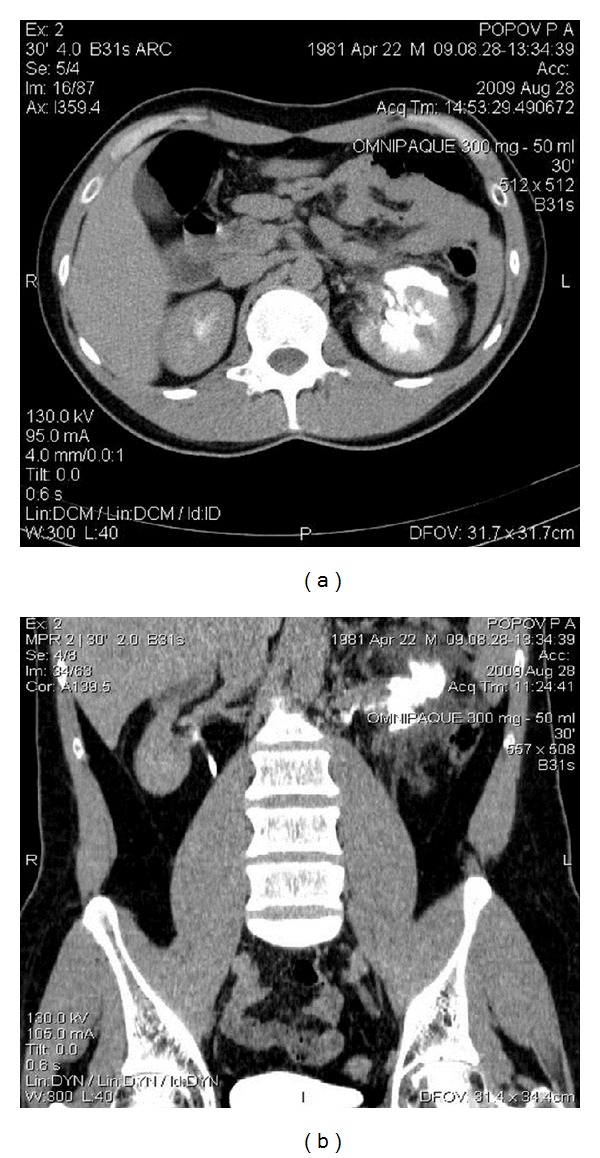
Computed tomography showing extravasation of the contrast medium through the gap in pelvis—spontaneous rupture. Axial (a) and sagittal (b) images.
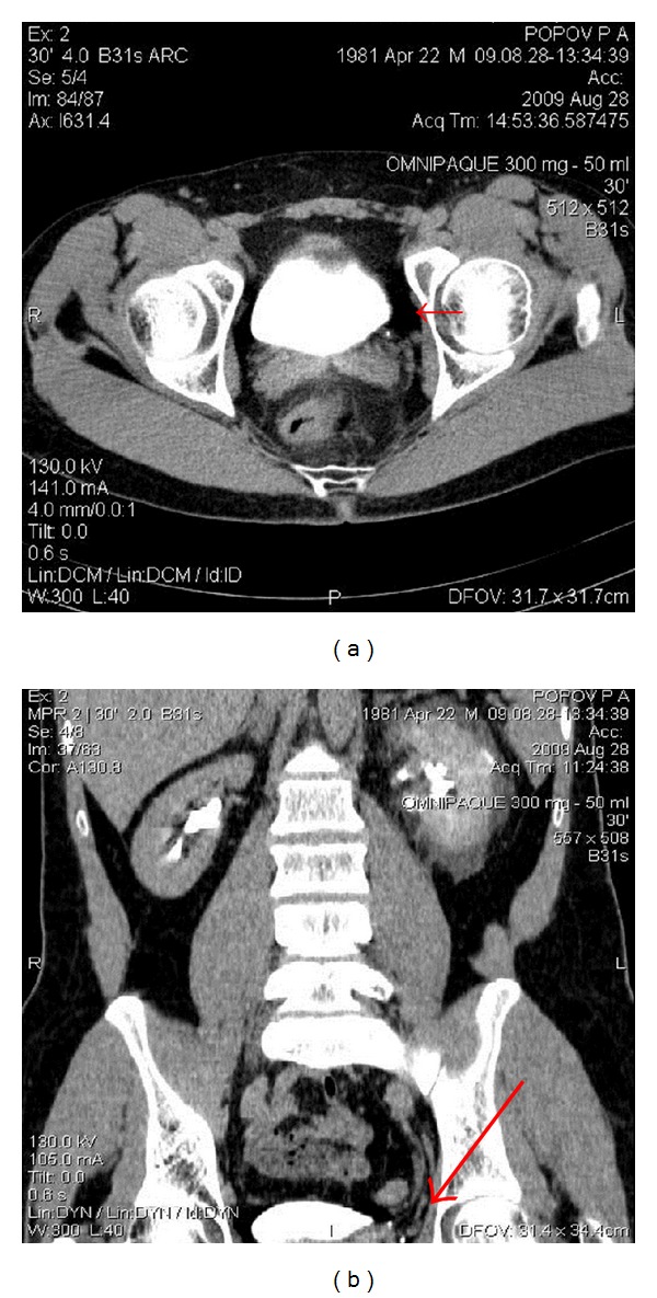
4 mm stone in the lower third of the left ureter (red arrow). Axial (a) and sagittal (b) images.
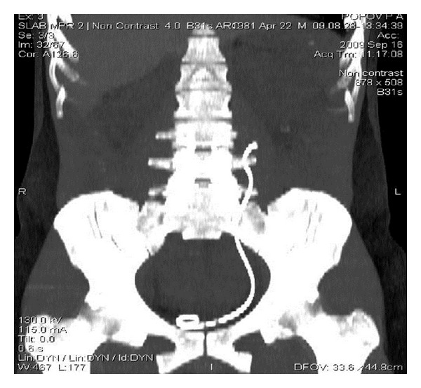
Distal migration of the ureteric stent.

Control CT (3 weeks after stenting and immediately after stent removal)—no extravasation of the contrast medium.

CT showing no stone in the ureter; the area of previous stone location is shown by red arrow.
References
-
- Fielding JR, Steele G, Fox LA, Heller H, Loughlin KR. Spiral computerized tomography in the evaluation of acute flank pain: a replacement for excretory urography. Journal of Urology. 1997;157(6):2071–2073. - PubMed
-
- Miller OF, Kane CJ. Time to stone passage for observed ureteral calculi: a guide for patient education. Journal of Urology. 1999;162(3, part 1):688–691. - PubMed
-
- Carter MR, Green BR. Renal calculi: emergency department diagnosis and treatment. Emergency Medicine Practice. 2011;13(7):1–17. - PubMed
-
- Segura JW, Preminger GM, Assimos DG, et al. Ureteral stones clinical guidelines panel summary report on the management of ureteral calculi. Journal of Urology. 1997;158(5):1915–1921. - PubMed
-
- Diaz ES, Buenrostro FG. Renal pelvis spontaneous rupture secondary to ureteral lithiasis. Case report and bibliographic review. Archivos Españoles de Urología. 2011;64(7):640–642. - PubMed
LinkOut - more resources
Full Text Sources
Other Literature Sources
