A large polypoid vascular ectasia removed by using a polypectomy with a detachable snare in an asymptomatic patient
- PMID: 23586013
- PMCID: PMC3624982
- DOI: 10.3393/ac.2013.29.1.31
A large polypoid vascular ectasia removed by using a polypectomy with a detachable snare in an asymptomatic patient
Abstract
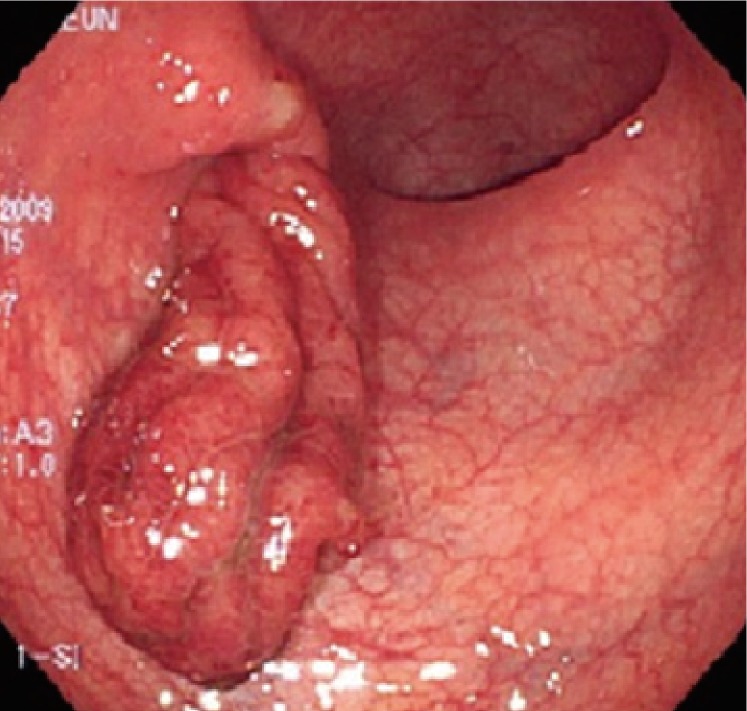
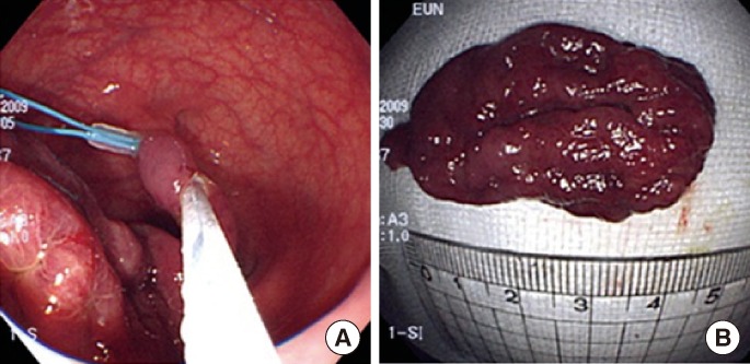
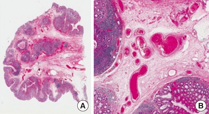
Vascular ectasia is a well-known cause of lower gastrointestinal bleeding in the elderly. Endoscopically, it usually appears as a flat or elevated bright red lesion. We report on an extremely rare case of a large, pedunculated, polypoid vascular ectasia in an asymptomatic patient. A large pedunculated polypoid mass in the sigmoid colon was observed on colonoscopy during a regular health check-up, and a polypectomy was performed using a detachable snare. In histology, vessels with massive dilation were found mainly in the submucosa, which was consistent with vascular ectasia.
Keywords: Detachable snare; Pedunculated polyp; Vascular ectasia.
Conflict of interest statement
No potential conflict of interest relevant to this article was reported.
Figures
Similar articles
-
Colonic Polypoid Vascular Ectasia in a Patient With Rectal Prolapse.Cureus. 2022 Jul 12;14(7):e26772. doi: 10.7759/cureus.26772. eCollection 2022 Jul. Cureus. 2022. PMID: 35967181 Free PMC article.
-
Colonic pedunculated polypoid vascular ectasia mimicking ileocolic intussusception: a rare case report.Ann Med Surg (Lond). 2023 May 26;85(7):3674-3678. doi: 10.1097/MS9.0000000000000913. eCollection 2023 Jul. Ann Med Surg (Lond). 2023. PMID: 37427223 Free PMC article.
-
Endoscopic resection of asymptomatic, colonic, polypoid arteriovenous malformations: Two case reports and a literature review.Saudi J Gastroenterol. 2017 Jan-Feb;23(1):67-70. doi: 10.4103/1319-3767.199111. Saudi J Gastroenterol. 2017. PMID: 28139503 Free PMC article. Review.
-
A case report of an unexpected colonic polyp: cavernous hemangioma.Ann Coloproctol. 2023 Jun;39(3):280-282. doi: 10.3393/ac.2020.00535.0076. Epub 2021 Jul 13. Ann Coloproctol. 2023. PMID: 34256425 Free PMC article.
-
A case of polypoid cavernous hemangioma of the sigmoid colon excised by colonoscopic polypectomy.Gastroenterol Jpn. 1993 Oct;28(5):712-8. doi: 10.1007/BF02806353. Gastroenterol Jpn. 1993. PMID: 8224623 Review.
Cited by
-
Colonic Polypoid Vascular Ectasia in a Patient With Rectal Prolapse.Cureus. 2022 Jul 12;14(7):e26772. doi: 10.7759/cureus.26772. eCollection 2022 Jul. Cureus. 2022. PMID: 35967181 Free PMC article.
-
Colonic pedunculated polypoid vascular ectasia mimicking ileocolic intussusception: a rare case report.Ann Med Surg (Lond). 2023 May 26;85(7):3674-3678. doi: 10.1097/MS9.0000000000000913. eCollection 2023 Jul. Ann Med Surg (Lond). 2023. PMID: 37427223 Free PMC article.
References
-
- Boley SJ, Sammartano R, Adams A, DiBiase A, Kleinhaus S, Sprayregen S. On the nature and etiology of vascular ectasias of the colon: degenerative lesions of aging. Gastroenterology. 1977;72(4 Pt 1):650–660. - PubMed
-
- Greenwald DA, Brandt LJ. Vascular abnormalities of the gastrointestinal tract. In: Feldman M, Scharschmidt BF, Sleisenger MH, editors. Sleisenger & Fordtran's gastrointestinal and liver disease: pathophysiology, diagnosis, management. 6th ed. Philadelphia: Saunders Co.; 1998. pp. 2024–2034.
-
- Koziara FJ, Brodmerkel GJ, Boylan JJ, Ciambotti GF, Agrawal RM. Bleeding from polypoid colonic arteriovenous malformations. Am J Gastroenterol. 1996;91:584–586. - PubMed
-
- Margulis AR, Heinbecker P, Bernard HR. Operative mesenteric arteriography in the search for the site of bleeding in unexplained gastrointestinal hemorrhage: a preliminary report. Surgery. 1960;48:534–539. - PubMed
-
- Skibba RM, Hartong WA, Mantz FA, Hinthorn DR, Rhodes JB. Angiodysplasia of the cecum: colonoscopic diagnosis. Gastrointest Endosc. 1976;22:177–179. - PubMed
LinkOut - more resources
Full Text Sources
Other Literature Sources