Review
. 2013:2013:123241.
doi: 10.1155/2013/123241. Epub 2013 Mar 19.

Inflammation and immunity in radiation damage to the gut mucosa

Affiliations
Review

Inflammation and immunity in radiation damage to the gut mucosa

Agnès François et al. Biomed Res Int. 2013.

Abstract

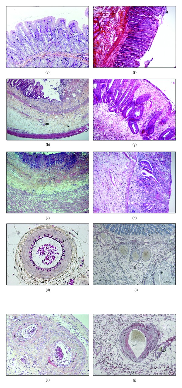
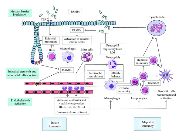
Erythema was observed on the skin of the first patients treated with radiation therapy. It is in particular to reduce this erythema, one feature of tissue inflammation, that prescribed dose to the tumor site started to be fractionated. It is now well known that radiation exposure of normal tissues generates a sustained and apparently uncontrolled inflammatory process. Radiation-induced inflammation is always observed, often described, sometimes partly explained, but still today far from being completely understood. The thing with the gut and especially the gut mucosa is that it is at the frontier between the external milieu and the organism, is in contact with a plethora of commensal and foreign antigens, possesses a dense-associated lymphoid tissue, and is particularly radiation sensitive because of a high mucosal turnover rate. All these characteristics make the gut mucosa a strong responsive organ in terms of radiation-induced immunoinflammation. This paper will focus on what has been observed in the normal gut and what remains to be done concerning the immunoinflammatory response following localized radiation exposure.


Figures

Figure 1
Figure 1
Left panel ((a) to (e)): Radiation-induced damage to the rectal wall following localized exposure to 27 Gy single dose in the rat. (a) Healthy mucosa. (b) Two weeks after exposure, tissue shows mucosal and submucosal inflammation, with mucosal ulceration (u) and submucosal (sm) oedema. Glandular recovering (arrow) alternates with ulcerated areas. (c) Height weeks after exposure, underlying the ulcerated areas, the entire rectal wall is pathologic. The mucosa and submucosa show still severe inflammation, with tissue necrosis at the luminal frontier (purple coloration). Inflammation reaches the oedematous and dystrophic external muscular layers. Note that the muscularis mucosa disappeared. (e) Eight weeks after exposure, severe epithelial, submucosal, and muscular damage is associated with dystrophic submucosal and mesenteric vessels. The elastic layer is dystrophic (arrow) compared to the healthy vessels ((d), arrow) and, neointimal hyperplasia reduces the vessel lumen (double arrow). HeS staining, original magnification ×100 ((a), (d), (e)) or ×40 ((b), (c)), pictures Agnès Francois. Right panel radiation-induced damage to the rectal wall in patients treated for rectal adenocarcinoma, 5 to 7 weeks following the end of radiation therapy (45 Gy). (f) Healthy rectal mucosa. (g): Epithelial atypia with mucosal oedema and inflammation. Crypt positioning is disorganized and some are bifid and show hyperplasia, both signing epithelial regeneration. (h) Severe mucosal ulceration with submucosal oedema and dense inflammatory infiltrate. Crypt number is drastically reduced. (j) Dystrophic submucosal arteriole, with collagen deposition in the vessel wall and neointimal hyperplasia reducing the vascular lumen. (f), (g), (h) HeS staining, (i), (j) Sirius Red staining. Original magnification ×40, pictures Agnès François.
Figure 2
Figure 2
Radiation exposure to the gut mucosa induces epithelial stem cells apoptosis and clonogenic cell death, endothelial cells death and activation, and mucosal barrier breakdown. Innate immunity: resident immune cells such as macrophages and mast cells are activated by DAMPs generated by dead cells and PAMPs coming from mucosal breakdown. Activated endothelial cells express adhesion molecules and cytokines favouring immune cells recruitment into the injured tissue. Activated macrophages increase neutrophil recruitment, which in turn emit signals favouring monocytes recruitment from the blood stream. The radiation-induced tissue M1/M2 balance is unknown. Adaptative immunity: Th1 lymphocytes can activate innate immune cells and favour cell-mediated immunity, whereas Th2 favour humoral immunity via B cells. The balance in irradiated gut tissue is in favour of a Th2 orientation. Treg maintain immune tolerance. Resident and recruited dendritic cells are activated and carry out the link with lymph nodes and the establishment of specific immune response. PAMPs are also detected by TLRs, whose activation can protect epithelium against radiation damage. ILCs play a role in epithelial homeostasis, but their role after radiation exposure is unknown.

References

    1. Denham JW, Hauer-Jensen M. The radiotherapeutic injury—a complex “wound”. Radiotherapy and Oncology. 2002;63(2):129–145. - PubMed
    1. Andreyev HJN, Wotherspoon A, Denham JW, Hauer-Jensen M. “Pelvic radiation disease”: new understanding and new solutions for a new disease in the era of cancer survivorship. Scandinavian Journal of Gastroenterology. 2011;46(4):389–397. - PubMed
    1. Andreyev HJN, Wotherspoon A, Denham JW, Hauer-Jensen M. Defining pelvic-radiation disease for the survivorship era. The Lancet Oncology. 2010;11(4):310–312. - PubMed
    1. Bjerknes M, Cheng H. Clonal analysis of mouse intestinal epithelial progenitors. Gastroenterology. 1999;116(1):7–14. - PubMed
    1. Bentzen SM. Preventing or reducing late side effects of radiation therapy: radiobiology meets molecular pathology. Nature Reviews Cancer. 2006;6(9):702–713. - PubMed

MeSH terms