Review
. 2013:2013:162513.
doi: 10.1155/2013/162513. Epub 2013 Mar 17.

Structures and properties of naturally occurring polyether antibiotics

Affiliations
Review

Structures and properties of naturally occurring polyether antibiotics

Jacek Rutkowski et al. Biomed Res Int. 2013.

Abstract

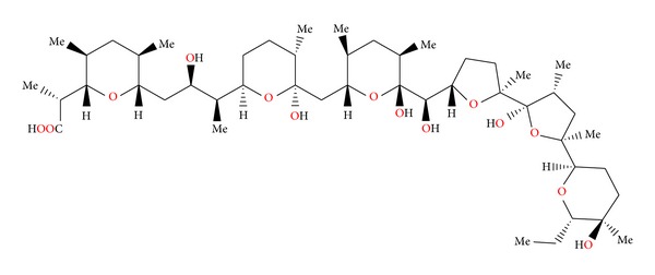
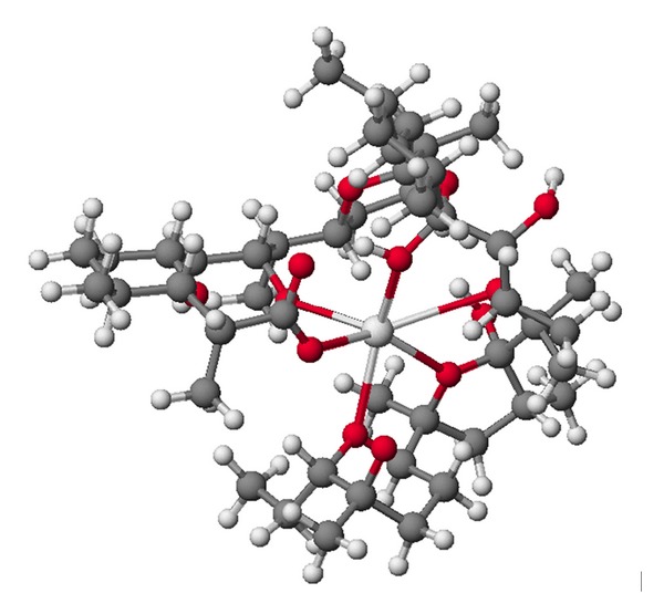
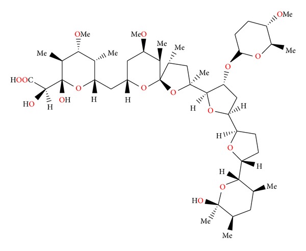
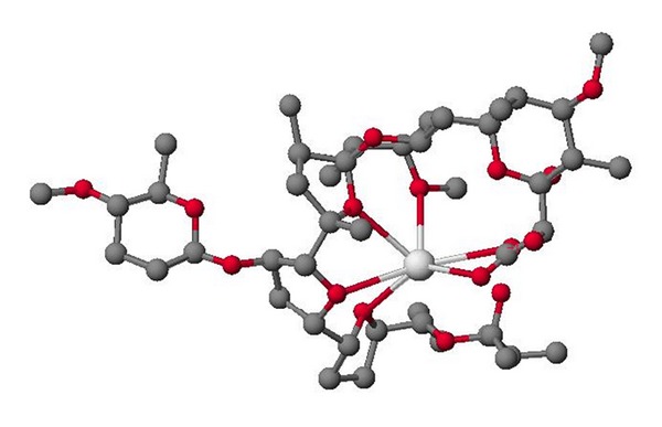
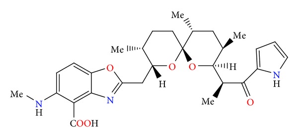
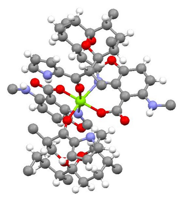
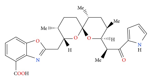
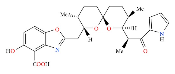
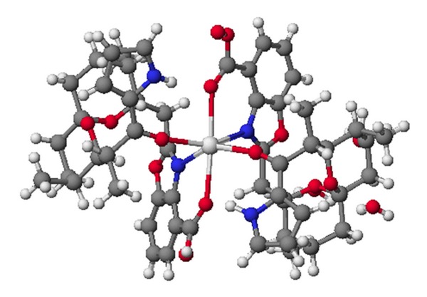
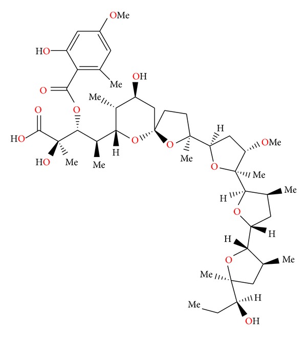
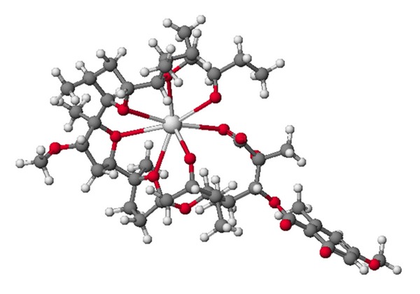
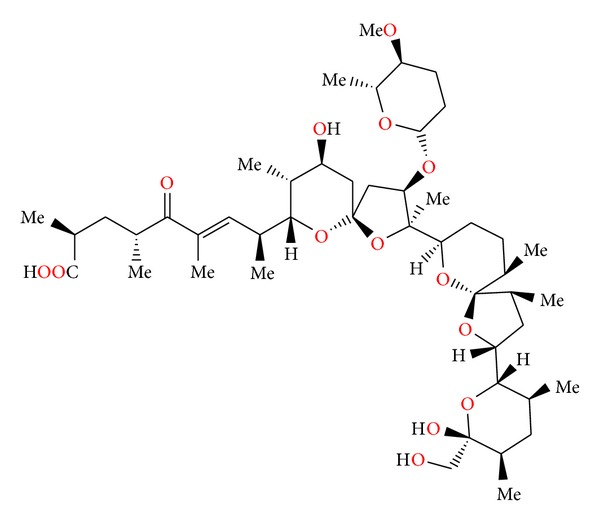
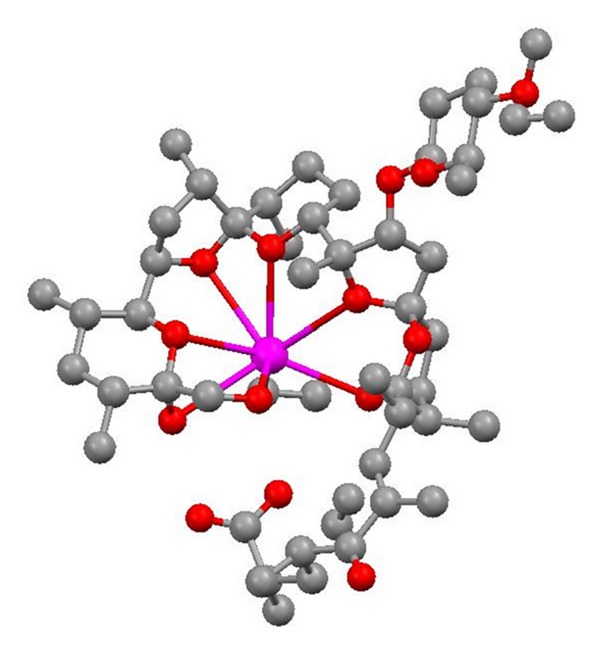
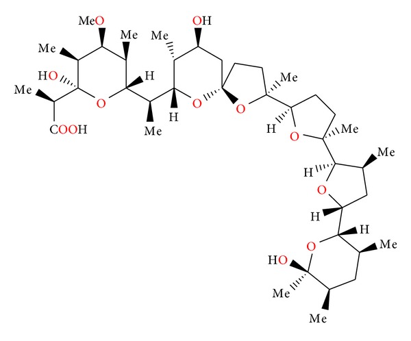
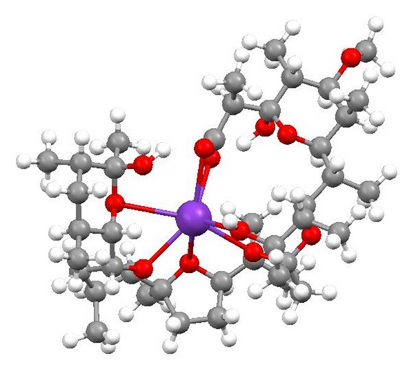
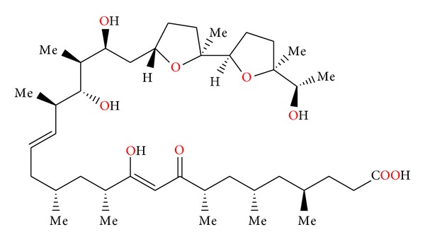
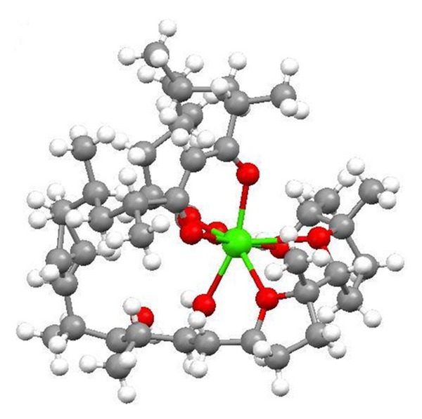
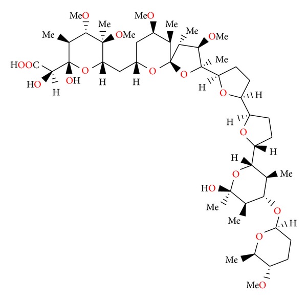
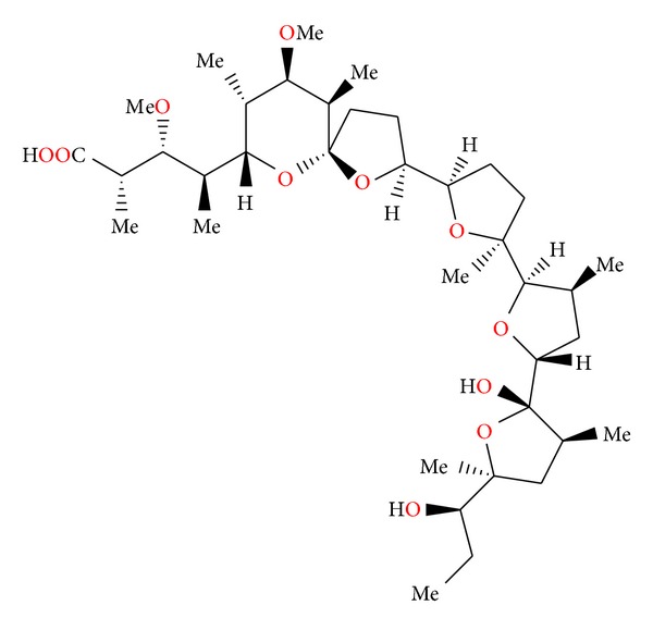
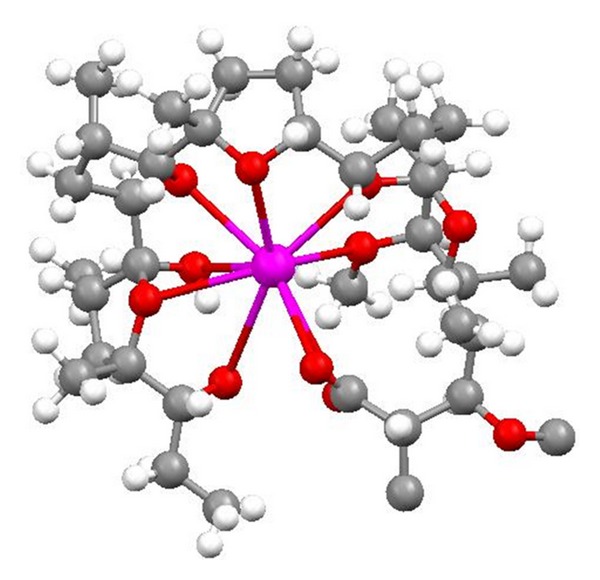
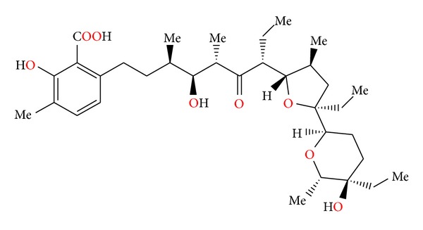
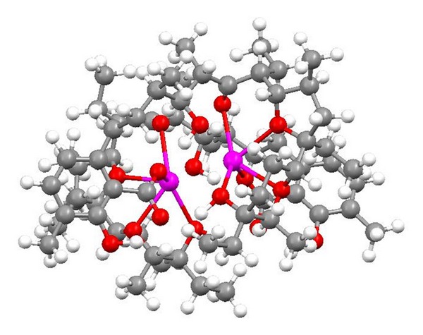
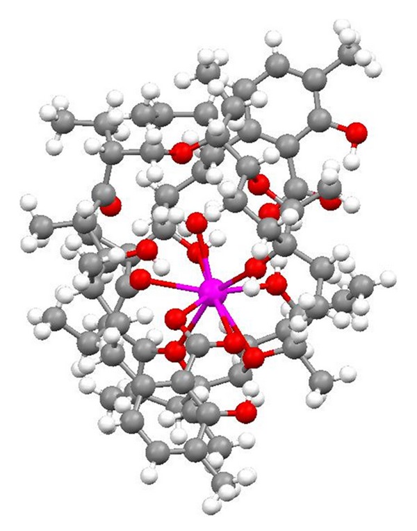
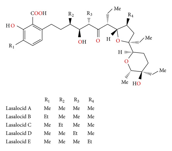
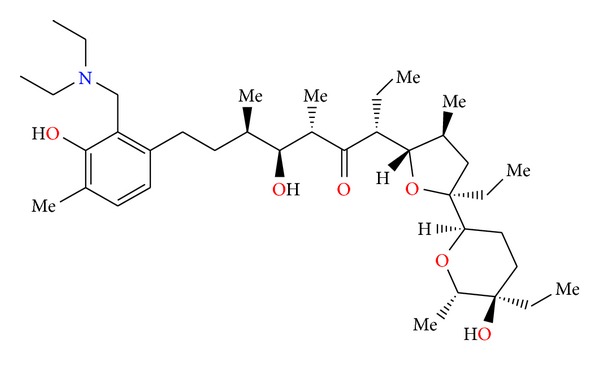
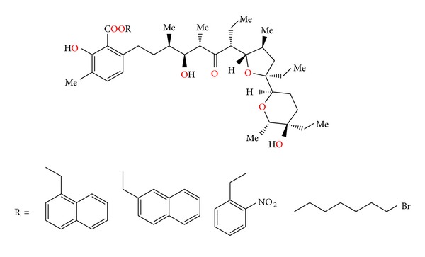
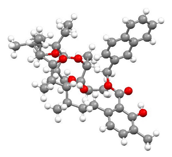
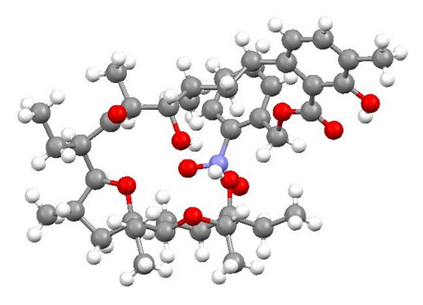
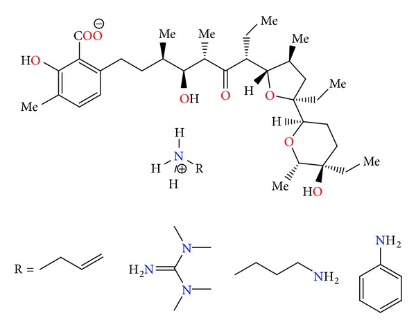
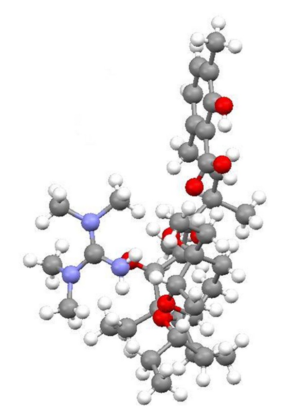
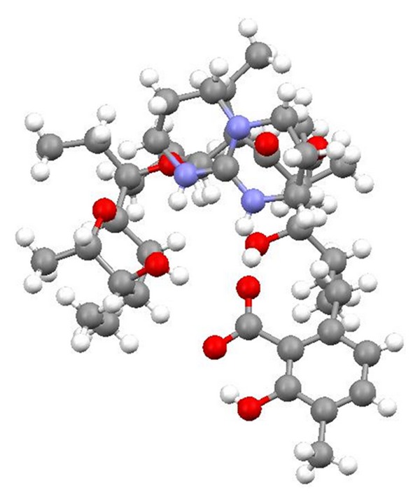
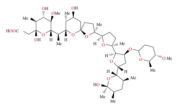
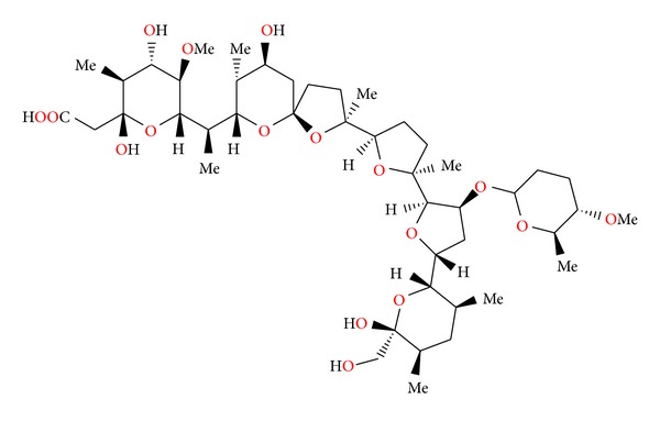
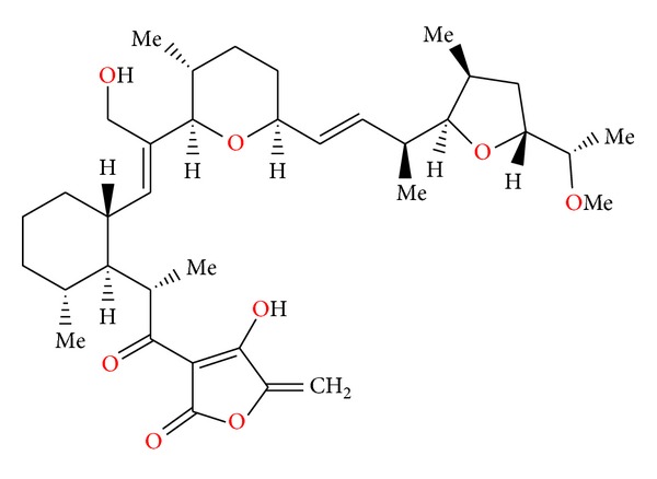
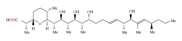
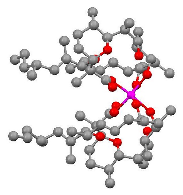
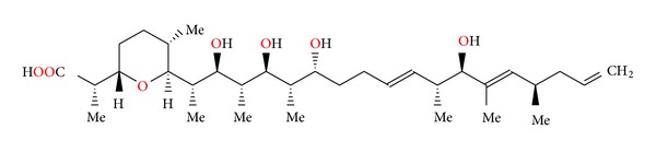
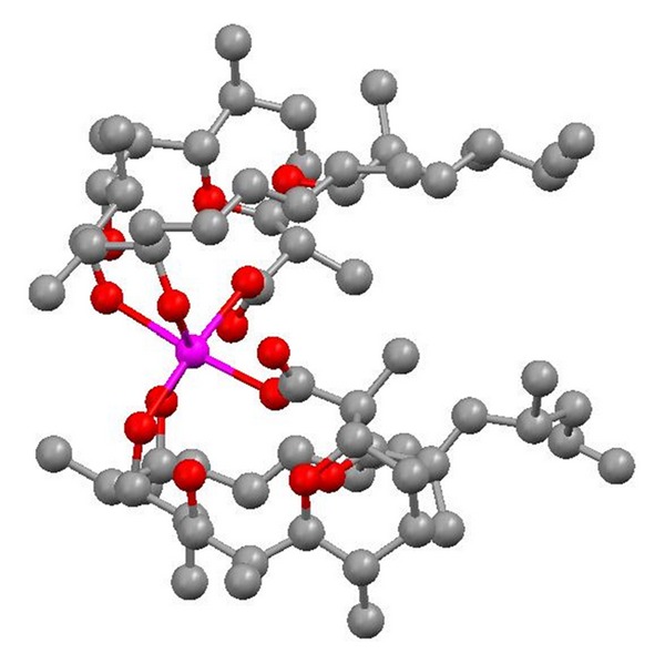
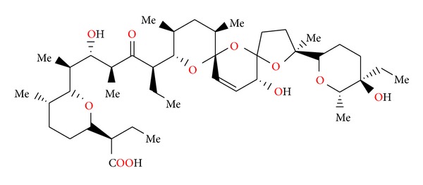
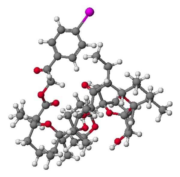
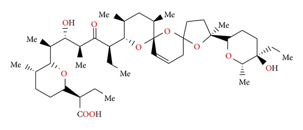
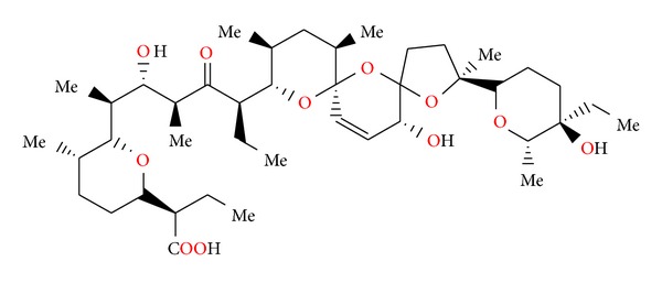
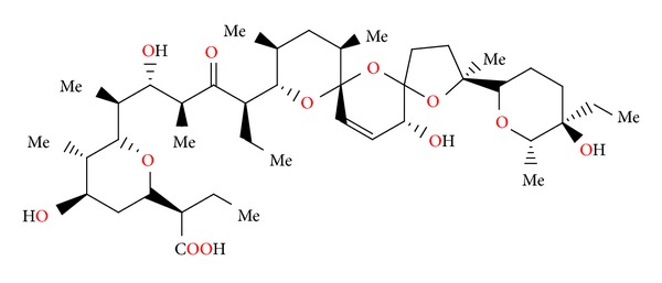
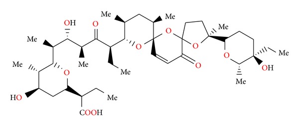
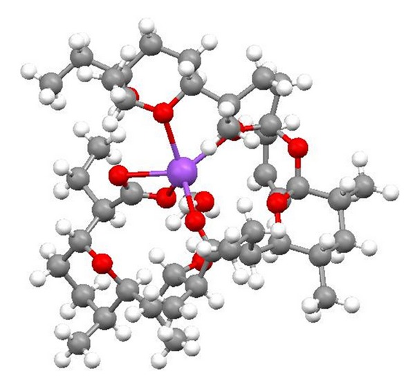
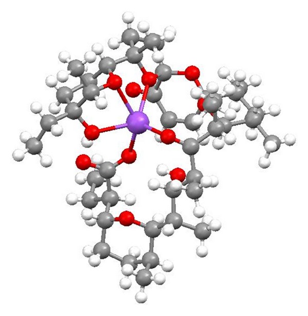
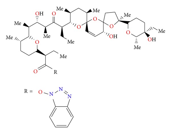
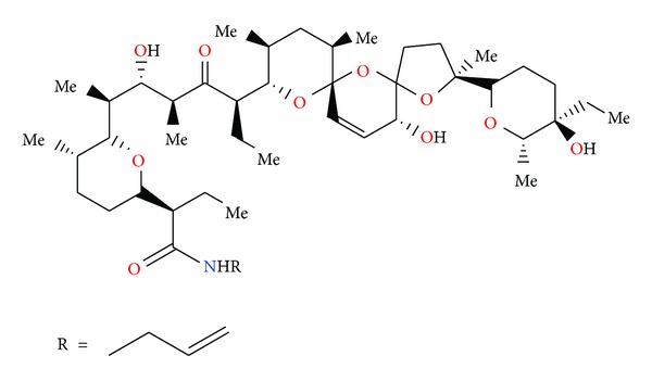
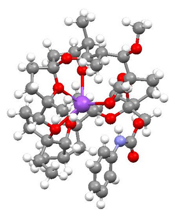
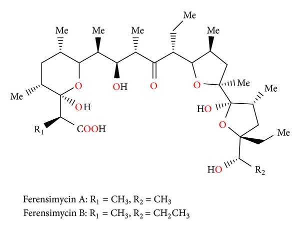
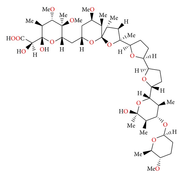
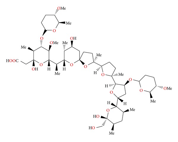
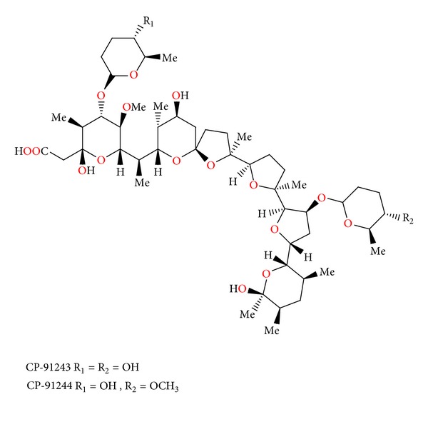
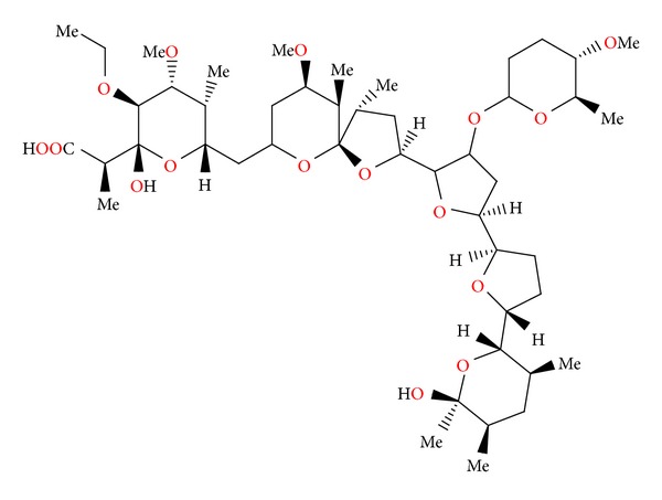
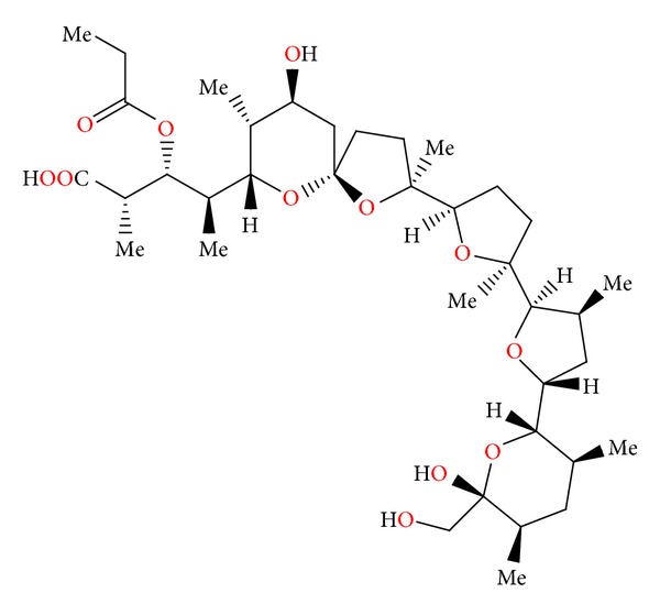
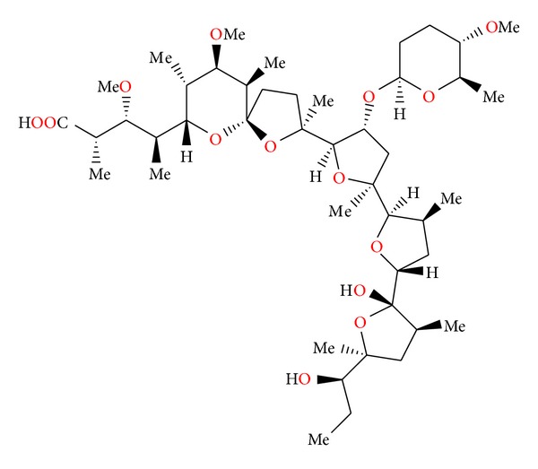
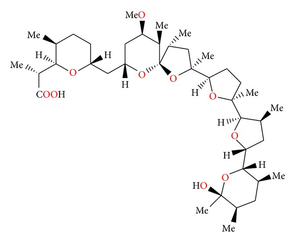
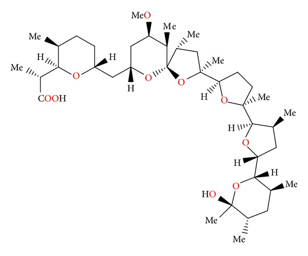
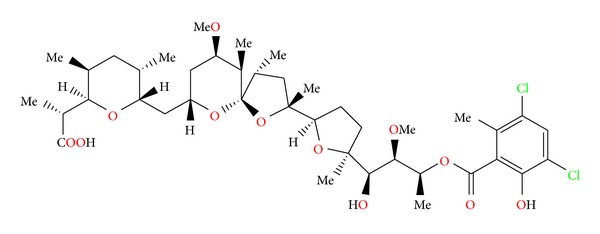
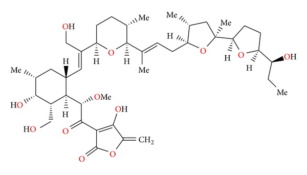
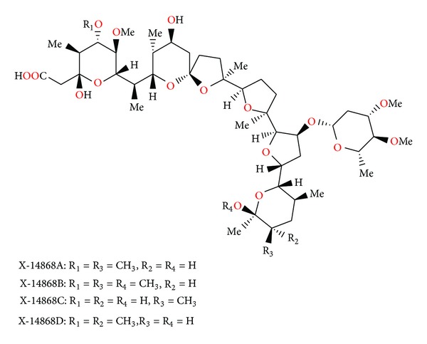
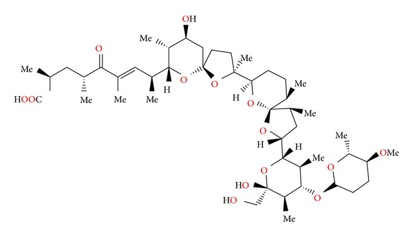
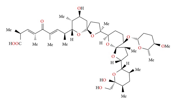
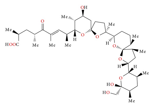
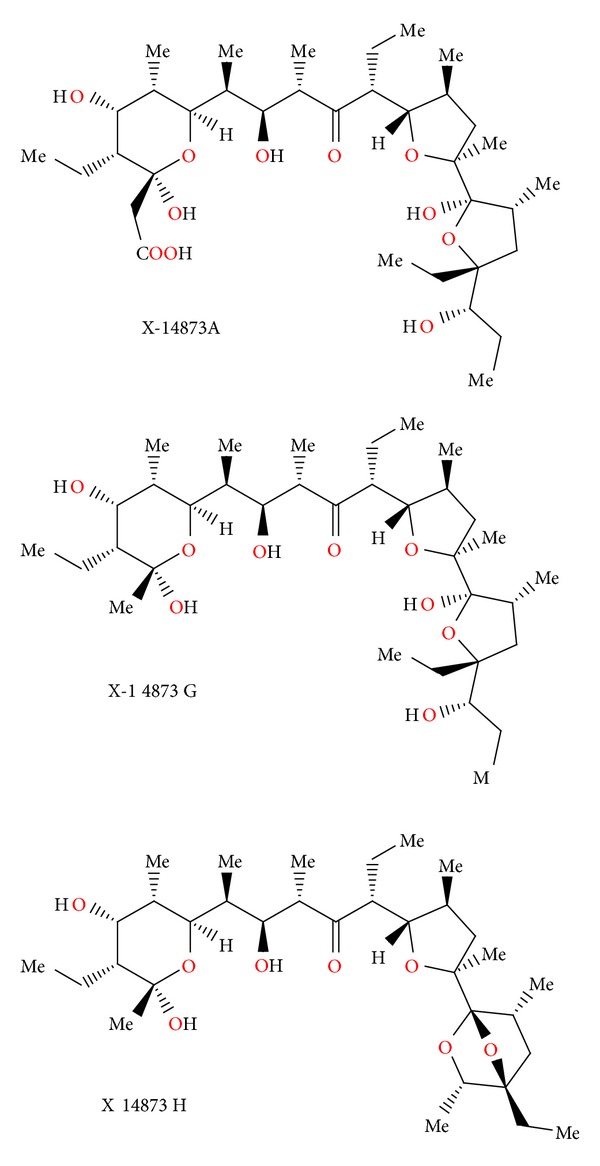
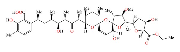
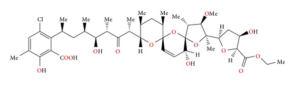
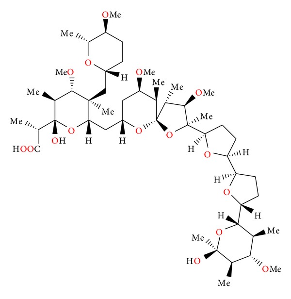
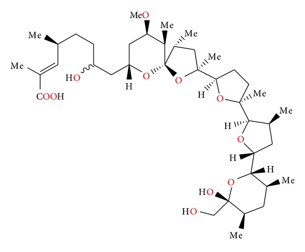
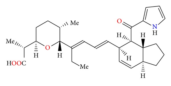
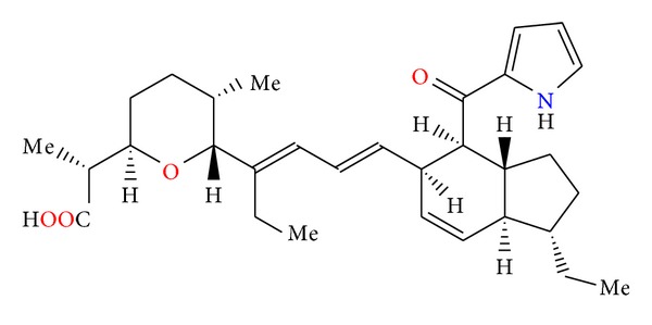
Polyether ionophores represent a large group of natural, biologically active substances produced by Streptomyces spp. They are lipid soluble and able to transport metal cations across cell membranes. Several of polyether ionophores are widely used as growth promoters in veterinary. Polyether antibiotics show a broad spectrum of bioactivity ranging from antibacterial, antifungal, antiparasitic, antiviral, and tumour cell cytotoxicity. Recently, it has been shown that some of these compounds are able to selectively kill cancer stem cells and multidrug-resistant cancer cells. Thus, they are recognized as new potential anticancer drugs. The biological activity of polyether ionophores is strictly connected with their molecular structure; therefore, the purpose of this paper is to present an overview of their formula, molecular structure, and properties.


Figures

Figure 1
Figure 1
Structure of alborixine.
Figure 2
Figure 2
Structure of 6-demethyl-alborixin complex with sodium cation.
Figure 3
Figure 3
Structure of antibiotic 6016.
Figure 4
Figure 4
Crystal structure of antibiotic 6016 thallium salt.
Figure 5
Figure 5
Structure of calcimycin.
Figure 6
Figure 6
Crystal structure of 2 : 1 complex of calcimycin with the magnesium cation.
Figure 7
Figure 7
Structure of cezomycin.
Figure 8
Figure 8
Structure of X-14885A.
Figure 9
Figure 9
Crystal structure of 2 : 1 complex of 11-demethyl-cezomycin complex with the sodium cation.
Figure 10
Figure 10
Structure of cationomycin.
Figure 11
Figure 11
Crystal structure of cationomycin thallium salt.
Figure 12
Figure 12
Structure of endusamycin.
Figure 13
Figure 13
Crystal structure of endusamycin rubidium salt.
Figure 14
Figure 14
Structure of mutalomycin.
Figure 15
Figure 15
Crystal structure of 28-epimutalomycin potassium salt.
Figure 16
Figure 16
Structure of ionomycin.
Figure 17
Figure 17
Crystal structure of ionomycin complex with the calcium cation.
Figure 18
Figure 18
Structure of K-41.
Figure 19
Figure 19
Structure of kijimicin.
Figure 20
Figure 20
Crystal structure of kijimicin rubidium salt.
Figure 21
Figure 21
Structure of lasalocid.
Figure 22
Figure 22
Crystal structure of lasalocid silver salt on 2 : 2 stoichiometry.
Figure 23
Figure 23
Crystal structure of 2 : 1 complex of lasalocid with the strontium cation.
Figure 24
Figure 24
Lasalocid acid analogues.
Figure 25
Figure 25
Structure of lasalocid acid Mannich base.
Figure 26
Figure 26
Structures of lasalocid acid esters.
Figure 27
Figure 27
Structure of lasalocid 2-naphthylmethyl ester.
Figure 28
Figure 28
Crystal structure of lasalocid orthonitrobenzyl ester.
Figure 29
Figure 29
Lasalocid acid complexes with several amines: allylamine, 1,1,3,3-tetramethylguanidine, N-butylamine, and phenylamine.
Figure 30
Figure 30
Crystal structure of lasalocid complex with tetramethylguanidine.
Figure 31
Figure 31
Crystal structure of lasalocid complex with TBD.
Figure 32
Figure 32
Structure of semduramicin.
Figure 33
Figure 33
Structure of CP-120509.
Figure 34
Figure 34
Structure of tetronasin.
Figure 35
Figure 35
Structure of zincophorin.
Figure 36
Figure 36
Crystal structure of zincophorin magnesium salt.
Figure 37
Figure 37
Structure of CP-78545.
Figure 38
Figure 38
Crystal structure of CP-78545 cadmium salt.
Figure 39
Figure 39
Structure of salinomycin.
Figure 40
Figure 40
Crystal structure of salinomycin p-iodophenacyl ester.
Figure 41
Figure 41
Structure of SY-1.
Figure 42
Figure 42
Structure of SY-2.
Figure 43
Figure 43
Structure of SY-4.
Figure 44
Figure 44
Structure of SY-9.
Figure 45
Figure 45
Crystal structure of SY-1 with the sodium cation.
Figure 46
Figure 46
Crystal structure of SY-9 with the sodium cation.
Figure 47
Figure 47
Structure of benzotriazole ester of salinomycin.
Figure 48
Figure 48
Structure of allyl amide of salinomycin.
Figure 49
Figure 49
Crystal structure of salinomycin benzotriazole ester acetonitrile solvate.
Figure 50
Figure 50
Crystal structure of salinomycin allyl amide acetonitrile solvate.
Figure 51
Figure 51
Structure of monensin.
Figure 52
Figure 52
Crystal structure of monensin sodium salt acetonitrile solvate.
Figure 53
Figure 53
Structure of monensin hydrate.
Figure 54
Figure 54
Crystal structure of monensin lithium salt acetonitrile solvate.
Figure 55
Figure 55
Structures of monensin ester amide and urethane derivatives.
Figure 56
Figure 56
Crystal structure of monensin 1-naphtylmethyl ester with the lithium perchlorate.
Figure 57
Figure 57
Crystal structure of monensin phenylurethane sodium salt.
Figure 58
Figure 58
Structures of ferensimycins A and B.
Figure 59
Figure 59
Structure of CP-96797.
Figure 60
Figure 60
Structure of octacyclomycin.
Figure 61
Figure 61
Structures of CP-91243 and CP-91244.
Figure 62
Figure 62
Structure of W341C.
Figure 63
Figure 63
Structure of laidlomycin.
Figure 64
Figure 64
Structure of CP-84657.
Figure 65
Figure 65
Structure of grisorixin.
Figure 66
Figure 66
Structure of epigrisorixin.
Figure 67
Figure 67
Structure of CP-54883.
Figure 68
Figure 68
Structure of SF-2487.
Figure 69
Figure 69
Structures of X-14868A, X-14868B, X-14868C, and X-14868D.
Figure 70
Figure 70
Structure of CP-80219.
Figure 71
Figure 71
Structure of moyukamycin.
Figure 72
Figure 72
Structure of X-14931A.
Figure 73
Figure 73
Structures of X-14873A, X-14873G, and X-14873H.
Figure 74
Figure 74
Structure of noboritomycin.
Figure 75
Figure 75
Structure of 6-chloronoboritomycin.
Figure 76
Figure 76
Structure of CP-82009.
Figure 77
Figure 77
Structure of abierixin.
Figure 78
Figure 78
Structure of A-83094A.
Figure 79
Figure 79
Structure of indanomycin.

References

    1. Dutton CJ, Banks BJ, Cooper CB. Polyether ionophores. Natural Product Reports. 1995;12(2):165–181. - PubMed
    1. Callaway TR, Edrington TS, Rychlik JL, et al. Ionophores: their use as ruminant growth promotants and impact on food safety. Current Issues in Intestinal Microbiology. 2003;4(2):43–51. - PubMed
    1. Rochefeuille S, Jimenez C, Tingry S, Seta P, Desfours JP. Mixed Langmuir-Blodgett monolayers containing carboxylic ionophores. Application to Na+ and Ca2+ ISFET-based sensors. Materials Science and Engineering C. 2002;21(1-2):43–46.
    1. Gabrielli C, Hemery P, Letellier P, et al. Investigation of ion-selective electrodes with neutral ionophores and ionic sites by EIS. II. Application to K+ detection. Journal of Electroanalytical Chemistry. 2004;570(2):291–304.
    1. Dobler M. Natural cation-binding agents. In: Gokel GW, editor. Comprehensive Supramolecular Chemistry: Molecular Recognition: Receptors for Cationic Guests. Vol. 1. New York, NY, USA: Pergamon; 2004. pp. 267–313.

MeSH terms