. 2013 Sep;126(6):792-804.
doi: 10.1111/jnc.12266. Epub 2013 Apr 30.

A HuD-ZBP1 ribonucleoprotein complex localizes GAP-43 mRNA into axons through its 3' untranslated region AU-rich regulatory element

Affiliations

A HuD-ZBP1 ribonucleoprotein complex localizes GAP-43 mRNA into axons through its 3' untranslated region AU-rich regulatory element

Soonmoon Yoo et al. J Neurochem. 2013 Sep.

Abstract

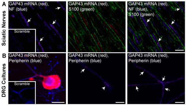
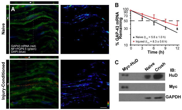
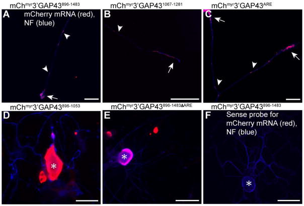
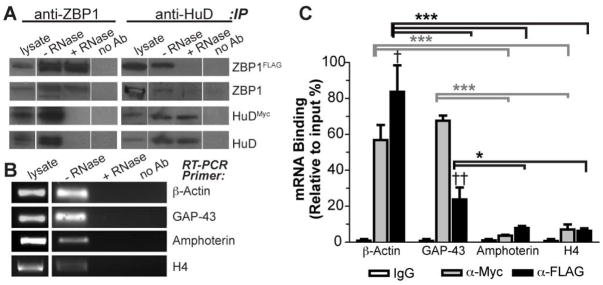
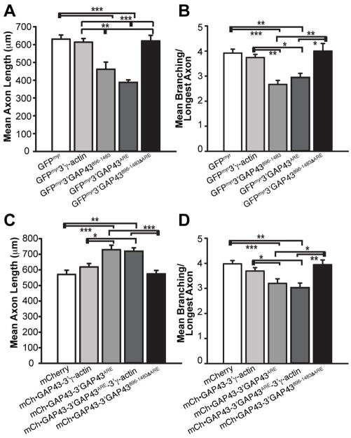
Localized translation of axonal mRNAs contributes to developmental and regenerative axon growth. Although untranslated regions (UTRs) of many different axonal mRNAs appear to drive their localization, there has been no consensus RNA structure responsible for this localization. We recently showed that limited expression of ZBP1 protein restricts axonal localization of both β-actin and GAP-43 mRNAs. β-actin 3'UTR has a defined element for interaction with ZBP1, but GAP-43 mRNA shows no homology to this RNA sequence. Here, we show that an AU-rich regulatory element (ARE) in GAP-43's 3'UTR is necessary and sufficient for its axonal localization. Axonal GAP-43 mRNA levels increase after in vivo injury, and GAP-43 mRNA shows an increased half-life in regenerating axons. GAP-43 mRNA interacts with both HuD and ZBP1, and HuD and ZBP1 co-immunoprecipitate in an RNA-dependent fashion. Reporter mRNA with the GAP-43 ARE competes with endogenous β-actin mRNA for axonal localization and decreases axon length and branching similar to the β-actin 3'UTR competing with endogenous GAP-43 mRNA. Conversely, over-expressing GAP-43 coding sequence with its 3'UTR ARE increases axonal elongation and this effect is lost when just the ARE is deleted from GAP-43's 3'UTR. We have recently found that over-expression of GAP-43 using an axonally targeted construct with the 3'UTRs of GAP-43 promoted elongating growth of axons, while restricting the mRNA to the cell body with the 3'UTR of γ-actin had minimal effect on axon length. In this study, we show that the ARE in GAP-43's 3'UTR is responsible for localization of GAP-43 mRNA into axons and is sufficient for GAP-43 protein's role in elongating axonal growth.

Keywords: GAP-43; HuD; RNA-immunoprecipitation; ZBP1; axon regeneration; mRNA transport.


Figures

Figure 1
Figure 1. GAP-43 mRNA localizes to peripheral axons
A, Representative images of single optical planes from adult rat sciatic nerve are shown for GAP-43 mRNA and indicated proteins; exposure matched image for scrambled probe (red) is shown in the inset panels. Images were taken at the center of the axoplasm for axons (arrows) to completely distinguish axonal and Schwann cell signals. Granular signals for GAP-43 mRNA overlap with only the NF positive axons [Scale bars = 5 μm]. B, Representative images of cultured DRG neurons are shown for GAP-43 mRNA and peripherin protein; exposure matched image for scrambled probe (red) is shown in the inset panels. The left panel shows a low magnification of neuron cell body; the middle and the right panels show higher power images of axon shaft (arrows) and growth cone (arrowhead) with GAP-43 mRNA extending into each [Scale bars: left panel = 50 μm, middle and right panels = 5 μm].
Figure 2
Figure 2. GAP-43 mRNA is stabilized in regenerating sciatic nerve
A, Matched exposure/post-processing confocal images of FISH/IF for 7 day crushed and naïve sciatic nerve are shown. Merged channels of confocal maximum projections for XYZ are shown in left column and RNA channel is shown in right column (RNA is shown as indicated intensity spectrum). Orthogonal XY and YZ projections at the levels indicated by grey dotted lines show that the GAP-43 mRNA signals completely overlap with the axons stained for NF and PGP 9.5 proteins (arrows) [Scale bars = 10 μm]. B, Relative stability of sciatic nerve GAP-43 mRNA was tested using naïve and 7 day injured nerve explants treated with 20 μM cyclosporin A to delay axonal degeneration. GAP-43 mRNA signals from RTqPCR were normalized to GAPDH mRNA and are expressed relative to time 0 to estimate of half-life of the nerve GAP-43 mRNA. Error bars represent standard deviation (n = 4). The slopes of the decay curves for GAP-43 mRNA were analyzed using nonlinear regression curves with a straight-line model (R2 =0.8855 for naïve and R2 =0.9837 for crush; p=0.0492). C, Representative immunoblot for axoplasm extracts prepared from the naïve and 7 day crushed sciatic nerves is shown. GAPDH signals show relatively equivalent loading between crush and naïve nerves. However, HuD signals were consistently higher in the crush nerve (1.98 ± 0.28 fold crush vs. naïve axoplasm; n = 3). To test HuD antibody specificity, lysates from F11 cells transfected with HuDMyc construct were included in these gels. Probing with anti-Myc antibody shows comparably migrating band as the anti-HuD antibody.
Figure 3
Figure 3. GAP-43 3′UTR is sufficient for translation of a reporter mRNA in axons
AD, Representative time-lapse images for FRAP analyses of DRG neurons transfected with the indicated constructs are shown. Laser intensity, gain and offset are matched for each image sequence. The boxed regions represent the ROIs that were photobleached. Arrows indicate growth cones [Scale bars = 50 μm]. EG, Quantifications for multiple time-lapse sequences as in A–D are shown as normalized average signal intensity relative to pre- and post-bleach ± SEM (n ≥ 7 neurons over > 3 separate transfection experiments for each construct; * = p ≤ 0.05, ** = p ≤ 0.01 and *** = p ≤ 0.001 for indicated points vs. t=0 min by repeated measures ANOVA with Bonferroni post-hoc comparisons).
Figure 4
Figure 4. TheARE in GAP-43’s 3′UTR is necessary and sufficient for axonal localization of a reporter mRNA
Representative exposure matched images of mCherry mRNA and NF protein are shown for DRG neurons expressing indicated constructs. Both the axons (arrowheads) and growth cones (arrows) of DRG neurons transfected with reporter constructs containing the ARE from GAP-43’s 3′UTR (nt 1211-1250) show mCherry mRNA signals (A, B and C). However, those lacking the ARE show mCherry mRNA in the cell body only (D and E, asterisks indicates cell body). Sense cRNA riboprobe for mCherry shows no signal in exposure-matched images of mChmyr3′GAP43896-1483 transfected DRGs (F) [Scale bars: A = 10 μm; B and C = 20 μm; D–F = 50 μm].
Figure 5
Figure 5. HuD and ZBP1 interact in an RNA-dependent fashion
A, ZBP1 and HuD were immunoprecipitated from mCh•ZBP1FLAG and HuDMyc co-transfected F11 cells and immunoblotted for indicated proteins; immunoprecipitation with mouse IgG was used as a control for non-specific binding (no Ab). HuD coimmunoprecipitates with ZBP1 and ZBP1 coimmunoprecipates with HuD (− RNase). However, the interaction of HuD and ZBP1 is near completely lost in lysates that were pretreated with RNase A (+ RNase). Note that gels were loaded with empty lanes separating lysate and no Ab samples from the immunoprecipitates to avoid any contamination from adjacent lanes (lanes shown were cropped from the same immunoblot exposure). B, Standard RT-PCR analysis of RNAs isolated from immunoprecipitates shows that several transcripts can be detected in ZBP1 and HuD immunoprecipitates performed as in A (− RNase). No RNAs were detected when coimmunoprecipitation was performed with mouse IgG (no Ab) or from samples pretreated with RNase (+ RNase). C, RTqPCR analysis was used to determine if mRNAs from B are enriched in the HuD (anti-Myc, grey columns) or ZBP1 (anti-FLAG, black columns) immunoprecipitates. Average ± S.D. for each mRNA normalized to input and then to no Ab control is shown ( = p<0.05 and †† = p<0.01 by Student’s T-test for HuDMyc vs. mCh•ZBP1FLAG precipitates; * = p<0.05, ** = p<0.01 and *** = p<0.001 by two-way ANOVA with Bonferroni post-hoc comparisons with grey symbols representing comparison between indicated HuD precipitates and black lines representing comparison between indicated ZBP1 precipitates).
Figure 6
Figure 6. Introduction of GAP-43’s 3′UTR attenuates axon outgrowth, but axonal GAP-43 coding sequence mRNA increases axonal outgrowth
A–B, Quantifications of axon length (A) and axon branching (B) in DRG neurons transfected with indicated constructs are shown. Transfection with the ARE-containing constructs (GFPmyr3′GAP43896-1483 and GFPmyr3′GAP43ARE) significantly decreased axon length and branching compared to the ARE deleted GFPmyr3′GAP43896-14833ARE and empty GFP constructs. C,D, Quantifications of axon length (A) and axon branching (B) in DRG neurons transfected with indicated constructs are shown. The ARE containing mCh•GAP43 fusion protein constructs (mCh•GAP43-3′GAP43ARE, mCh•GAP43-3′GAP43ARE-3′γ-actin) significantly increased axon length and decreased axon branch numbers compared to those lacking the GAP-43 mRNA ARE (mCh•GAP43-3′γ-actin, GAP43•mCh-3′GAP43896-1483ΔARE) (N ≥ 30 neurons for each construct over 3 separate experiments; * = p<0.05, ** = p < 0.001 and *** = p < 0.001 by one-way ANOVA with Bonferroni post-hoc comparisions).

References

    1. Aakalu G, Smith WB, Nguyen N, Jiang C, Schuman EM. Dynamic visualization of local protein synthesis in hippocampal neurons. Neuron. 2001;30:489–502. - PubMed
    1. Akten B, Kye MJ, Hao le T, Wertz MH, Singh S, Nie D, Huang J, Merianda TT, Twiss JL, Beattie CE, Steen JA, Sahin M. Interaction of survival of motor neuron (SMN) and HuD proteins with mRNA cpg15 rescues motor neuron axonal deficits. Proc Natl Acad Sci U S A. 2011;108:10337–10342. - PMC - PubMed
    1. Andreassi C, Riccio A. To localize or not to localize: mRNA fate is in 3′UTR ends. Trends Cell Biol. 2009;19:465–474. - PubMed
    1. Aronov S, Marx R, Ginzburg I. Identification of 3′UTR region implicated in tau mRNA stabilization in neuronal cells. J Mol Neurosci. 1999;12:131–145. - PubMed
    1. Atlas R, Behar L, Elliott E, Ginzburg I. The insulin-like growth factor mRNA binding-protein IMP-1 and the Ras-regulatory protein G3BP associate with tau mRNA and HuD protein in differentiated P19 neuronal cells. J Neurochem. 2004;89:613–626. - PubMed

Publication types

MeSH terms

LinkOut - more resources