. 2013 Aug;38(3):2445-52.
doi: 10.1111/ejn.12217. Epub 2013 Apr 16.

Early stimulation treatment provides complete sensory-induced protection from ischemic stroke under isoflurane anesthesia

Affiliations

Early stimulation treatment provides complete sensory-induced protection from ischemic stroke under isoflurane anesthesia

Christopher C Lay et al. Eur J Neurosci. 2013 Aug.

Abstract

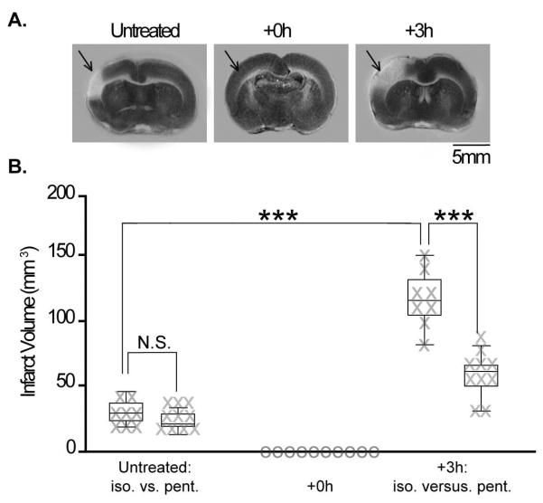
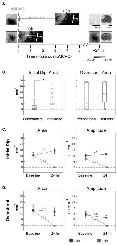
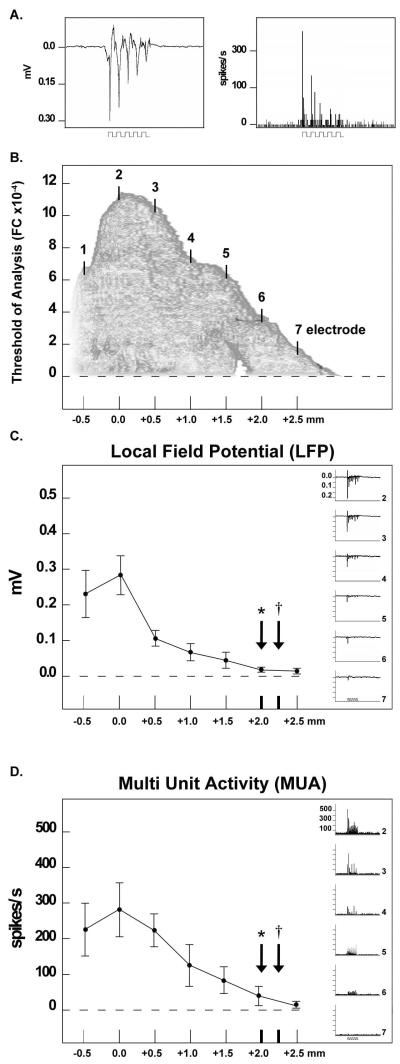
Using a rodent model of ischemia [permanent middle cerebral artery occlusion (pMCAO)], previous studies demonstrated that whisker stimulation treatment completely protects the cortex from impending stroke when initiated within 2 h following pMCAO. When initiated 3 h post-pMCAO, the identical treatment exacerbates stroke damage. Rats in these studies, however, were anesthetised with sodium pentobarbital, whereas human stroke patients are typically awake. To overcome this drawback, our laboratory has begun to use the anesthetic isoflurane, which allows rats to rapidly recover from pMCAO within minutes, to test stimulation treatment in awake rats and to determine whether isoflurane has an effect upon the pMCAO stroke model. We found no difference in infarct volume between pMCAO in untreated controls under either sodium pentobarbital or isoflurane, and the primary finding was that rats that received treatment immediately post-pMCAO maintain cortical function and no stroke damage, whereas rats that received treatment 3 h post-pMCAO exhibited eliminated cortical activity and extensive stroke damage. The only difference between anesthetics was the broad extent of evoked cortical activity observed during both functional imaging and electrophysiological recording, suggesting that the extent of evoked activity evident under isoflurane anesthesia is supported by underlying neuronal activity. Given the high degree of similarity with previous data, we conclude that the pMCAO stroke model is upheld with the use of isoflurane. This study demonstrated that the isoflurane-anesthetised rat pMCAO model can be used for cerebrovascular studies, and allows for highly detailed investigation of potential novel treatments for ischemic stroke using awake, behaving animals.

Keywords: isoflurane; protection; rat; rodent model; stimulation treatment; stroke.


Figures

Fig. 1
Fig. 1
Schematic of the experimental design. For both experimental groups, vertical lines indicate the time of pMCAO, time of treatment, and the following 24 h reassessment.
Fig. 2
Fig. 2
Box-and-whisker plots, with individual data plotted, of the volume of infarct sustained by animals that underwent: pMCAO (no-stimulation controls), pMCAO and single whisker stimulation immediately (+0 h), or 3 h (+3 h) post-occlusion as assessed via 2,3,5-triphenyltetrazolium chloride assay for infarct. No-stimulation controls never received whisker stimulation, yet underwent anesthetic and surgical procedures (including pMCAO) that were identical to those of the experimental groups. Significant difference in infarct volume between groups (***p<0.0001).
Fig. 3
Fig. 3
Isoflurane is an effective alternative to sodium pentobarbital for use in sensory-induced neuroprotective studies. (A) Representative data from ISOI of the initial dip for +0 h and +3 h groups, and from 2,3,5-triphenyltetrazolium chloride (TTC) staining of +0 h and +3 h groups for comparison. All +0 h subjects regained whisker functional representation and did not sustain infarct, whereas +3 h rats never demonstrated any post-pMCAO cortical activity and sustained infarct larger than that of untreated controls (staining indicates healthy tissue, lack of staining would indicate ischemic infarct). Linear grayscale bar indicates intrinsic signal strength ×10−4. Scale bar below imaging data indicates 2 mm, the bar below TTC indicates 5 mm, and arrows indicate approximate region vulnerable to pMCAO infarct. (B) Box-and-whisker plots, with individual data plotted, for quantitative comparison of whisker functional representation observed at baseline in +0 h animals anesthetized with isoflurane (black) or sodium pentobarbital (gray). Both the area of the initial dip (left) and overshoot (right) are included for comparison. Significant difference between the area of the initial dip evoked while under sodium pentobarbital vs. isoflurane anesthesia (*p<0.05). Quantitative analysis of whisker functional representation in terms of area (left) and amplitude (right) of the initial dip (C) and overshoot (D). Group baseline and 24 h post-pMCAO data are plotted in each graph. A value of zero indicates no response. Means and SEs are provided for the area and amplitude of the whisker functional representation. Significant differences between group baseline and 24 h values (***p<0.001).
Fig. 4
Fig. 4
The whisker functional representation is quantified at a higher threshold of analysis under isoflurane anesthesia. Each phase of the whisker functional representation may be rendered three-dimensionally by plotting FC along the z-axis and its two-dimensional areal extent may be visualized and quantified at incrementally higher thresholds. The threshold used in this study is 5.0 × 10⁻⁴ FC (used to quantify the area of activity). Although previous work has utilized 2.5 × 10⁻⁴ FC, the higher 5.0 × 10⁻⁴ FC threshold was chosen here to achieve areal extent values that were comparable to the previous studies. Outlined in white, a +0 h subject's initial dip is visualized at 2.5 × 10⁻⁴ FC under sodium pentobarbital anesthesia (left), and a +0 h subject's initial dip is visualized at 5.0 × 10⁻⁴ FC under isoflurane (right).
Fig. 5
Fig. 5
Evoked neuronal activity underlies the whisker functional representation. (A) Representative LFP (measured in mV) and MUA (measured in spikes/s) responses recorded from a control rat anesthetized with isoflurane. Stepping function indicates stimulus delivery. Note the fast adaptation of both the LFP and MUA response to whisker stimulation. (B) Approximate placement of the seven-electrode array superimposed upon the initial dip. The X-axis represents the distance from the peak of the initial dip. The Y-axis represents the amplitude of whisker-evoked initial dip activity at a given distance from the peak. In this example, the initial dip is an average of the five rats used for extracellular recording. Evoked LFP (C) and MUA (D) activity plotted along the electrode array. Mean and SE are plotted for each recording site. The X-axis represents the distance from the peak of whisker-evoked activity, and arrows along the X-axis represent the average radius of the initial dip (arrows with asterisks) and overshoot (arrows with daggers) observed in recording subjects during ISOI mapping. Insets: representative LFP and MUA data from recording electrodes 2 (peak activity) to 7. Stepping function indicates stimulus delivery.

References

    1. Alkire MT. Probing the mind: anesthesia and neuroimaging. Clin Pharmacol Ther. 2008;84:149–152. - PubMed
    1. Alkire MT, Hudetz AG, Tononi G. Consciousness and anesthesia. Science. 2008;322:876–880. - PMC - PubMed
    1. Baughman VL, Hoffman WE, Miletich DJ, Albrecht RF, Thomas C. Neurologic outcome in rats following incomplete cerebral ischemia during halothane, isoflurane, or N2O. Anesthesiology. 1988;69:192–198. - PubMed
    1. Bederson JB, Pitts LH, Tsuji M, Nishimura MC, Davis RL, Bartkowski H. Rat middle cerebral artery occlusion: evaluation of the model and development of a neurologic examination. Stroke. 1986;17:472–476. - PubMed
    1. Berger T, Borgdorff A, Crochet S, Neubauer FB, Lefort S, Fauvet B, Ferezou I, Carleton A, Luscher HR, Petersen CC. Combined voltage and calcium epifluorescence imaging in vitro and in vivo reveals subthreshold and suprathreshold dynamics of mouse barrel cortex. J Neurophysiol. 2007;97:3751–3762. - PubMed

Publication types

LinkOut - more resources