

Elastic fibres are broadly distributed in tendon and highly localized around tenocytes
- PMID: 23587025
- PMCID: PMC3666236
- DOI: 10.1111/joa.12048
Elastic fibres are broadly distributed in tendon and highly localized around tenocytes
Abstract
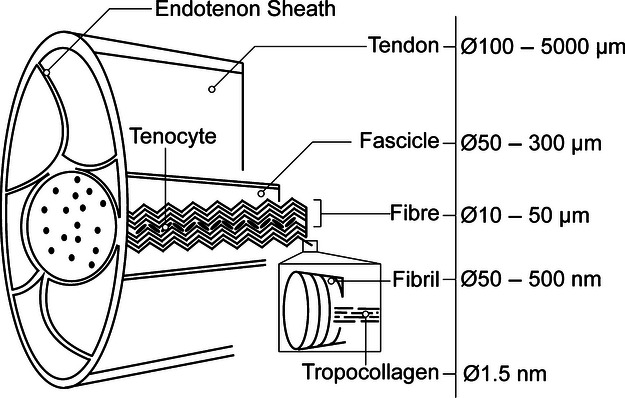
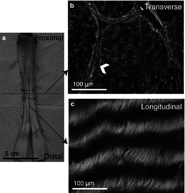
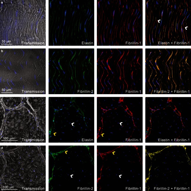
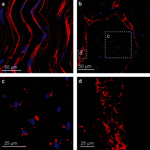
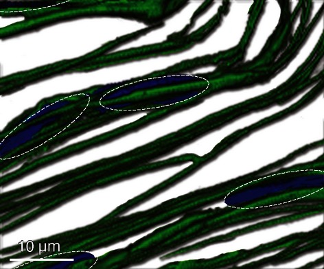
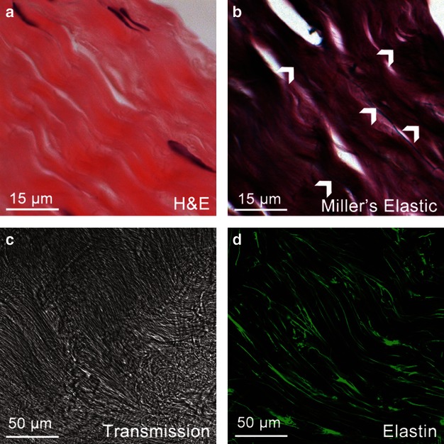
Elastic fibres have the unique ability to withstand large deformations and are found in numerous tissues, but their organization and structure have not been well defined in tendon. The objective of this study was to characterize the organization of elastic fibres in tendon to understand their function. Immunohistochemistry was used to visualize elastic fibres in bovine flexor tendon with fibrillin-1, fibrillin-2 and elastin antibodies. Elastic fibres were broadly distributed throughout tendon, and highly localized longitudinally around groups of cells and transversely between collagen fascicles. The close interaction of elastic fibres and cells suggests that elastic fibres are part of the pericellular matrix and therefore affect the mechanical environment of tenocytes. Fibres present between fascicles are likely part of the endotenon sheath, which enhances sliding between adjacent collagen bundles. These results demonstrate that elastic fibres are highly localized in tendon and may play an important role in cellular function and contribute to the tissue mechanics of the endotenon sheath.
© 2013 Anatomical Society.
Figures
Similar articles
-
The organisation of elastin and fibrillins 1 and 2 in the cruciate ligament complex.J Anat. 2011 Jun;218(6):600-7. doi: 10.1111/j.1469-7580.2011.01374.x. Epub 2011 Apr 6. J Anat. 2011. PMID: 21466551 Free PMC article.
-
Distribution of the elastic fiber and associated proteins in flexor tendon reflects function.Anat Rec. 2002 Dec 1;268(4):430-40. doi: 10.1002/ar.10175. Anat Rec. 2002. PMID: 12420291
-
Contributions of collagen and elastin to elastic behaviours of tendon fascicle.Acta Biomater. 2024 Mar 1;176:334-343. doi: 10.1016/j.actbio.2024.01.014. Epub 2024 Jan 17. Acta Biomater. 2024. PMID: 38237712
-
Structure of the tendon connective tissue.Scand J Med Sci Sports. 2000 Dec;10(6):312-20. doi: 10.1034/j.1600-0838.2000.010006312.x. Scand J Med Sci Sports. 2000. PMID: 11085557 Review.
-
Basic components of connective tissues and extracellular matrix: elastin, fibrillin, fibulins, fibrinogen, fibronectin, laminin, tenascins and thrombospondins.Adv Exp Med Biol. 2014;802:31-47. doi: 10.1007/978-94-007-7893-1_3. Adv Exp Med Biol. 2014. PMID: 24443019 Review.
Cited by
-
Achilles and tail tendons of perlecan exon 3 null heparan sulphate deficient mice display surprising improvement in tendon tensile properties and altered collagen fibril organisation compared to C57BL/6 wild type mice.PeerJ. 2018 Jun 29;6:e5120. doi: 10.7717/peerj.5120. eCollection 2018. PeerJ. 2018. PMID: 30042881 Free PMC article.
-
Elastic fibers in orthopedics: Form and function in tendons and ligaments, clinical implications, and future directions.J Orthop Res. 2020 Nov;38(11):2305-2317. doi: 10.1002/jor.24695. Epub 2020 Apr 28. J Orthop Res. 2020. PMID: 32293749 Free PMC article. Review.
-
Fibrillins in Tendon.Front Aging Neurosci. 2016 Oct 20;8:237. doi: 10.3389/fnagi.2016.00237. eCollection 2016. Front Aging Neurosci. 2016. PMID: 27812333 Free PMC article. Review.
-
Hierarchical ultrastructure: An overview of what is known about tendons and future perspective for tendon engineering.Bioact Mater. 2021 Jun 29;8:124-139. doi: 10.1016/j.bioactmat.2021.06.007. eCollection 2022 Feb. Bioact Mater. 2021. PMID: 34541391 Free PMC article. Review.
-
Elastase treatment of tendon specifically impacts the mechanical properties of the interfascicular matrix.Acta Biomater. 2021 Mar 15;123:187-196. doi: 10.1016/j.actbio.2021.01.030. Epub 2021 Jan 26. Acta Biomater. 2021. PMID: 33508509 Free PMC article.
References
-
- Butler D, Grood E, Noyes F, et al. Biomechanics of ligaments and tendons. Exerc Sport Sci Rev. 1978;6:125–181. - PubMed
-
- Caldini EG, Caldini N, De-Pasquale V, et al. Distribution of elastic system fibres in the rat tail tendon and its associated sheaths. Acta Anat. 1990;139:341–348. - PubMed
-
- Carvalho HF, Felisbino SL, Keene DR, et al. Identification, content, and distribution of type VI collagen in bovine tendons. Cell Tissue Res. 2006;325:315–324. - PubMed
MeSH terms
Substances
LinkOut - more resources
Full Text Sources
Other Literature Sources
