Review
. 2013 May;23(3):342-9.
doi: 10.1111/bpa.12044.

"Prion-like" templated misfolding in tauopathies

Affiliations
Review

"Prion-like" templated misfolding in tauopathies

Florence Clavaguera et al. Brain Pathol. 2013 May.

Abstract

The soluble microtubule-associated protein tau forms hyperphosphorylated, insoluble and filamentous inclusions in a number of neurodegenerative diseases referred to as "tauopathies." In Alzheimer's disease, tau pathology develops in a stereotypical manner, with the first lesions appearing in the locus coeruleus and entorhinal cortex, from where they appear to spread to the hippocampus and neocortex. Propagation of tau pathology is also a characteristic of argyrophilic grain disease, where the tau lesions spread throughout the limbic system. Significantly, isoform composition and morphology of tau filaments can differ between tauopathies, suggesting the existence of distinct tau strains. Extensive experimental findings indicate that prion-like mechanisms underly the pathogenesis of tauopathies.


Conflict of interest statement

The authors declare that they have no conflict of interest related to this article.

Figures

Figure 1
Figure 1
Schematic representation of MAPT and the six tau isoforms expressed in adult human brain. Human MAPT consists of 16 exons (E). Alternative mRNA splicing of E2 (pink), E3 (green) and E10 (yellow) gives rise to the six tau isoforms (352–441 amino acids). E1, E4, E5, E7, E9, E11, E12 and E13 (blue) are constitutively spliced exons. E0, which is part of the promoter, and E14, are non‐coding (white). E6 and E8 (violet), as well as E4a (orange) are not transcribed in human brain. The core regions of the repeats of tau are shown as black bars. Three isoforms have four repeats each (4R hTau) and three isoforms have three repeats each (3R hTau). Similar levels of 4R and 3R tau isoforms are expressed in normal adult human brain. The exons and introns are not drawn to scale.
Figure 2
Figure 2
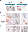
Tau transgenic mouse models and experimental induction of filamentous tauopathy. A. Left: Mice expressing the 383 amino acid 4R human tau with the P301S mutation under the control of the murine Thy1 promoter develop abundant hyperphosphorylated (AT8‐immunoreactive) and Gallyas–Braak silver‐positive filamentous tau inclusions in various brain regions, including the brainstem, from which extracts were prepared for injection into the brains of ALZ17 mice. Right: ALZ17 mice express the 441‐amino acid 4R human tau isoform under the control of the murine Thy1 promoter. These mice develop tau that is immunoreactive with the phosphorylation‐dependent antibody AT8, but Gallyas–Braak silver‐negative as shown here for the hippocampus. These mice also lack tau filaments. The sections were counterstained with hematoxylin. Scale bar: 50 μm (the same magnification in all panels). B. Staining of the hippocampal CA3 region from 18‐month‐old ALZ17 mice with AT8, Gallyas–Braak silver and phosphorylation‐dependent anti‐tau antibody AT100, 15 months after no injection (upper panel) or after the injection with brain extract from 6‐month‐old P301S tau mice (lower panel). The injection of brain extracts induced the formation of AT100‐ and Gallyas–Braak silver‐positive filamentous inclusions made of wild‐type human tau. No such inclusions were found in ALZ17 mice injected with extracts from control mice 19. The sections were counterstained with hematoxylin. Scale bar: 50 μm (the same magnification in all panels). C. Different types of filamentous tau pathology in ALZ17 brains after the injection with brain extracts from mice transgenic for human P301S tau: Gallyas–Braak silver impregnation shows a neurofibrillary tangle (left), neuropil threads (middle, arrows) and coiled bodies (right, arrows). The sections were counterstained with hematoxylin. Scale bar: 50 μm (the same magnification in all panels).
Figure 3
Figure 3
Injection of synthetic tau filaments into the hippocampus induces the formation of tau inclusions in mice transgenic for human mutant P301S tau. A. Gallyas–Braak silver stained section of the hippocampus from a 4‐month‐old homozygous mouse transgenic for human mutant P301S tau that had been injected with 7.5 μg assembled recombinant human P301S tau (four‐repeat isoform lacking amino‐terminal inclusions) at the age of 3 months. For assembly, recombinant human P301S tau (3 mg/mL) was incubated with heparin (400 μg/mL) for 48 h at 37°C. B. Gallyas–Braak silver stained section of the hippocampus from a 4‐month‐old non‐injected mouse transgenic for human mutant P301S tau. Scale bar: 50 μm.

References

    1. Akoury E, Gajda M, Pickhardt M, Biernat J, Soraya P, Griesinger C et al (2013) Inhibition of tau filament formation by conformational modulation. J Am Chem Soc 35:2853–2862. - PubMed
    1. Allen B, Ingram E, Takao M, Smith MJ, Jakes R, Virdee K et al (2002) Abundant tau filaments and nonapoptotic neurodegeneration in transgenic mice expressing human P301S tau protein. J Neurosci 22:9340–9351. - PMC - PubMed
    1. Andreadis A, Brown WM, Kosik KS (1992) Structure and novel exons of the human tau gene. Biochemistry 31:10626–10633. - PubMed
    1. Arai T, Ikeda K, Akiyama H, Nonaka T, Hasegawa M, Ishiguro K et al (2004) Identification of amino‐terminally cleaved tau fragments that distinguish progressive supranuclear palsy from corticobasal degeneration. Ann Neurol 55:72–79. - PubMed
    1. Arnold CS, Johnson GV, Cole RN, Dong DL, Lee M, Hart GW (1996) The microtubule‐associated protein tau is extensively modified with O‐linked N‐acetylglucosamine. J Biol Chem 271:28741–28744. - PubMed

Publication types

MeSH terms