Review
. 2013 Apr 16:21:29.
doi: 10.1186/1757-7241-21-29.

Thrombelastography (TEG®): practical considerations on its clinical use in trauma resuscitation

Affiliations
Review

Thrombelastography (TEG®): practical considerations on its clinical use in trauma resuscitation

Luis Teodoro da Luz et al. Scand J Trauma Resusc Emerg Med. .

Abstract

Background: Thrombelastography is a laboratorial test that measures viscoelastic changes of the entire clotting process. There is growing interest in its clinical use in trauma resuscitation, particularly for managing acute coagulopathy of trauma and assisting decision making concerning transfusion. This review focuses on the clinical use of thrombelastography in trauma, with practical points to consider on its use in civilian and military settings.

Methods: A search in the literature using the terms "thrombelastography AND trauma" was performed in PUBMED database. We focused the review on the main clinical aspects of this viscoelastic method in diagnosing and treating patients with acute coagulopathy of trauma during initial resuscitation.

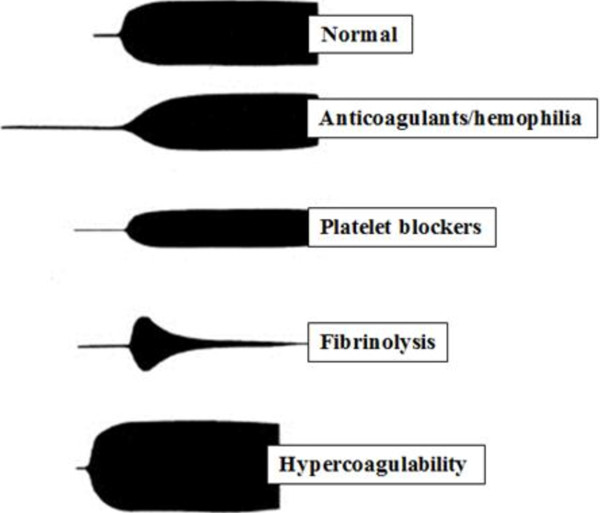
Results: Thrombelastography is not a substitute for conventional laboratorial tests such as INR and aPTT but offers additional information and may guide blood transfusion. Thrombelastography can be used as a point of care test but requires multiple daily calibrations, should be performed by trained personnel and its technique requires standardization. While useful partial results may be available in minutes, the whole test may take as long as other conventional tests. The most important data provided by thrombelastography are clot strength and fibrinolysis. Clot strength measure can establish whether the bleeding is due to coagulopathy or not, and is the key information in thrombelastography-based transfusion algorithms. Thrombelastography is among the few tests that diagnose and quantify fibrinolysis and thus guide the use of anti-fibrinolytic drugs and blood products such as cryoprecipitate and fibrinogen concentrate. It may also diagnose platelet dysfunction and hypercoagulability and potentially prevent inappropriate transfusions of hemostatic blood products to non-coagulopathic patients.

Conclusions: Thrombelastography has characteristics of an ideal coagulation test for use in early trauma resuscitation. It has limitations, but may prove useful as an additional test. Future studies should evaluate its potential to guide blood transfusion and the understanding of the mechanisms of trauma coagulopathy.


Figures

Figure 1
Figure 1
The Principles of the Thromboelastography. The viscoelastic properties of the clot are measured. Whole blood is inserted in the cup. A torsion wire suspends a pin immersed in the cup and connects with a mechanical–electrical transducer. The cup rotates through 4°45 to imitate sluggish venous flow and activate coagulation. The speed and strength of clot formation is measured in various ways (now usually by computer), and depends on the activity of the plasmatic coagulation system, platelet function and fibrinolysis.
Figure 2
Figure 2
A representative signature waveform of a normal TEG® tracing. R(seconds) = time of latency from start of test to initial fibrin formation; K (seconds) = time taken to achieve a certain level of clot strength (amplitude of 20mm); α angle (degrees) = measures the speed at which fibrin build up and cross linking takes place, hence assesses the rate of clot formation; MA (millimeters) = represents the ultimate strength of the fibrin clot; LY30 (%) = percentage decrease in amplitude at 30 minutes post-MA and gives measure of degree of fibrinolysis.
Figure 3
Figure 3
Examples of normal and abnormal tracings on TEG®. Normal (R, K, alpha and MA are normal); patient in use of anticoagulants or with hemophilia – deficit of coagulation factors (R, K are prolonged and alpha and MA are decreased); platelet dysfunction (R is normal, K is prolonged and MA is decreased); fibrinolysis (R is normal and MA is continuously decreases) and hypercoagulability (R, K are decreased and alpha and MA are increased).

References

    1. Greer SE, Rhynhart KK, Gupta R, Corwin HL. New developments in massive transfusion in trauma. Curr Opin Anesthesiol. 2010;23(2):246–250. doi: 10.1097/ACO.0b013e328336ea59. - DOI - PubMed
    1. Johansson PI, Ostrowski SR, Secher NH. Management of major blood loss: an update. Acta Anaesthesiol Scand. 2010;54:1039–1049. doi: 10.1111/j.1399-6576.2010.02265.x. - DOI - PubMed
    1. Scarpelini S, Rhind GS, Nascimento B, Tien H, Shek PN, Peng HT, Huang H, Pinto R, Speers V, Reis M, Rizoli SB. Normal range values for thromboelastography in healthy adult volunteers. Braz J Med Biol Res. 2009;42(12):1210–1217. doi: 10.1590/S0100-879X2009007500002. - DOI - PubMed
    1. Hartert H. Blutgerinnungsstudien mit der thrombelastographie, enemneuen untersuchungsverfahren. Klin Wochensogr. 1948;26:577–583. doi: 10.1007/BF01697545. - DOI - PubMed
    1. Nielsen VG. A comparison of the thromboelastograph and the ROTEM®. Blood Coagul Fibrinolysis. 2007;18:247–252. doi: 10.1097/MBC.0b013e328092ee05. - DOI - PubMed

Publication types

MeSH terms