. 2013 Apr 15:8:61.
doi: 10.1186/1746-1596-8-61.

The Th17/Treg balance and the expression of related cytokines in Uygur cervical cancer patients

Affiliations

The Th17/Treg balance and the expression of related cytokines in Uygur cervical cancer patients

Zhifang Chen et al. Diagn Pathol. .

Abstract

Background: The fine balance of Th17/Treg is crucial for maintenance of immune homeostasis. The objective of this study was to investigate the balance of Th17/Treg and the expression of related cytokines in Uighur cervical cancer patients.

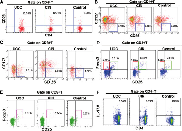
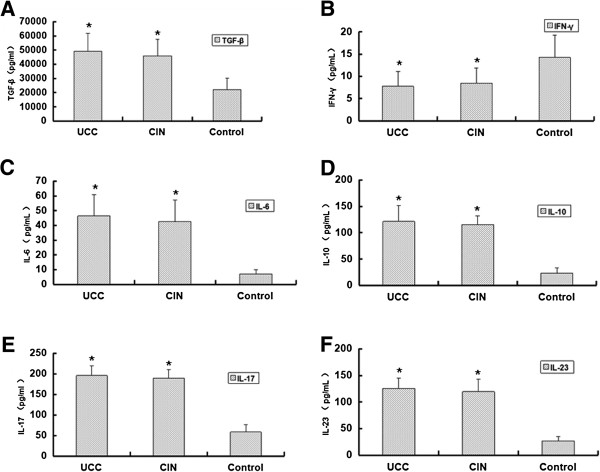
Methods: Peripheral blood was collected from 65 cases of cervical cancer patients, 42 cases of cervical CIN patients and 40 healthy people. Flow cytometry was used to detect the percentages of T cell subsets, including CD3+ T cells, CD4+ T cells, CD8+ T cells, Treg cells and Th17 cells. ELISA assay was conducted to detect expression levels of TGF-β, IL-6, IL-10, IL-17, IL-23 and IFN-γ.

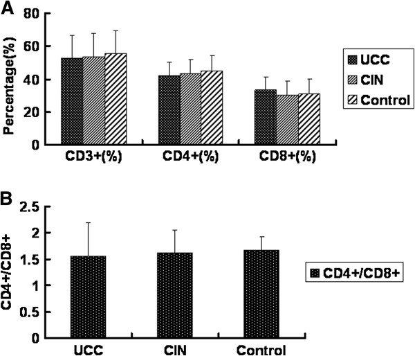
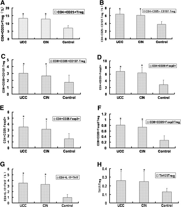
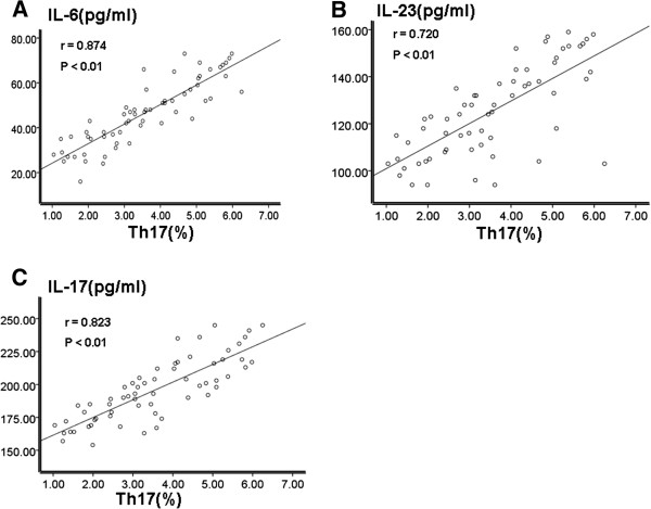
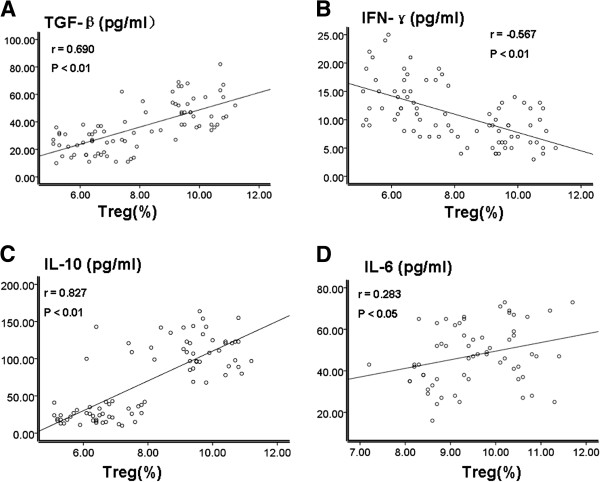
Results: There were no significant difference in the levels of CD3+ T cells, CD4+ T cells, CD8+ T cells, and the ratio of CD4+/CD8+ among the cervical cancer group, the CIN group and the healthy control group. However, compared with the healthy control group, the percentages of CD4+ CD25+ Treg, CD4+CD25+CD127- Treg, CD4+IL17+ Th17, CD4+CD25+Foxp3+, CD4+CD25- Foxp3+, CD8+CD25+CD127-Treg and CD8+CD25+Foxp3 were significantly higher in the cervical cancer group and the CIN group. Similar results were also found in the Th17/Treg ratio and the related cytokines. There was no significant difference between the cervical cancer group and the CIN group. Additionally, Th17 cell levels were positively correlated with IL-6, IL-23 and IL-17. Also, Treg cell levels were positively correlated with TGF-β, IL-10 and IL-6. Contrarily, Treg cell levels and IFN-γ were negatively correlated.

Conclusions: Our data indicated that the Th17/Treg balance was broken in peripheral blood of cervical cancer patients. Analysis of Th17/Treg balance may have a significant implication in diagnosing cervical cancer.

Virtual slides: The virtual slide for this article can be found here: http://www.diagnosticpathology.diagnomx.eu/vs/1813823795931511.


Figures

Figure 1
Figure 1
Analysis of CD3+ T cells, CD4+ T cells, CD8+ T cells and the CD4+/CD8+ ratio in the peripheral blood of three groups. Peripheral blood samples were collected from 65 cases of cervical cancer patients, 42 cases of cervical CIN patients and 40 cases of healthy people. Flow cytometry was used to conduct this experiment. (A) Percentages of CD3+ T cells, CD4+ T cells and CD8+ T cells; (B) The ratio of CD4+/CD8+ in the UCC group, the cervical CIN group and the healthy control group. p > 0.05.
Figure 2
Figure 2
Flow cytometric analysis of Treg cells and Th17 cells in the peripheral blood of three groups. Peripheral blood samples were collected from 65 cases of cervical cancer patients, 42 cases of cervical CIN patients and 40 cases of healthy people. Representative flow cytometric results were shown. The indicated percentages in each figure represented the levels of the analyzed cell subsets. (A) Percentages of CD4+CD25+Treg cells in CD4+T cells; (B) Percentages of CD4+CD25+CD127lowTreg cells in CD4+T cells; (C) Percentages of CD8+CD25+CD127lowTreg cells in CD8+T cells; (D) Percentages of CD4+CD25+ FOXP3+Treg cells and CD4+CD25-FOXP3+Treg in CD4+T cells; (E) Percentages of CD8+CD25+ FOXP3+Treg cells in CD8+T cells; (F) Percentages of CD4+IL-17+Th17 cells in CD4+T cells.
Figure 3
Figure 3
Quantitative analysis of Treg cells and Th17 cells in the peripheral blood of three groups. Peripheral blood samples were collected from 65 cases of cervical cancer patients, 42 cases of cervical CIN patients and 40 cases of healthy people. Quantitative results of the flow cytometric analysis were shown. (A) Percentages of CD4+CD25+Treg cells in CD4+T cells; (B) Percentages of CD4+CD25+CD127lowTreg cells in CD4+T cells; (C) Percentages of CD8+CD25+CD127lowTreg cells in CD8+T cells; (D) Percentages of CD4+CD25+ FOXP3+Treg cells in CD4+T cells; (E) Percentages of CD4+CD25-FOXP3+Treg in CD4+T cells; (F) Percentages of CD8+CD25+ FOXP3+Treg cells in CD8+T cells; (G) Percentages of CD4+IL-17+Th17 cells in CD4+T cells. (H) Ratio of Th17/ Treg in three groups. Each experiment was performed three times. Percentages of cells in the UCC group and the cervical group were compared with those in the healthy control group. *p < 0.05.
Figure 4
Figure 4
Detection of related cytokines. Peripheral blood samples were collected from 65 cases of cervical cancer patients, 42 cases of cervical CIN patients and 40 cases of healthy people. ELISA assay was performed to detect the expression levels of related cytokines in the serum of the three groups. (A) levels of TGF-β; (B) levels of IFN-γ; (C) levels of IL-6; (D) levels of IL-10; (E) levels of IL-17; (F) levels of IL-23. Each experiment was performed three times. Levels of cytokines in the UCC group and the cervical group were compared with those in the healthy control group. *p < 0.05.
Figure 5
Figure 5
Correlation analysis of levels of Th17 cells and levels of IL-6, IL-23 and IL-17 in the UCC group. Correlation assay was conducted using the Spearman rank correlation test. (A) Correlation analysis between Th17 and IL-6; (B) Correlation analysis between Th17 and IL-23; (C) Correlation analysis between Th17 and IL-17.
Figure 6
Figure 6
Correlation analysis of levels of Treg cells and levels of TGF-β, IFN-γ, IL-10 and IL-6 in the UCC group. Correlation analysis was performed by the Spearman rank correlation test. (A) Correlation analysis between Treg and TGF-β; (B) Correlation analysis between Treg and IFN-γ; (C) Correlation analysis between Treg and IL-10; (D) Correlation analysis between Treg and IL-6.

References

    1. Shibuya K, Mathers CD, Boschi-Pinto C, Lopez AD, Murray CJ. Global and regional estimates of cancer mortality and incidence by site: II. Results for the global burden of disease 2000. BMC Cancer. 2002;2:37. doi: 10.1186/1471-2407-2-37. - DOI - PMC - PubMed
    1. Guzhalinuer A, Chen JX, Mick R. HPV spectroscopy study of Xinjiang Uygur cancer women. Tumor. 2007;27:379–382.
    1. Liu H, Liu S, Wang H. Genomic amplification of the human telomerase gene (hTERC) associated with human papillomavirus is related to the progression of uterine cervical dysplasia to invasive cancer. Diagn Pathol. 2012;7:147. doi: 10.1186/1746-1596-7-147. - DOI - PMC - PubMed
    1. Chen S, Yang Z, Zhang Y. Genomic amplification patterns of human telomerase RNA gene and C-MYC in liquid-based cytological specimens used for the detection of high-grade cervical intraepithelial neoplasia. Diagn Pathol. 2012;7:40. doi: 10.1186/1746-1596-7-40. - DOI - PMC - PubMed
    1. Han H, Yang Y, Lu Z. Decreased D2-40 and increased p16INK4A immunoreactivities correlate with higher grade of cervical intraepithelial neoplasia. Diagn Pathol. 2011;6:59. doi: 10.1186/1746-1596-6-59. - DOI - PMC - PubMed

Publication types

MeSH terms