

Brucella abortus induces TNF-α-dependent astroglial MMP-9 secretion through mitogen-activated protein kinases
- PMID: 23587438
- PMCID: PMC3637408
- DOI: 10.1186/1742-2094-10-47
Brucella abortus induces TNF-α-dependent astroglial MMP-9 secretion through mitogen-activated protein kinases
Abstract
Background: Central nervous system (CNS) invasion by bacteria of the genus Brucella results in an inflammatory disorder called neurobrucellosis. We have recently demonstrated that B. abortus infects microglia and astrocytes, eliciting the production of a variety of pro-inflammatory cytokines which contribute to CNS damage. Matrix metalloproteinases (MMP) have been implicated in inflammatory tissue destruction in a range of pathological situations in the CNS. Increased MMP secretion is induced by pro-inflammatory cytokines in a variety of CNS diseases characterized by tissue-destructive pathology.
Methods: In this study, the molecular mechanisms that regulate MMP secretion from Brucella-infected astrocytes in vitro were investigated. MMP-9 was evaluated in culture supernatants by ELISA, zymography and gelatinolytic activity. Involvement of mitogen-activated protein kinases (MAPK) signaling pathways was evaluated by Western blot and using specific inhibitors. The role of TNF-α was evaluated by ELISA and by assays with neutralizing antibodies.
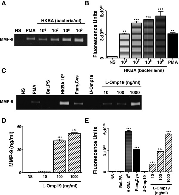
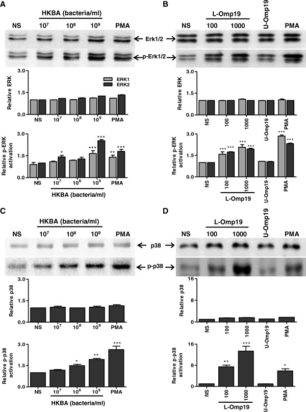
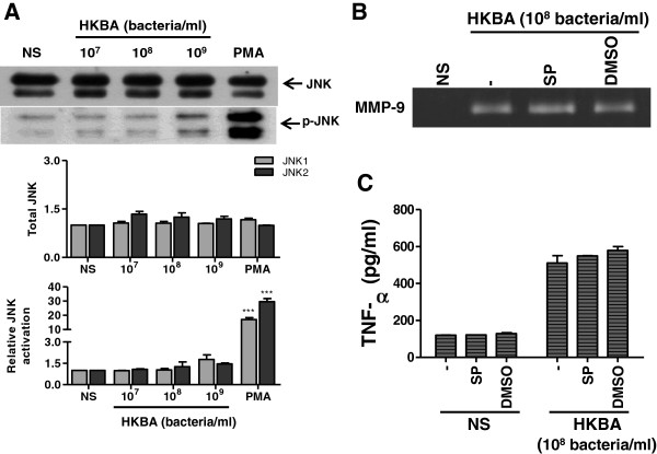
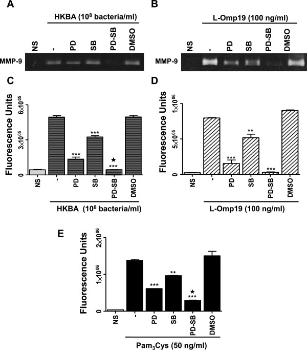
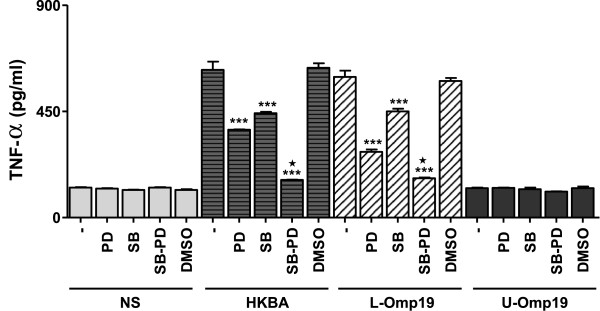
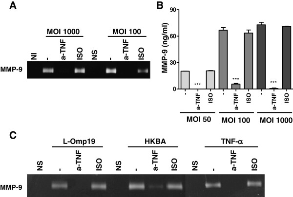
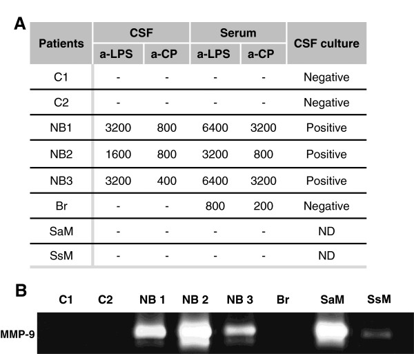
Results: B. abortus infection induced the secretion of MMP-9 from murine astrocytes in a dose-dependent fashion. The phenomenon was independent of bacterial viability and was recapitulated by L-Omp19, a B. abortus lipoprotein model, but not its LPS. B. abortus and L-Omp19 readily activated p38 and Erk1/2 MAPK, thus enlisting these pathways among the kinase pathways that the bacteria may address as they invade astrocytes. Inhibition of p38 or Erk1/2 significantly diminished MMP-9 secretion, and totally abrogated production of this MMP when both MAPK pathways were inhibited simultaneously. A concomitant abrogation of B. abortus- and L-Omp19-induced TNF-α production was observed when p38 and Erk1/2 pathways were inhibited, indicating that TNF-α could be implicated in MMP-9 secretion. MMP-9 secretion induced by B. abortus or L-Omp19 was completely abrogated when experiments were conducted in the presence of a TNF-α neutralizing antibody. MMP-9 activity was detected in cerebrospinal fluid (CSF) samples from patients suffering from neurobrucellosis.
Conclusions: Our results indicate that the inflammatory response elicited by B. abortus in astrocytes would lead to the production of MMP-9 and that MAPK may play a role in this phenomenon. MAPK inhibition may thus be considered as a strategy to control inflammation and CNS damage in neurobrucellosis.
Figures

Similar articles
-
Brucella abortus induces the secretion of proinflammatory mediators from glial cells leading to astrocyte apoptosis.Am J Pathol. 2010 Mar;176(3):1323-38. doi: 10.2353/ajpath.2010.090503. Epub 2010 Jan 21. Am J Pathol. 2010. PMID: 20093491 Free PMC article.
-
Potential role of fibroblast-like synoviocytes in joint damage induced by Brucella abortus infection through production and induction of matrix metalloproteinases.Infect Immun. 2011 Sep;79(9):3619-32. doi: 10.1128/IAI.05408-11. Epub 2011 Jul 5. Infect Immun. 2011. PMID: 21730088 Free PMC article.
-
Pathogenesis of Lyme neuroborreliosis: mitogen-activated protein kinases Erk1, Erk2, and p38 in the response of astrocytes to Borrelia burgdorferi lipoproteins.Neurosci Lett. 2005 Aug 12-19;384(1-2):112-6. doi: 10.1016/j.neulet.2005.04.069. Neurosci Lett. 2005. PMID: 15893422
-
The role of NLRP3 and AIM2 in inflammasome activation during Brucella abortus infection.Semin Immunopathol. 2017 Feb;39(2):215-223. doi: 10.1007/s00281-016-0581-1. Epub 2016 Jul 12. Semin Immunopathol. 2017. PMID: 27405866 Free PMC article. Review.
-
Neurobrucellosis: Brief Review.Neurologist. 2021 Nov 4;26(6):248-252. doi: 10.1097/NRL.0000000000000348. Neurologist. 2021. PMID: 34734902 Review.
Cited by
-
Microglia at the Crossroads of Pathogen-Induced Neuroinflammation.ASN Neuro. 2022 Jan-Dec;14:17590914221104566. doi: 10.1177/17590914221104566. ASN Neuro. 2022. PMID: 35635133 Free PMC article. Review.
-
Brucella Downregulates Tumor Necrosis Factor-α to Promote Intracellular Survival via Omp25 Regulation of Different MicroRNAs in Porcine and Murine Macrophages.Front Immunol. 2018 Jan 17;8:2013. doi: 10.3389/fimmu.2017.02013. eCollection 2017. Front Immunol. 2018. PMID: 29387067 Free PMC article.
-
Administration of SB203580, a p38 MAPK Inhibitor, Reduced the Expression of MMP9, and Relieved Neurologic Severity in the Experimental Autoimmune Neuritis (EAN) in Rats.Neurochem Res. 2015 Jul;40(7):1410-20. doi: 10.1007/s11064-015-1608-z. Epub 2015 May 22. Neurochem Res. 2015. PMID: 25998885
-
The research trend on neurobrucellosis over the past 30 years (1993-2023): a bibliometric and visualization analysis.Front Neurol. 2024 Sep 23;15:1349530. doi: 10.3389/fneur.2024.1349530. eCollection 2024. Front Neurol. 2024. PMID: 39381075 Free PMC article.
-
Brucella abortus-Stimulated Platelets Activate Brain Microvascular Endothelial Cells Increasing Cell Transmigration through the Erk1/2 Pathway.Pathogens. 2020 Aug 27;9(9):708. doi: 10.3390/pathogens9090708. Pathogens. 2020. PMID: 32867217 Free PMC article.
References
-
- Gimsa U, ORen A, Pandiyan P, Teichmann D, Bechmann I, Nitsch R, Brunner-Weinzierl MC. Astrocytes protect the CNS: antigen-specific T helper cell responses are inhibited by astrocyte-induced upregulation of CTLA-4 (CD152) J Mol Med (Berl) 2004;82:364–372. doi: 10.1007/s00109-004-0531-6. - DOI - PubMed
Publication types
MeSH terms
Substances
LinkOut - more resources
Full Text Sources
Other Literature Sources
Miscellaneous
