

Chronic nicotine exposure stimulates biliary growth and fibrosis in normal rats
- PMID: 23587498
- PMCID: PMC3800482
- DOI: 10.1016/j.dld.2013.02.023
Chronic nicotine exposure stimulates biliary growth and fibrosis in normal rats
Abstract
Background: Epidemiological studies have indicated smoking to be a risk factor for the progression of liver diseases. Nicotine is the chief addictive substance in cigarette smoke and has powerful biological properties throughout the body. Nicotine has been implicated in a number of disease processes, including increased cell proliferation and fibrosis in several organ systems.
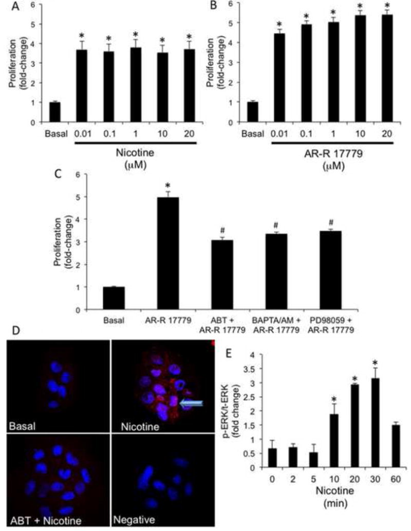
Aims: The aim of this study was to evaluate the effects of chronic administration of nicotine on biliary proliferation and fibrosis in normal rats.
Methods: In vivo, rats were treated with nicotine by osmotic minipumps for two weeks. Proliferation, α7-nicotinic receptor and profibrotic expression were evaluated in liver tissue, cholangiocytes and a polarized cholangiocyte cell line (normal rat intrahepatic cholangiocyte). Nicotine-dependent activation of the Ca(2+)/IP3/ERK 1/2 intracellular signalling pathway was also evaluated in normal rat intrahepatic cholangiocyte.
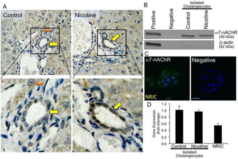
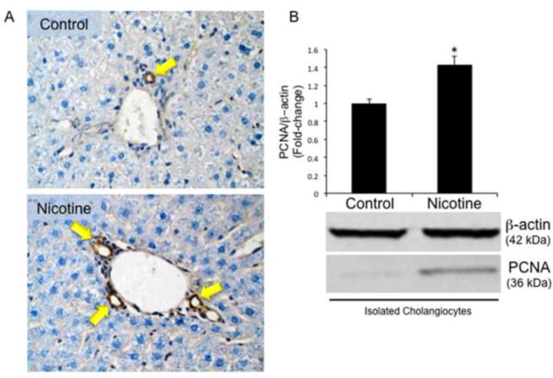
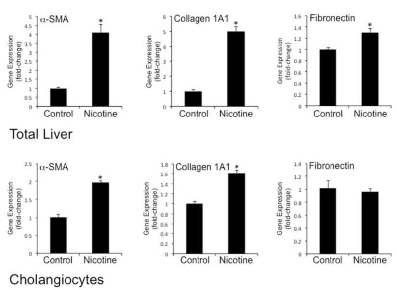
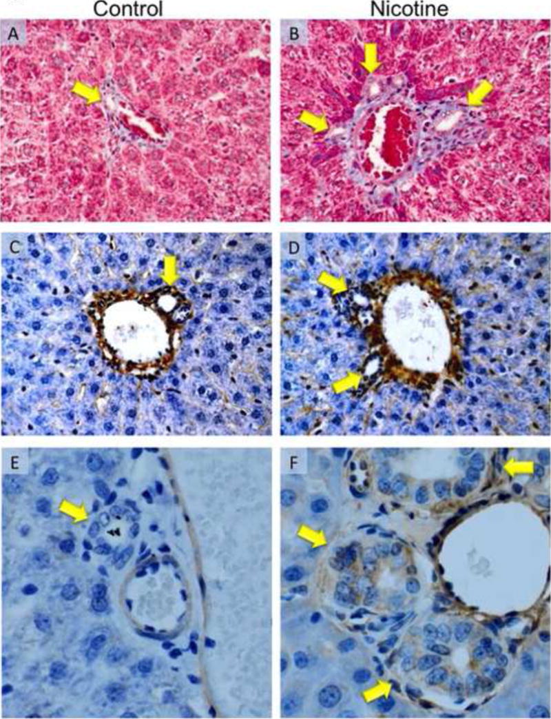
Results: Cholangiocytes express α7-nicotinic receptor. Chronic administration of nicotine to normal rats stimulated biliary proliferation and profibrotic gene and protein expression such as alpha-smooth muscle actin and fibronectin 1. Activation of α7-nicotinic receptor stimulated Ca(2+)/ERK1/2-dependent cholangiocyte proliferation.
Conclusion: Chronic exposure to nicotine contributes to biliary fibrosis by activation of cholangiocyte proliferation and expression of profibrotic genes. Modulation of α7-nicotinic receptor signalling axis may be useful for the management of biliary proliferation and fibrosis during cholangiopathies.
Keywords: Biliary epithelia; Cholangiocytes; Fibrosis; Nicotinic receptors; Proliferation.
Published by Elsevier Ltd.
Figures
Similar articles
-
Inhibition of integrin alphavbeta6 on cholangiocytes blocks transforming growth factor-beta activation and retards biliary fibrosis progression.Gastroenterology. 2008 Aug;135(2):660-70. doi: 10.1053/j.gastro.2008.04.009. Epub 2008 Apr 16. Gastroenterology. 2008. PMID: 18538673 Free PMC article.
-
Nicotine aggravates liver fibrosis via α7 nicotinic acetylcholine receptor expressed on activated hepatic stellate cells in mice.Hepatol Commun. 2024 Jun 5;8(6):e0457. doi: 10.1097/HC9.0000000000000457. eCollection 2024 Jun 1. Hepatol Commun. 2024. PMID: 38836815 Free PMC article.
-
H3 histamine receptor agonist inhibits biliary growth of BDL rats by downregulation of the cAMP-dependent PKA/ERK1/2/ELK-1 pathway.Lab Invest. 2007 May;87(5):473-87. doi: 10.1038/labinvest.3700533. Epub 2007 Mar 5. Lab Invest. 2007. PMID: 17334413 Free PMC article.
-
Cholangiocyte proliferation and liver fibrosis.Expert Rev Mol Med. 2009 Feb 25;11:e7. doi: 10.1017/S1462399409000994. Expert Rev Mol Med. 2009. PMID: 19239726 Free PMC article. Review.
-
Mechanisms of cholangiocyte responses to injury.Biochim Biophys Acta Mol Basis Dis. 2018 Apr;1864(4 Pt B):1262-1269. doi: 10.1016/j.bbadis.2017.06.017. Epub 2017 Jun 23. Biochim Biophys Acta Mol Basis Dis. 2018. PMID: 28648950 Free PMC article. Review.
Cited by
-
Regulators of Cholangiocyte Proliferation.Gene Expr. 2017 Feb 10;17(2):155-171. doi: 10.3727/105221616X692568. Epub 2016 Jul 12. Gene Expr. 2017. PMID: 27412505 Free PMC article. Review.
-
Is exposure to tobacco associated with extrahepatic cholangiocarcinoma epidemics? A retrospective proportional mortality study in China.BMC Cancer. 2019 Apr 11;19(1):348. doi: 10.1186/s12885-019-5484-9. BMC Cancer. 2019. PMID: 30975121 Free PMC article.
-
Effects of nicotine on microRNA-124 expression in bile duct ligation-induced liver fibrosis in rats.BMC Pharmacol Toxicol. 2024 Mar 28;25(1):27. doi: 10.1186/s40360-024-00749-3. BMC Pharmacol Toxicol. 2024. PMID: 38549169 Free PMC article.
-
Differential effect of visfatin inhibition on the testicular androgen and estrogen receptors expression in early pubertal mice.Endocrine. 2024 Jun;84(3):1216-1228. doi: 10.1007/s12020-024-03692-9. Epub 2024 Jan 26. Endocrine. 2024. PMID: 38273138
-
Nicotine alters the proteome of two human pancreatic duct cell lines.JOP. 2014 Sep 28;15(5):465-74. doi: 10.6092/1590-8577/2559. JOP. 2014. PMID: 25262714 Free PMC article.
References
-
- LeSage G, Benedetti A, Glaser S, et al. Acute carbon tetrachloride feeding selectively damages large, but not small, cholangiocytes from normal rat liver. Hepatology. 1999;29:307–319. - PubMed
-
- LeSage G, Glaser S, Gubba S, et al. Regrowth of the rat biliary tree after 70% partial hepatectomy is coupled to increased secretin-induced ductal secretion. Gastroenterology. 1996;111:1633–1644. - PubMed
-
- Alpini G, Prall RT, LaRusso NF. The pathobiology of biliary epithelia. In: Arias IM, Boyer JL, Chisari FV, Fausto N, Jakoby W, Schachter D, Shafritz DA, editors. The Liver; Biology & Pathobiology, 4E. Philadelphia, PA: Lippincott Williams & Wilkins; 2001. pp. 421–435.
-
- Alpini G, McGill JM, Larusso NF. The pathobiology of biliary epithelia. Hepatology. 2002;35:1256–1268. - PubMed
Publication types
MeSH terms
Substances
Grants and funding
LinkOut - more resources
Full Text Sources
Other Literature Sources
Medical
Miscellaneous
