. 2013 May 14;127(19):1957-67.
doi: 10.1161/CIRCULATIONAHA.112.001219. Epub 2013 Apr 15.

Divergent mitochondrial biogenesis responses in human cardiomyopathy

Affiliations

Divergent mitochondrial biogenesis responses in human cardiomyopathy

Preeti Ahuja et al. Circulation. .

Abstract

Background: Mitochondria are key players in the development and progression of heart failure (HF). Mitochondrial (mt) dysfunction leads to diminished energy production and increased cell death contributing to the progression of left ventricular failure. The fundamental mechanisms that underlie mt dysfunction in HF have not been fully elucidated.

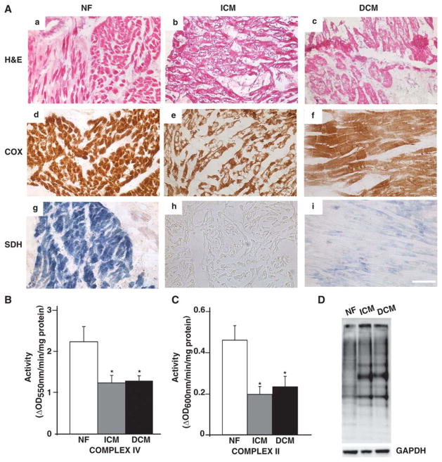
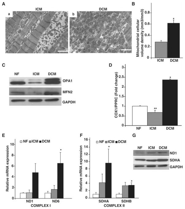
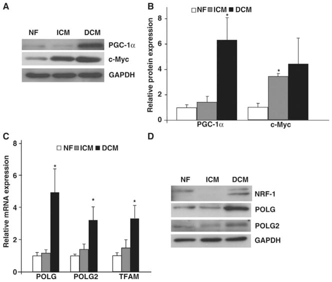
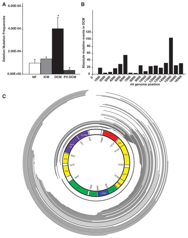
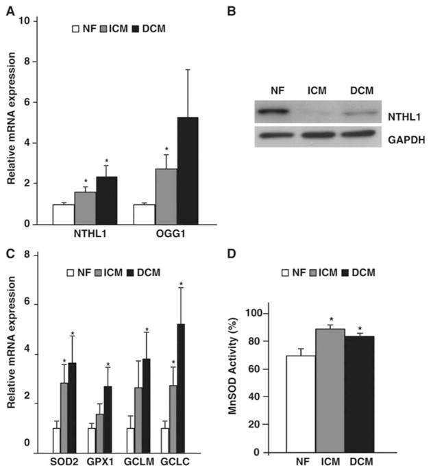
Methods and results: To characterize mt morphology, biogenesis, and genomic integrity in human HF, we investigated left ventricular tissue from nonfailing hearts and end-stage ischemic (ICM) or dilated (DCM) cardiomyopathic hearts. Although mt dysfunction was present in both types of cardiomyopathy, mt were smaller and increased in number in DCM compared with ICM or nonfailing hearts. mt volume density and mtDNA copy number was increased by ≈2-fold (P<0.001) in DCM hearts in comparison with ICM hearts. These changes were accompanied by an increase in the expression of mtDNA-encoded genes in DCM versus no change in ICM. mtDNA repair and antioxidant genes were reduced in failing hearts, suggestive of a defective repair and protection system, which may account for the 4.1-fold increase in mtDNA deletion mutations in DCM (P<0.05 versus nonfailing hearts, P<0.05 versus ICM).

Conclusions: In DCM, mt dysfunction is associated with mtDNA damage and deletions, which could be a consequence of mutating stress coupled with a peroxisome proliferator-activated receptor γ coactivator 1α-dependent stimulus for mt biogenesis. However, this maladaptive compensatory response contributes to additional oxidative damage. Thus, our findings support further investigations into novel mechanisms and therapeutic strategies for mt dysfunction in DCM.

Keywords: DNA, mitochondrial; cardiomyopathy, dilated; heart failure; mitochondrial turnover.


Conflict of interest statement

Conflict of Interest Disclosures: None.

Figures

Figure 1
Figure 1. Mitochondrial functional integrity and respiratory dysfunction in failing hearts
(A) H&E (a–c), COX (d–f) and SDH (g–i) stained frozen sections from NF, ICM and DCM left ventricular myocardium (n=3/group) showing cardiac myocyte degeneration and reduced functional state of electron transport chain complexes IV and II. Scale bar represents 50μm. Enzymatic activity of electron transport chain complexes (B) II and (C) IV on mt rich fractions obtained from NF, ICM and DCM hearts (*P<0.05 vs NF; n=8/group). (D) Representative micrograph of measurement of oxidized proteins in the failing hearts using oxyblot.
Figure 2
Figure 2. Mitochondrial morphology and biogenesis in failing hearts
(A) Transmission electron microscopy performed on heart sections from (a) ICM and (b) DCM myocardium demonstrating abnormal mt biogenesis in failing hearts. Scale bar represents 1μm. (B) Quantitative morphometric measurement of mt cellular volume density (μm3/μm3) based on analysis of electron micrographs from ICM and DCM ventricles (*P<0.001 vs NF; n=4/group). (C) Total ventricular protein lysates from indicated phenotypes were probed with antibodies against mt fusion proteins (OPA1 and MFN2) along with GAPDH as a loading control (n=4/group). (D) Quantitative real-time PCR on the mt gene COX1, along with the nuclear gene PPRC as an internal control from NF, ICM and DCM hearts showing increase in mtDNA content in DCM hearts. (*P<0.001, **P<0.05 vs NF; n=8/group). (E) Quantification of mt encoded Complex I ND1 and ND6 genes on NF, ICM and DCM hearts. (n=8/group, *P<0.05 vs NF). (F) Quantification of nuclear encoded Complex II SDHA and SDHB genes on NF, ICM and DCM hearts. (*P<0.05 vs NF). (G) Total ventricular protein lysates from indicated phenotypes probed with antibodies against electron transport Complex I and II proteins (ND1 and SDHA) along with GAPDH as a loading control (n=4/group).
Figure 3
Figure 3. Mitochondrial biogenesis in failing hearts
Total ventricular protein from NF, ICM and DCM ventricles assayed by (A) immunoblot and (B) quantified for mt biogenesis regulators PGC-1α and c-Myc. (*P<0.05 vs NF; (n=4/group). (C) Real-time PCR from NF and failing ventricles for mt biogenesis genes POLG, POLG2 and TFAM. (*P<0.05, **P<0.05 vs NF; n=8/group). (D) Total ventricular lysates from NF, ICM and DCM probed with antibodies against NRF-1, POLG and POLG2 along with GAPDH as a loading control (n=4/group).
Figure 4
Figure 4. Mitochondrial DNA deletion mutations in failing and in reverse remodeled hearts
(A) Deletions were PCR-amplified and quantified using RMC assay on DNA obtained from NF, ICM, DCM and Post-VAD supported hearts demonstrating increased frequency of mtDNA mutations in DCM hearts (*P<0.05, vs normal; n=8/group). (B) Histogram showing frequency of deletion mutations observed across the mt genome in one of the DCM failing heart. (C) Schematic representation of the human mtDNA-deletion mutations obtained from an end-stage DCM failing heart. mtDNA deletion mutations were determined by Solexa sequencing of mtDNA and breakpoints were determined by DNA sequence analysis. Arcs represent the deleted regions of the genome.
Figure 5
Figure 5. Mitochondrial DNA repair in failing hearts
Total ventricular RNA (A) and protein (B) from NF, ICM and DCM ventricles assayed by real-time PCR and immunoblot for mt DNA repair genes NTHL1 and OGG1. (*P<0.05 vs NF; n=8/group). (C) Real-time PCR from NF and failing ventricles for mitochondrial anti-oxidant genes SOD2, GPX1, GCLM and GCLC. (*P<0.05 vs NF; n=8/group. (D) Total ventricular protein lysates from NF, ICM and DCM determined for MnSOD activity (n=8/group).

Comment in

References

    1. Capetanaki Y. Desmin cytoskeleton: A potential regulator of muscle mitochondrial behavior and function. Trends Cardiovasc Med. 2002;12:339–348. - PubMed
    1. Marin-Garcia J, Goldenthal MJ. Mitochondrial centrality in heart failure. Heart Fail Rev. 2008;13:137–150. - PubMed
    1. Marin-Garcia J, Goldenthal MJ. the mitochondrial organelle and the heart. Rev Esp Cardiol. 2002;55:1293–1310. - PubMed
    1. Attardi G, Schatz G. Biogenesis of mitochondria. Annu Rev Cell Biol. 1988;4:289–333. - PubMed
    1. Huss JM, Kelly DP. Mitochondrial energy metabolism in heart failure: A question of balance. J Clin Invest. 2005;115:547–555. - PMC - PubMed

Publication types

Substances