

Protective effect of hydroferrate fluid, MRN-100, against lethality and hematopoietic tissue damage in γ-radiated Nile tilapia, Oreochromis niloticus
- PMID: 23589025
- PMCID: PMC3766301
- DOI: 10.1093/jrr/rrt029
Protective effect of hydroferrate fluid, MRN-100, against lethality and hematopoietic tissue damage in γ-radiated Nile tilapia, Oreochromis niloticus
Abstract
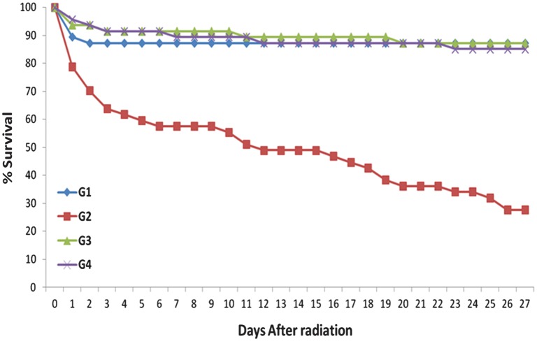
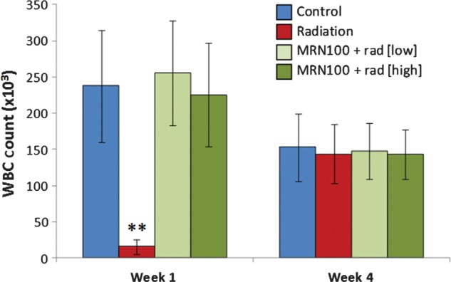
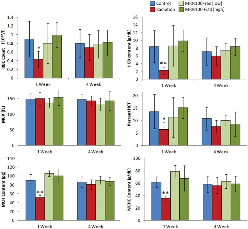
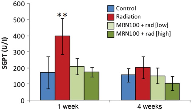
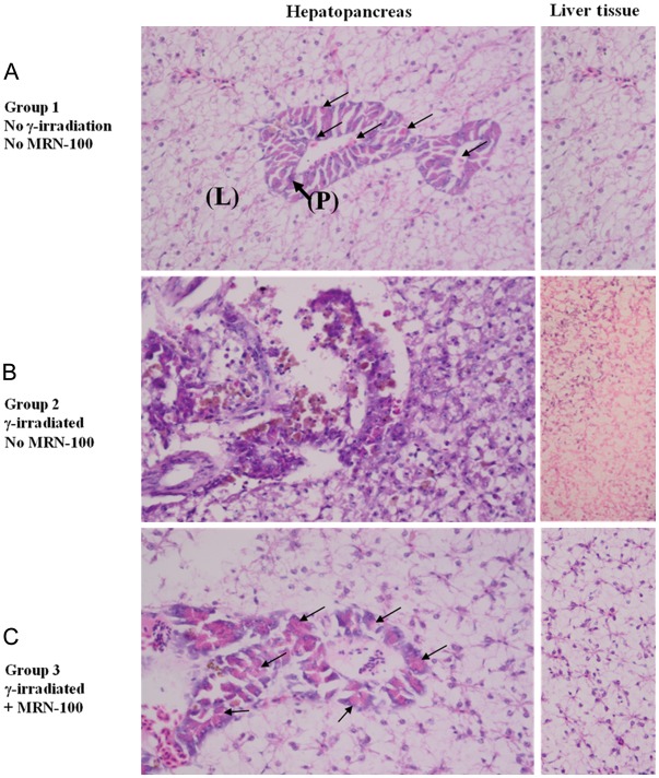
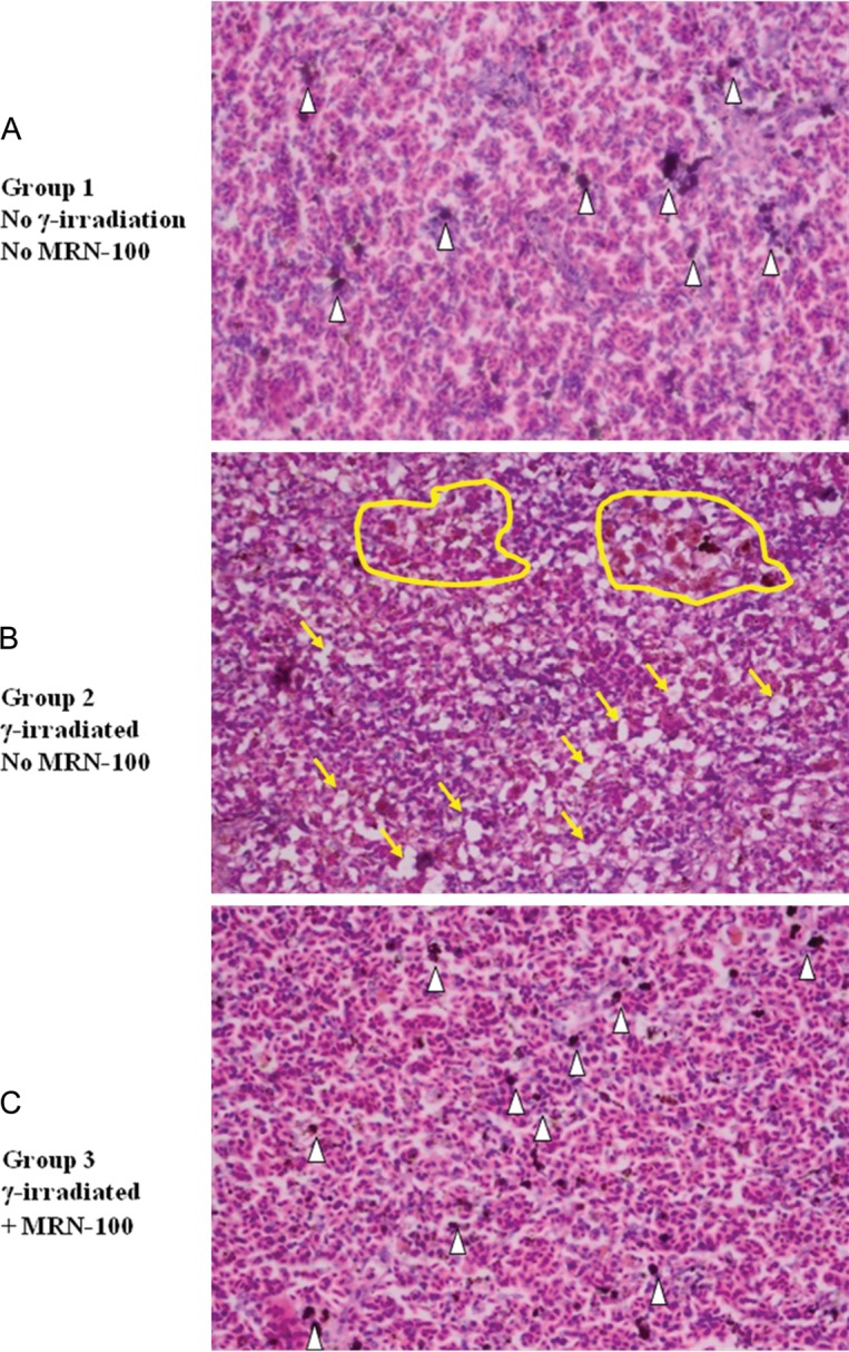
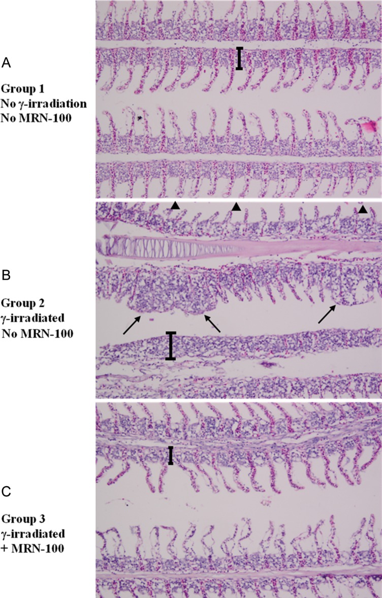
Hydroferrate fluid, MRN-100, an iron-based compound derived from bivalent and trivalent ferrates, is a potent antioxidant compound. Therefore, we examined the protective effect of MRN-100 against γ-radiation-induced lethality and damage to hematopoietic tissues in fish. A total of 216 Nile tilapia fish (Oreochromis niloticus) were randomly divided into four groups. Group 1 served as a control that was administered no radiation and no MRN-100 treatment. Group 2 was exposed only to γ-radiation (15 Gy). Groups 3 and 4 were pre-treated with MRN-100 at doses of either 1 ml/l or 3 ml/l in water for 1 week, and subsequently exposed to radiation while continuing to receive MRN-100 for 27 days. The survival rate was measured, and biochemical and histopathological analyses of hematopoietic tissues were performed for the different treatment groups at 1 and 4 weeks post-radiation. Exposure to radiation reduced the survival rate to 27.7%, while treatment with MRN-100 maintained the survival rate at 87.2%. In addition, fish exposed to γ-radiation for 1 week showed a significant decrease in the total number of white blood cells (WBCs) and red blood cells (RBCs) series. However, treatment with MRN-100 protected the total WBC count and the RBCs series when compared with irradiated fish. Furthermore, significant histological lesions were observed in the hepatopancreas, spleen and gills of irradiated fish. However, treatment with MRN-100 protected the histopathology of various organs. We conclude that MRN-100 is a radioprotective agent in fish and may be useful as an adjuvant treatment to counteract the adverse side effects associated with radiation exposure.
Keywords: Oreochromis niloticus; hydroferrate fluid; radiation; survival.
Figures
Similar articles
-
Hydroferrate fluid, MRN-100, provides protection against chemical-induced gastric and esophageal cancer in Wistar rats.Int J Biol Sci. 2015 Jan 28;11(3):295-303. doi: 10.7150/ijbs.10586. eCollection 2015. Int J Biol Sci. 2015. PMID: 25678848 Free PMC article.
-
Anti-radiation effect of MRN-100: a hydro-ferrate fluid, in vivo.J Radiat Res. 2024 Mar 22;65(2):145-158. doi: 10.1093/jrr/rrad095. J Radiat Res. 2024. PMID: 38247158 Free PMC article.
-
Arabinoxylan rice bran (MGN-3/Biobran) provides protection against whole-body γ-irradiation in mice via restoration of hematopoietic tissues.J Radiat Res. 2013 May;54(3):419-29. doi: 10.1093/jrr/rrs119. Epub 2013 Jan 3. J Radiat Res. 2013. PMID: 23287771 Free PMC article.
-
Impact of Abbreviated Filgrastim Schedule on Survival and Hematopoietic Recovery after Irradiation in Four Mouse Strains with Different Radiosensitivity.Radiat Res. 2017 Jun;187(6):659-671. doi: 10.1667/RR14555.1. Epub 2017 Mar 31. Radiat Res. 2017. PMID: 28362168 Free PMC article.
-
The Multiple Faces of the MRN Complex: Roles in Medulloblastoma and Beyond.Cancers (Basel). 2023 Jul 13;15(14):3599. doi: 10.3390/cancers15143599. Cancers (Basel). 2023. PMID: 37509263 Free PMC article. Review.
Cited by
-
Mitigating effect of biotin against irradiation-induced cerebral cortical and hippocampal damage in the rat brain tissue.Environ Sci Pollut Res Int. 2019 May;26(13):13441-13452. doi: 10.1007/s11356-019-04806-x. Epub 2019 Mar 25. Environ Sci Pollut Res Int. 2019. PMID: 30911963
-
Hydroferrate fluid, MRN-100, provides protection against chemical-induced gastric and esophageal cancer in Wistar rats.Int J Biol Sci. 2015 Jan 28;11(3):295-303. doi: 10.7150/ijbs.10586. eCollection 2015. Int J Biol Sci. 2015. PMID: 25678848 Free PMC article.
-
Potential role of MRN-100, an iron-based compound, in upregulating production of cytokine IL-10 in human dendritic cells to promote an anti-inflammatory response in vitro.Int J Immunopathol Pharmacol. 2019 Jan-Dec;33:2058738419844932. doi: 10.1177/2058738419844932. Int J Immunopathol Pharmacol. 2019. PMID: 30994016 Free PMC article.
-
Anti-radiation effect of MRN-100: a hydro-ferrate fluid, in vivo.J Radiat Res. 2024 Mar 22;65(2):145-158. doi: 10.1093/jrr/rrad095. J Radiat Res. 2024. PMID: 38247158 Free PMC article.
References
-
- Uma DP. Radiosensitivity of the developing haemopoietic system in mammals and its adult consequences: animal studies. Br J Radiol. 2003;76:366–72. - PubMed
-
- Fliedner TM, Graessle D-H. Hematopoietic cell renewal systems: mechanisms of coping and failing after chronic exposure to ionizing radiation. Radiat Environ Biophys. 2008;47:63–9. - PubMed
-
- Kalpana KB, Devipriya N, Srinivasan M, et al. Evaluating the radioprotective effect of hesperidin in the liver of Swiss albino mice. Eur J Pharmacol. 2011;658:206–12. - PubMed
-
- Ghoneum M, Badr El-Din NK, Abdel Fattah SM, et al. Arabinoxylan rice bran (MGN-3/Biobran) provides protection against whole-body gamma-irradiation in mice via restoration of hematopoietic tissues. J Radiat Res, (3 January 2013) 10.1093/jrr/rrs119. - DOI - PMC - PubMed
Publication types
MeSH terms
Substances
Grants and funding
LinkOut - more resources
Full Text Sources
Other Literature Sources
Medical
