A prospective clinical trial of tumor hypoxia imaging with 18F-fluoromisonidazole positron emission tomography and computed tomography (F-MISO PET/CT) before and during radiation therapy
- PMID: 23589026
- PMCID: PMC3823770
- DOI: 10.1093/jrr/rrt033
A prospective clinical trial of tumor hypoxia imaging with 18F-fluoromisonidazole positron emission tomography and computed tomography (F-MISO PET/CT) before and during radiation therapy
Abstract
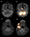
To visualize intratumoral hypoxic areas and their reoxygenation before and during fractionated radiation therapy (RT), (18)F-fluoromisonidazole positron emission tomography and computed tomography (F-MISO PET/CT) were performed. A total of 10 patients, consisting of four with head and neck cancers, four with gastrointestinal cancers, one with lung cancer, and one with uterine cancer, were included. F-MISO PET/CT was performed twice, before RT and during fractionated RT of approximately 20 Gy/10 fractions, for eight of the 10 patients. F-MISO maximum standardized uptake values (SUVmax) of normal muscles and tumors were measured. The tumor-to-muscle (T/M) ratios of F-MISO SUVmax were also calculated. Mean SUVmax ± standard deviation (SD) of normal muscles was 1.25 ± 0.17, and SUVmax above the mean + 2 SD (≥1.60 SUV) was regarded as a hypoxic area. Nine of the 10 tumors had an F-MISO SUVmax of ≥1.60. All eight tumors examined twice showed a decrease in the SUVmax, T/M ratio, or percentage of hypoxic volume (F-MISO ≥1.60) at approximately 20 Gy, indicating reoxygenation. In conclusion, accumulation of F-MISO of ≥1.60 SUV was regarded as an intratumoral hypoxic area in our F-MISO PET/CT system. Most human tumors (90%) in this small series had hypoxic areas before RT, although hypoxic volume was minimal (0.0-0.3%) for four of the 10 tumors. In addition, reoxygenation was observed in most tumors at two weeks of fractionated RT.
Keywords: 18F-misonidazole; PET/CT; reoxygenation; tumor hypoxia.
Figures


Similar articles
-
The reoxygenation of hypoxia and the reduction of glucose metabolism in head and neck cancer by fractionated radiotherapy with intensity-modulated radiation therapy.Eur J Nucl Med Mol Imaging. 2016 Nov;43(12):2147-2154. doi: 10.1007/s00259-016-3431-4. Epub 2016 Jun 1. Eur J Nucl Med Mol Imaging. 2016. PMID: 27251644 Clinical Trial.
-
Simultaneous positron emission tomography (PET) assessment of metabolism with ¹⁸F-fluoro-2-deoxy-d-glucose (FDG), proliferation with ¹⁸F-fluoro-thymidine (FLT), and hypoxia with ¹⁸fluoro-misonidazole (F-miso) before and during radiotherapy in patients with non-small-cell lung cancer (NSCLC): a pilot study.Radiother Oncol. 2011 Jan;98(1):109-16. doi: 10.1016/j.radonc.2010.10.011. Epub 2010 Nov 4. Radiother Oncol. 2011. PMID: 21056487
-
Comparison of Hypermetabolic and Hypoxic Volumes Delineated on [18F]FDG and [18F]Fluoromisonidazole PET/CT in Non-small-cell Lung Cancer Patients.Mol Imaging Biol. 2020 Jun;22(3):764-771. doi: 10.1007/s11307-019-01422-6. Mol Imaging Biol. 2020. PMID: 31432388
-
Hyperaccumulation of (18)F-FDG in order to differentiate solid pseudopapillary tumors from adenocarcinomas and from neuroendocrine pancreatic tumors and review of the literature.Hell J Nucl Med. 2013 May-Aug;16(2):97-102. doi: 10.1967/s002449910084. Epub 2013 May 20. Hell J Nucl Med. 2013. PMID: 23687644 Review.
-
The promise and pitfalls of positron emission tomography and single-photon emission computed tomography molecular imaging-guided radiation therapy.Semin Radiat Oncol. 2011 Apr;21(2):88-100. doi: 10.1016/j.semradonc.2010.11.004. Semin Radiat Oncol. 2011. PMID: 21356477 Free PMC article. Review.
Cited by
-
Cellular Uptake of the ATSM-Cu(II) Complex under Hypoxic Conditions.ChemistryOpen. 2021 Apr;10(4):486-492. doi: 10.1002/open.202100044. ChemistryOpen. 2021. PMID: 33908707 Free PMC article.
-
Biological imaging in clinical oncology-introduction.Int J Clin Oncol. 2016 Aug;21(4):617-618. doi: 10.1007/s10147-016-0999-4. Epub 2016 Jun 14. Int J Clin Oncol. 2016. PMID: 27300172 Review. No abstract available.
-
F-18 fluoromisonidazole for imaging tumor hypoxia: imaging the microenvironment for personalized cancer therapy.Semin Nucl Med. 2015 Mar;45(2):151-62. doi: 10.1053/j.semnuclmed.2014.10.006. Semin Nucl Med. 2015. PMID: 25704387 Free PMC article. Review.
-
Current Radiopharmaceuticals for Positron Emission Tomography of Brain Tumors.Brain Tumor Res Treat. 2018 Oct;6(2):47-53. doi: 10.14791/btrt.2018.6.e13. Brain Tumor Res Treat. 2018. PMID: 30381916 Free PMC article. Review.
-
Imaging radiation response in tumor and normal tissue.Am J Nucl Med Mol Imaging. 2015 Jun 15;5(4):317-32. eCollection 2015. Am J Nucl Med Mol Imaging. 2015. PMID: 26269771 Free PMC article. Review.
References
-
- Vaupel P, Mayer A. Hypoxia in cancer: significance and impact on clinical outcome. Cancer Metastasis Rev. 2007;26:225–39. - PubMed
-
- Asquith JC, Watts ME, Patel K, et al. Electron affinic sensitization. V. Radiosensitization of hypoxic bacteria and mammalian cells in vitro by some nitroimidazoles and nitropyrazoles. Radiat Res. 1974;60:108–18. - PubMed
-
- Chapman JD, Baer K, Lee J. Characteristics of the metabolism-induced binding of misonidazole to hypoxic mammalian cells. Cancer Res. 1983;43:1523–8. - PubMed
-
- Koh WJ, Rasey JS, Evans ML, et al. Imaging of hypoxia in human tumors with [F-18]fluoromisonidazole. Int J Radiat Oncol Biol Phys. 1991;22:199–212. - PubMed
-
- Zimny M, Gagel B, DiMartino E, et al. FDG—a marker of tumour hypoxia? A comparison with [18F]fluoromisonidazole and pO2-polarography in metastatic head and neck cancer. Eur J Nucl Med Mol Imaging. 2006;33:1426–31. - PubMed
Publication types
MeSH terms
Substances
LinkOut - more resources
Full Text Sources
Other Literature Sources
Medical

