. 2013 May 31;288(22):15913-25.
doi: 10.1074/jbc.M113.453928. Epub 2013 Apr 15.

An autoinhibited structure of α-catenin and its implications for vinculin recruitment to adherens junctions

Affiliations

An autoinhibited structure of α-catenin and its implications for vinculin recruitment to adherens junctions

Noboru Ishiyama et al. J Biol Chem. .

Abstract

α-Catenin is an actin- and vinculin-binding protein that regulates cell-cell adhesion by interacting with cadherin adhesion receptors through β-catenin, but the mechanisms by which it anchors the cadherin-catenin complex to the actin cytoskeleton at adherens junctions remain unclear. Here we determined crystal structures of αE-catenin in the autoinhibited state and the actin-binding domain of αN-catenin. Together with the small-angle x-ray scattering analysis of full-length αN-catenin, we deduced an elongated multidomain assembly of monomeric α-catenin that structurally and functionally couples the vinculin- and actin-binding mechanisms. Cellular and biochemical studies of αE- and αN-catenins show that αE-catenin recruits vinculin to adherens junctions more effectively than αN-catenin, partly because of its higher affinity for actin filaments. We propose a molecular switch mechanism involving multistate conformational changes of α-catenin. This would be driven by actomyosin-generated tension to dynamically regulate the vinculin-assisted linkage between adherens junctions and the actin cytoskeleton.

Keywords: Adherens Junction; Cadherins; Catenin; Cell Adhesion; Cytoskeleton; Vinculin; α-Catenin.


Figures
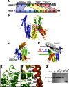
FIGURE 1.
FIGURE 1.
Crystal structure of αEcatNM in the autoinhibited conformation. A, α-catenin architecture. α-Catenin contains three major domains: N, M, and C. Structured regions are indicated by colored boxes with αE-catenin residue numbers shown below. Vinculin contains five domains, D1–D5. α-Catenin shares ∼30% sequence identity with vinculin at Vinculin Homology regions, VH1, VH2, and VH3. B, 6.5-Å structure of αEcatNM. The VBS consists of the second and third α-helices (indicated by single and double asterisks, respectively) of the MI bundle. C, homodimer structure of the αEcatNM. One protomer is outlined by a cyan dashed line. Two chains of αE-catenin form an asymmetric dimer with two juxtaposed MI bundles. D, the unfurled structure of the MI region bound to the D1 domain of vinculin (PDB code 4EHP; MI in black and D1 in gray) (30) is aligned to the fourth helix of the MI bundle (green) in the autoinhibited state. E, close-up view of the four-helix bundle formed by the MI region. Amino acid residues in the hydrophobic core of the MI bundle are shown as sticks. F, numerous salt bridges formed between charged residues (sticks) from different regions are involved in the stabilization of the MI-MIII and MII-MIII interfaces. The location of these interfaces is indicated by a magenta box in B. G, GST-vinculin-D1/GST pulldown assay with wild type αE-catenin or its single residue mutants, M371W, E521A, and R551A. The M371W mutant was designed to directly disrupt the MI bundle (Fig. 1E).
FIGURE 2.
FIGURE 2.
Crystal structures of the αE-catenin MII-III fragment. A, crystal structure of the MII-III fragment within αEcatNM. Angles between the MII and MIII bundles are shown in A–C. B, crystal structure of the MII-III fragment within the M fragment residues 377–633 (PDB code 1H6G) (25). C, crystal structure of the MII-III fragment within the M fragment residues 385–651 (PDB code 1L7C) (28). The location of Arg551, which is in close proximity with Asp503 to form salt bridges at the MII-MIII interface in the αEcatNM structure (Fig. 1F), is shown as cyan sticks. D, previously determined structures of MII-III fragments (PDB codes 1H6G and 1L7C) are superposed onto the MII-III structure of αEcatNM by aligning the MII bundles. The MIII bundles in PDB codes 1H6G and 1L7C are further rotated 124° and 93°, respectively, away from where the MIII bundle is located in αEcatNM, possibly due to the absence of N domain and MI bundle. E, superposition of the M domain structure of αEcatNM and the D3–D4 domain structure of vinculin (PDB code 1ST6).
FIGURE 3.
FIGURE 3.
Junctional organization in R2/7 cells stably transfected with various αE-catenin (A) or αN-catenin (B) mutants tagged with EGFP, or transfected with EGFP only as a control. Schematic figures represent a series of mutant constructs used. Cells were triple-immunostained for GFP, vinculin, and ZO-1 (a tight junction protein). αEcatFL, full-length αE-catenin; αEcatNMI, αE-catenin-NMI fragment; αEcatNMI-III, αE-catenin-NMI-III fragment; and αEcatNMI-III/αNcatC, a chimera consisting of αE-catenin-NMI-III fragment fused to αN-catenin-C fragment. αNcatFL, full-length αN-catenin; αNcatNMI, αN-catenin-NMI fragment; αNcatNMI-III, αN-catenin-NMI-III fragment; and αNcat NMI-III/αEcatC, a chimera consisting of αN-catenin-NMI-III fragment fused to the αE-catenin-C fragment. Scale bars, 10 μm.
FIGURE 4.
FIGURE 4.
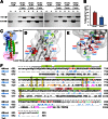
Crystal structure of the C domain of αN-catenin. A, actin pelleting assays with αE-catenin C domain (αEcatC) and αN-catenin C domain (αNcatC). Supernatant (S) and pellet (P) fractions are indicated. B, the average percentage (and S.D.) of total protein cosedimenting with F-actin in three independent experiments was: αEcatC, 20.0(±2.6)%; αNcatC, 12.0(±2.0)%. C, crystal structure of αNcatC consists of six α-helices and a short β-hairpin and forms a five-helix bundle. Disordered N and C termini extensions are indicated by dashed lines. Regions equivalent to upper and lower actin-binding sites of vinculin (59)(supplemental Fig. S7B) are indicated. D and E, close-up views of the “upper site” (D) and “lower site” (E) of αN-catenin. Side chains of non-identical residues between αE-catenin (blue) and αN-catenin (green for conserved residues and red for non-conserved residues) are shown as sticks. F, structural alignment of actin-binding domain sequences of αN-catenin, αE-catenin, and vinculin (PDB code 1ST6). αE- and αN-catenins share 87% identical and 93% similar residues within their C domain sequences. αE-catenin and vinculin residues that are identical to αN-catenin residues are indicated by dots. Non-identical residues of αE-catenin are shown in blue, whereas αN-catenin residues in green and red indicate conserved and non-conserved substitutions found between αE- and αN-catenins. Vinculin residues that constitute the upper and lower sites (59) are shown as orange and magenta, respectively. Secondary structure information (green bar for α-helix and red arrow for β-strand) based on the crystal structures of αNcatC is shown.
FIGURE 5.
FIGURE 5.
Full-length structure of αN-catenin. A, sedimentation equilibrium analysis of αN-catenin. αN-catenin exists as a monodisperse species with an average molecular mass of 97.4 kDa, which is approximately the size expected of a monomer. In contrast, αE-catenin exists in a monomer-dimer equilibrium (Kd = 73 μm) with an average molecular mass of 132.3 kDa. B, actin pelleting assay with full-length αN-catenin (αNcat). C, experimental scattering curve (red) of αN-catenin and a fitted theoretical scattering curve (blue) (χ2 = 1.4) of its full-length structure model (supplemental Fig. S9A). D, docked crystal structures of αEcatNM and αNcatC within the ab initio molecular envelope of full-length αN-catenin (gray). E, pairwise distance-distribution function of αN-catenin scattering data. F, comparison of the Rg and Dmax values obtained for αN-catenin to values previously reported for other α-catenins and vinculin (60, 61).
FIGURE 6.
FIGURE 6.
Comparison of the multidomain structures of vinculin and αN-catenin. Three individual domains (N, M, and C) of α-catenin share high structural homology with their equivalent domains of vinculin (D1, D3–D4, and D5) (supplemental Fig. S3A, S2E, and S7A). A, the elongated structure of full-length αN-catenin. B, the full-length crystal structure of vinculin (PDB code 1ST6) in the globular state. The actin-binding D5 domain forms interdomain interactions with the vinculin head region consisting of D1–D4 domains in a “pincer-shape” (56). C, superposition of αN-catenin and vinculin (gray). The D2 domain of vinculin is shown as a transparent ribbon to help visualize other structural features. The spatial arrangements of the N and M domains of α-catenin highly resemble the “pincer”-shaped arrangement of D1, D3, and D4 domains required for clamping the D5 domain and partially occluding the actin-binding sites (56).
FIGURE 7.
FIGURE 7.
Model for α-catenin-mediated localization of vinculin at AJs. A, model of tension-dependent activation of α-catenin involving multistate conformational changes. α-Catenin acts as a “molecular switch” to modulate the connection between the cadherin-catenin complex and the actin cytoskeleton. In the no-tension state, the VBS within the MI region remains closed (OFF) as there is no force being sensed by the C domain. In the high-tension state, the actomyosin-generated contractile force is detected by the C domain and helps to trigger “open to closed” conformational changes (black arrows) within the M domain. The VBS in the open (ON) conformation allows the activated vinculin to localize with the cadherin-catenin complex and engage in F-actin binding. B, model of the tension-dependent AJ-actin cytoskeleton connections regulated by cadherin-catenin-vinculin complexes.

References

    1. Meng W., Takeichi M. (2009) Adherens junction. Molecular architecture and regulation. Cold Spring Harbor Perspect. Biol. 1, a002899 - PMC - PubMed
    1. Tamada M., Perez T. D., Nelson W. J., Sheetz M. P. (2007) Two distinct modes of myosin assembly and dynamics during epithelial wound closure. J. Cell Biol. 176, 27–33 - PMC - PubMed
    1. Vasioukhin V., Fuchs E. (2001) Actin dynamics and cell-cell adhesion in epithelia. Curr. Opin. Cell Biol. 13, 76–84 - PubMed
    1. Takeichi M. (1995) Morphogenetic roles of classic cadherins. Curr. Opin. Cell Biol. 7, 619–627 - PubMed
    1. Ishiyama N., Lee S.-H., Liu S., Li G.-Y., Smith M. J., Reichardt L. F., Ikura M. (2010) Dynamic and static interactions between p120 catenin and E-cadherin regulate the stability of cell-cell adhesion. Cell 141, 117–128 - PubMed

Publication types

MeSH terms

LinkOut - more resources