. 2013 May 24;288(21):15148-53.
doi: 10.1074/jbc.M113.474502. Epub 2013 Apr 15.

Substrate selectivity of the acid-activated glutamate/γ-aminobutyric acid (GABA) antiporter GadC from Escherichia coli

Affiliations

Substrate selectivity of the acid-activated glutamate/γ-aminobutyric acid (GABA) antiporter GadC from Escherichia coli

Dan Ma et al. J Biol Chem. .

Abstract

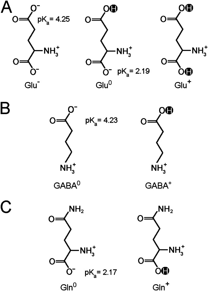
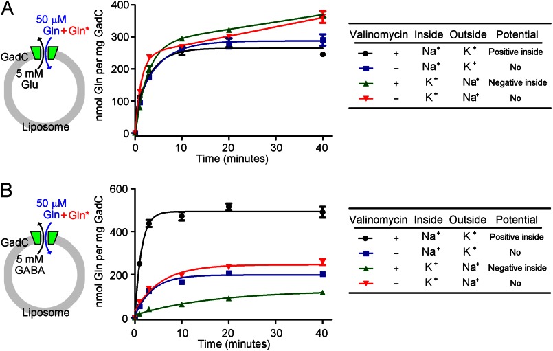
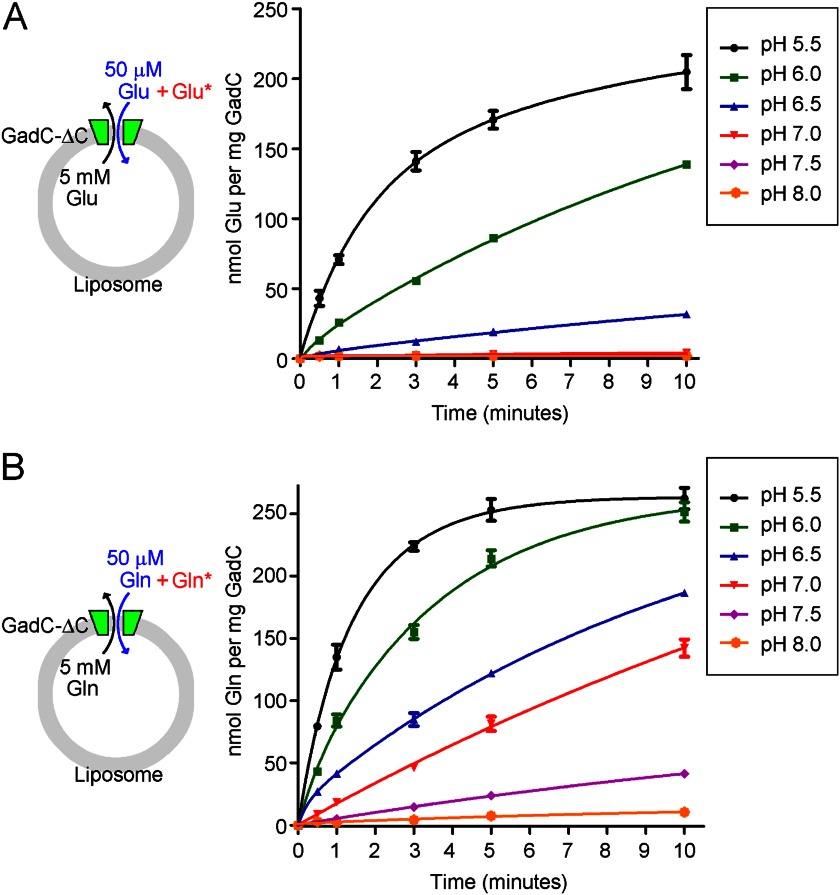
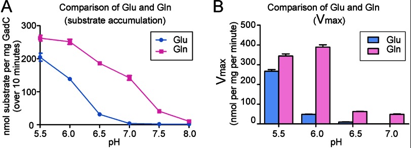
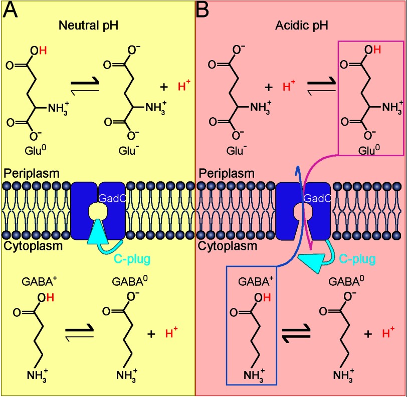
GadC, a central component of the Escherichia coli acid resistance system, is a Glu/GABA antiporter. A previous structural study and biochemical characterization showed that GadC exhibits a stringent pH dependence for substrate transport, with no detectable activity at pH values above 6.5. However, the substrate selectivity and the mechanism of pH-dependent transport activity of GadC remain enigmatic. In this study, we demonstrate that GadC selectively transports Glu with no net charge and GABA with a positive charge. A C-plug-truncated variant of GadC (residues 1-470) transported Gln (a mimic of Glu with no net charge), but not Glu, even at pH 8.0. The pH-dependent transport of Gln by this GadC variant was shifted ~1 unit toward a higher pH compared with Glu transport. Taken together, the results identify the substrate selectivity for GadC and show that the protonation states of substrates are crucial for transport.

Keywords: Acid Resistance; Amino Acid Transport; GadC; Glutamate; Glutamine; Membrane Antiporter; Membrane Proteins; Membrane Transporter Reconstitution; Protonation/Deprotonation; Substrate Selectivity.


Figures

FIGURE 1.
FIGURE 1.
Glu, Gln, and GABA display more than one charge state at pH 5.5. A, Glu may carry one negative charge (Glu), no net charge (Glu0), or one positive charge (Glu+). B, GABA exists in two states: with no net charge (GABA0) and with one positive charge (GABA+). C, Gln exists in two potential charged states: with no net charge (Gln0) and with one positive charge (Gln+).
FIGURE 2.
FIGURE 2.
Influence of membrane potential on substrate transport by GadC. A, influence of membrane potential on the exchange of Gln and Glu. Substrate transport by WT GadC was measured in a proteoliposome-based assay at pH 5.5 in the presence or absence of valinomycin, which allows selective passage of K+ (but not Na+) ions. In this assay, the proteoliposomes were loaded with 5 mm Glu, and the external buffer contained 50 μm unlabeled Gln and 0.2 μm [3H]Gln. With 120 mm Na+ in the proteoliposome and 120 mm K+ in the external buffer, valinomycin allowed influx of K+ into the proteoliposome, generating a positive potential inside the proteoliposome. Such a potential had little effect on the influx of Gln and the efflux of Glu (compare black and blue lines). Conversely, with 120 mm K+ in the proteoliposome and 120 mm Na+ in the external buffer, valinomycin allowed efflux of K+ into the external buffer, resulting in a negative potential inside the proteoliposome. Such treatment had little effect on substrate transport by GadC (compare green and red lines). These results indicate that there is no net charge difference between the transport substrates Gln and Glu. B, influence of membrane potential on exchange of Gln and GABA. The experimental design used here was similar to that described for A, except that the proteoliposomes were loaded with 5 mm GABA. The positive potential inside the proteoliposome resulted in markedly enhanced substrate transport by GadC, i.e. increased influx of Gln and efflux of GABA (compare black and blue lines). Conversely, the negative potential inside the proteoliposome led to significantly decreased substrate transport by GadC (compare green and red lines). These results indicate that, compared with the charge state of Gln, GABA should be at least +1.
FIGURE 3.
FIGURE 3.
GadC-ΔC exhibits pH-dependent transport of Glu and Gln. A, the transport of Glu was robust at pH 5.5 and rapidly decreased with increasing pH values, with no detectable transport activity at pH 7.0 and above. The transport of Glu by the C-plug-deleted GadC variant (GadC-ΔC) was measured in a proteoliposome-based assay at different pH values. In this assay, the proteoliposomes were loaded with 5 mm Glu, and the external buffer contained 50 μm unlabeled Glu and 0.2 μm [3H]Glu. B, the transport of Gln was still measurable at pH 8.0. The transport of Gln by GadC-ΔC decreased with increasing pH values, but the rate of decrease was markedly slower than that for transport of Glu.
FIGURE 4.
FIGURE 4.
pH-dependent transport of Gln by GadC-ΔC is shifted ∼1 pH unit toward a higher pH compared with that of Glu. A, comparison of the substrate accumulation of Glu and Gln. For Glu, GadC-ΔC exhibited no detectable transport activity at pH above 7.0. In contrast, GadC-ΔC could transport Gln even at pH 8.0. B, comparison of Vmax values of GadC-ΔC for Glu and Gln at different pH values.
FIGURE 5.
FIGURE 5.
Proposed mechanism of pH-dependent substrate transport by GadC. A, GadC is inactive at neutral pH. Most of the substrate molecules (Glu and GABA) are deprotonated, thus are unfavorable for transport. In addition, the substrate transport path is blocked by the C-plug of GadC. Under these conditions, no substrate exchange occurs. B, GadC is activated at acidic pH. When the extracellular pH is extremely acidic (pH 2∼3), the cytoplasmic pH in E. coli drops to a value between 3.5 and 5.0. In the cytoplasm, GABA exists in two forms, with no net charge (GABA0) and with one positive charge (GABA+), whereas the majority of Glu in the extracellular space carries no net charge (Glu0). At acidic pH, the C-plug of GadC may be displaced, allowing influx of Glu0 and efflux of GABA+.

References

    1. Donnenberg M. S. (2000) Pathogenic strategies of enteric bacteria. Nature 406, 768–774 - PubMed
    1. Audia J. P., Webb C. C., Foster J. W. (2001) Breaking through the acid barrier: an orchestrated response to proton stress by enteric bacteria. Int. J. Med. Microbiol. 291, 97–106 - PubMed
    1. Foster J. W. (2004) Escherichia coli acid resistance: tales of an amateur acidophile. Nat. Rev. Microbiol. 2, 898–907 - PubMed
    1. Lin J., Lee I. S., Frey J., Slonczewski J. L., Foster J. W. (1995) Comparative analysis of extreme acid survival in Salmonella typhimurium, Shigella flexneri, and Escherichia coli. J. Bacteriol. 177, 4097–4104 - PMC - PubMed
    1. Lin J., Smith M. P., Chapin K. C., Baik H. S., Bennett G. N., Foster J. W. (1996) Mechanisms of acid resistance in enterohemorrhagic Escherichia coli. Appl. Environ. Microbiol. 62, 3094–3100 - PMC - PubMed

Publication types

MeSH terms

LinkOut - more resources