

The hyaluronic acid receptor CD44 coordinates normal and metaplastic gastric epithelial progenitor cell proliferation
- PMID: 23589310
- PMCID: PMC3668764
- DOI: 10.1074/jbc.M112.445551
The hyaluronic acid receptor CD44 coordinates normal and metaplastic gastric epithelial progenitor cell proliferation
Abstract
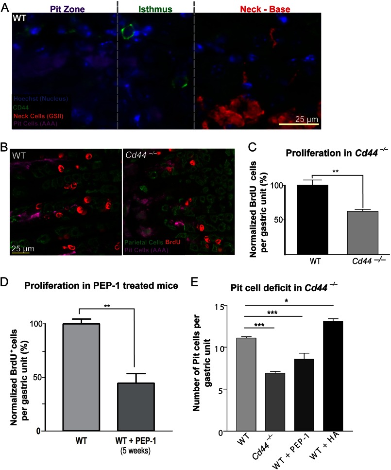
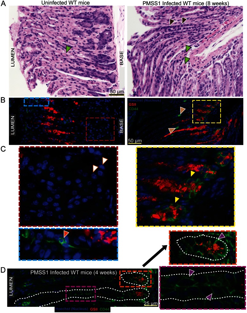
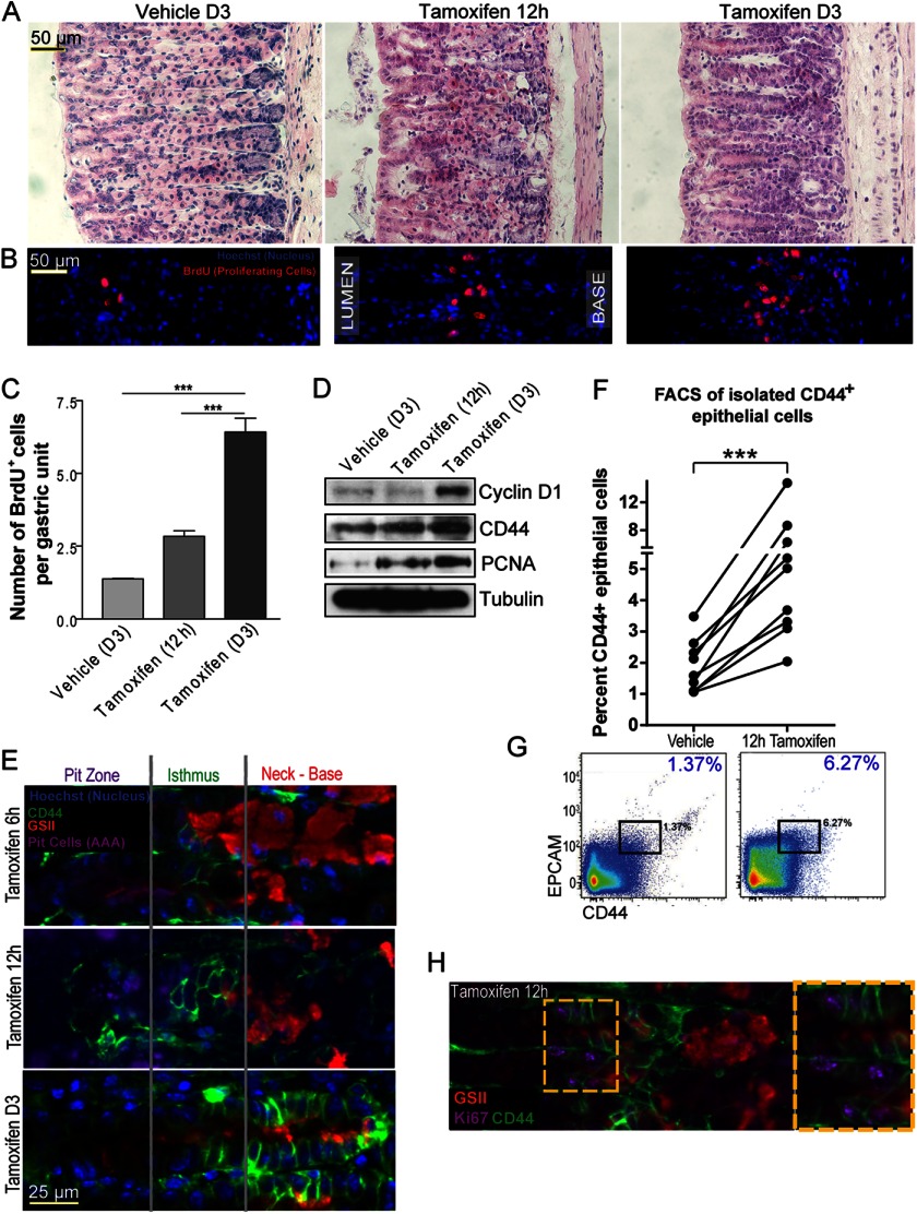
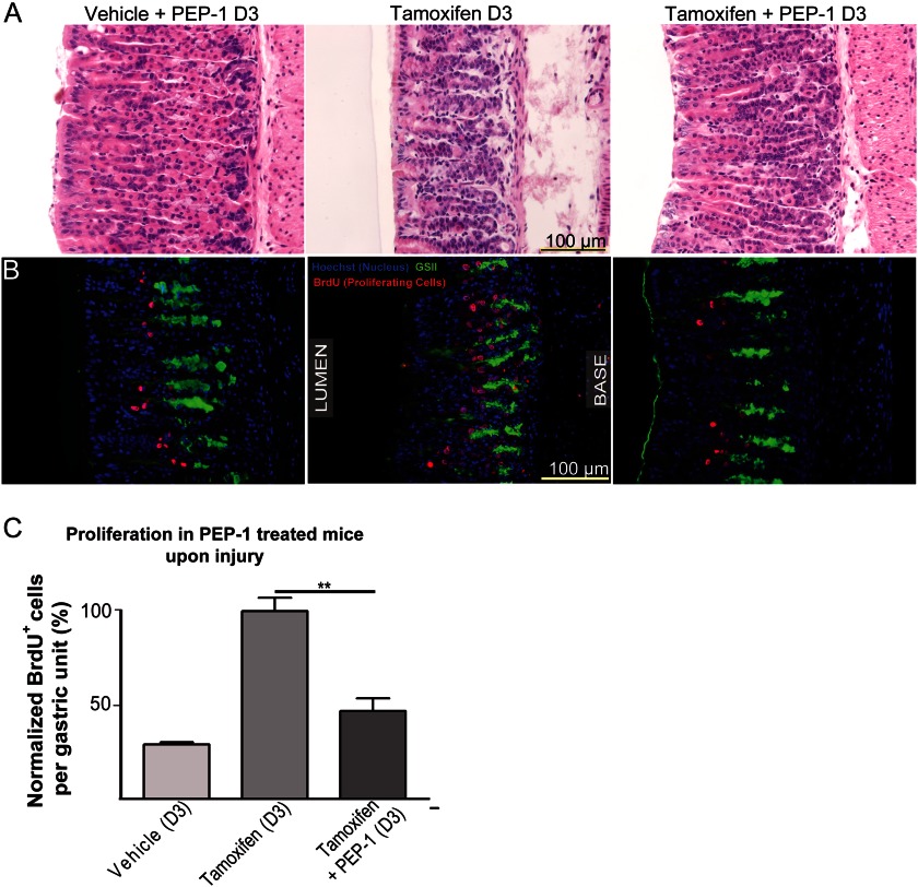
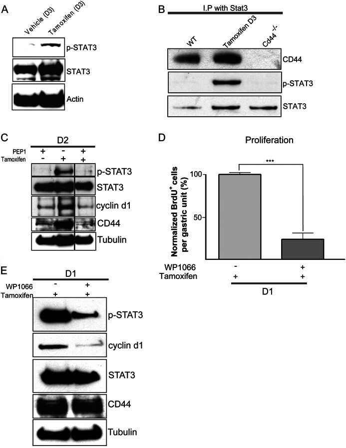
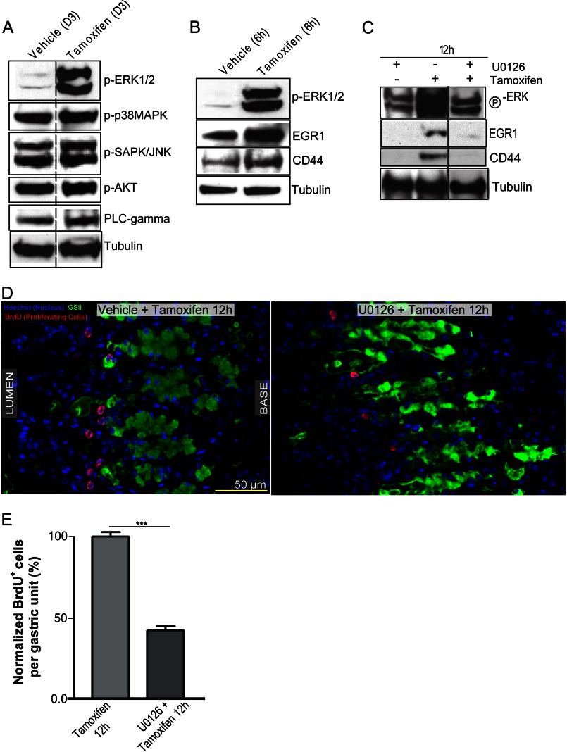
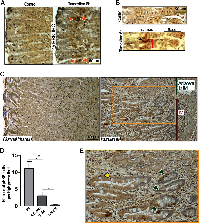
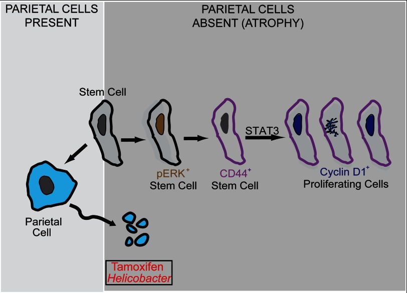
The stem cell in the isthmus of gastric units continually replenishes the epithelium. Atrophy of acid-secreting parietal cells (PCs) frequently occurs during infection with Helicobacter pylori, predisposing patients to cancer. Atrophy causes increased proliferation of stem cells, yet little is known about how this process is regulated. Here we show that CD44 labels a population of small, undifferentiated cells in the gastric unit isthmus where stem cells are known to reside. Loss of CD44 in vivo results in decreased proliferation of the gastric epithelium. When we induce PC atrophy by Helicobacter infection or tamoxifen treatment, this CD44(+) population expands from the isthmus toward the base of the unit. CD44 blockade during PC atrophy abrogates the expansion. We find that CD44 binds STAT3, and inhibition of either CD44 or STAT3 signaling causes decreased proliferation. Atrophy-induced CD44 expansion depends on pERK, which labels isthmal cells in mice and humans. Our studies delineate an in vivo signaling pathway, ERK → CD44 → STAT3, that regulates normal and atrophy-induced gastric stem/progenitor-cell proliferation. We further show that we can intervene pharmacologically at each signaling step in vivo to modulate proliferation.
Keywords: Cd44; ERK; Gastric Cancer; Hyaluronan; PEP-1; Signaling; Stem Cells; Tamoxifen; U0126; WP1066.
Figures
Similar articles
-
Gastric Metaplasia Induced by Helicobacter pylori Is Associated with Enhanced SOX9 Expression via Interleukin-1 Signaling.Infect Immun. 2015 Dec 7;84(2):562-72. doi: 10.1128/IAI.01437-15. Print 2016 Feb. Infect Immun. 2015. PMID: 26644382 Free PMC article.
-
CD44 plays a functional role in Helicobacter pylori-induced epithelial cell proliferation.PLoS Pathog. 2015 Feb 6;11(2):e1004663. doi: 10.1371/journal.ppat.1004663. eCollection 2015 Feb. PLoS Pathog. 2015. PMID: 25658601 Free PMC article.
-
CD44 and TLR4 mediate hyaluronic acid regulation of Lgr5+ stem cell proliferation, crypt fission, and intestinal growth in postnatal and adult mice.Am J Physiol Gastrointest Liver Physiol. 2015 Dec 1;309(11):G874-87. doi: 10.1152/ajpgi.00123.2015. Epub 2015 Oct 1. Am J Physiol Gastrointest Liver Physiol. 2015. PMID: 26505972 Free PMC article.
-
Differentiation of the gastric mucosa III. Animal models of oxyntic atrophy and metaplasia.Am J Physiol Gastrointest Liver Physiol. 2006 Dec;291(6):G999-1004. doi: 10.1152/ajpgi.00187.2006. Am J Physiol Gastrointest Liver Physiol. 2006. PMID: 17090722 Review.
-
Dysregulation of stem cell signaling network due to germline mutation, SNP, Helicobacter pylori infection, epigenetic change and genetic alteration in gastric cancer.Cancer Biol Ther. 2007 Jun;6(6):832-9. doi: 10.4161/cbt.6.6.4196. Epub 2007 Mar 26. Cancer Biol Ther. 2007. PMID: 17568183 Review.
Cited by
-
Hepatocyte nuclear factor 4α is required for cell differentiation and homeostasis in the adult mouse gastric epithelium.Am J Physiol Gastrointest Liver Physiol. 2016 Aug 1;311(2):G267-75. doi: 10.1152/ajpgi.00195.2016. Epub 2016 Jun 23. Am J Physiol Gastrointest Liver Physiol. 2016. PMID: 27340127 Free PMC article.
-
Chronic tamoxifen use is associated with a decreased risk of intestinal metaplasia in human gastric epithelium.Dig Dis Sci. 2014 Jun;59(6):1244-54. doi: 10.1007/s10620-013-2994-1. Epub 2013 Dec 25. Dig Dis Sci. 2014. PMID: 24368421 Free PMC article.
-
Gastric intestinal metaplasia: progress and remaining challenges.J Gastroenterol. 2024 Apr;59(4):285-301. doi: 10.1007/s00535-023-02073-9. Epub 2024 Jan 19. J Gastroenterol. 2024. PMID: 38242996 Review.
-
The use of murine-derived fundic organoids in studies of gastric physiology.J Physiol. 2015 Apr 15;593(8):1809-27. doi: 10.1113/jphysiol.2014.283028. Epub 2015 Feb 19. J Physiol. 2015. PMID: 25605613 Free PMC article.
-
Glioblastoma Spheroid Invasion through Soft, Brain-Like Matrices Depends on Hyaluronic Acid-CD44 Interactions.Adv Healthc Mater. 2023 Jun;12(14):e2203143. doi: 10.1002/adhm.202203143. Epub 2023 Feb 8. Adv Healthc Mater. 2023. PMID: 36694362 Free PMC article.
References
-
- Ferlay J., Shin H. R., Bray F., Forman D., Mathers C., Parkin D. M. (2010) Estimates of worldwide burden of cancer in 2008. GLOBOCAN 2008. Int. J. Cancer 127, 2893–2917 - PubMed
-
- Roder D. M. (2002) The epidemiology of gastric cancer. Gastric Cancer 5, 5–11 - PubMed
-
- Correa P., Houghton J. (2007) Carcinogenesis of Helicobacter pylori. Gastroenterology 133, 659–672 - PubMed
-
- Nomura S., Baxter T., Yamaguchi H., Leys C., Vartapetian A. B., Fox J. G., Lee J. R., Wang T. C., Goldenring J. R. (2004) Spasmolytic polypeptide expressing metaplasia to preneoplasia in H. felis-infected mice. Gastroenterology 127, 582–594 - PubMed
Publication types
MeSH terms
Substances
Grants and funding
- R01 CA077955/CA/NCI NIH HHS/United States
- DK079798-4/DK/NIDDK NIH HHS/United States
- F32 CA153539/CA/NCI NIH HHS/United States
- F32 CA 153539/CA/NCI NIH HHS/United States
- R01 DK079798/DK/NIDDK NIH HHS/United States
- R01 DK055753/DK/NIDDK NIH HHS/United States
- R01 DK033165/DK/NIDDK NIH HHS/United States
- DK55753/DK/NIDDK NIH HHS/United States
- R01 DK58587/DK/NIDDK NIH HHS/United States
- R01 DK094989/DK/NIDDK NIH HHS/United States
- DK079798-3/DK/NIDDK NIH HHS/United States
- 2P30 DK052574-12/DK/NIDDK NIH HHS/United States
- P30 DK 508404/DK/NIDDK NIH HHS/United States
- DK33165/DK/NIDDK NIH HHS/United States
- R01 CA 77955/CA/NCI NIH HHS/United States
- P01 116037/PHS HHS/United States
- R37 DK033165/DK/NIDDK NIH HHS/United States
- R01 DK058587/DK/NIDDK NIH HHS/United States
- P30 DK052574/DK/NIDDK NIH HHS/United States
LinkOut - more resources
Full Text Sources
Other Literature Sources
Medical
Molecular Biology Databases
Miscellaneous
