. 2013 Apr 15;201(2):249-61.
doi: 10.1083/jcb.201207139.

Cycling of the signaling protein phospholipase D through cilia requires the BBSome only for the export phase

Affiliations

Cycling of the signaling protein phospholipase D through cilia requires the BBSome only for the export phase

Karl F Lechtreck et al. J Cell Biol. .

Abstract

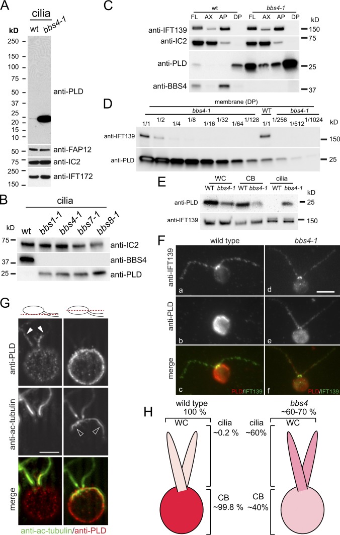
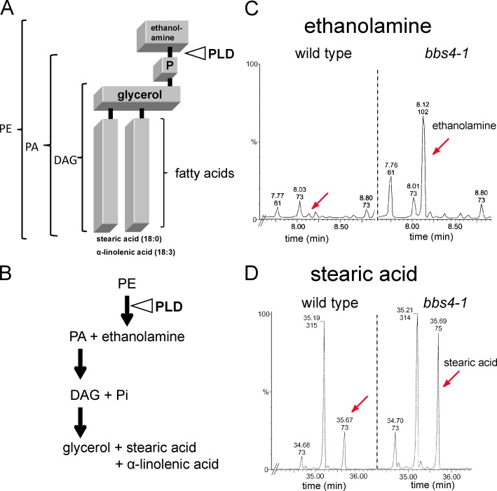
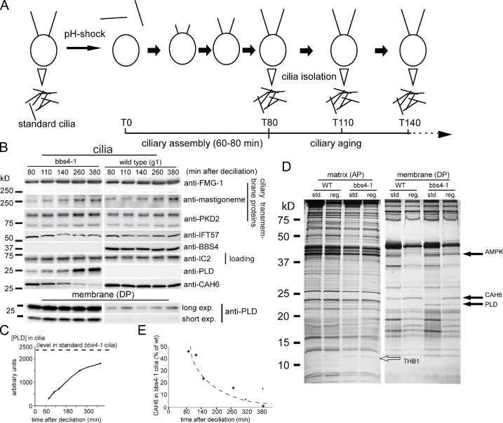
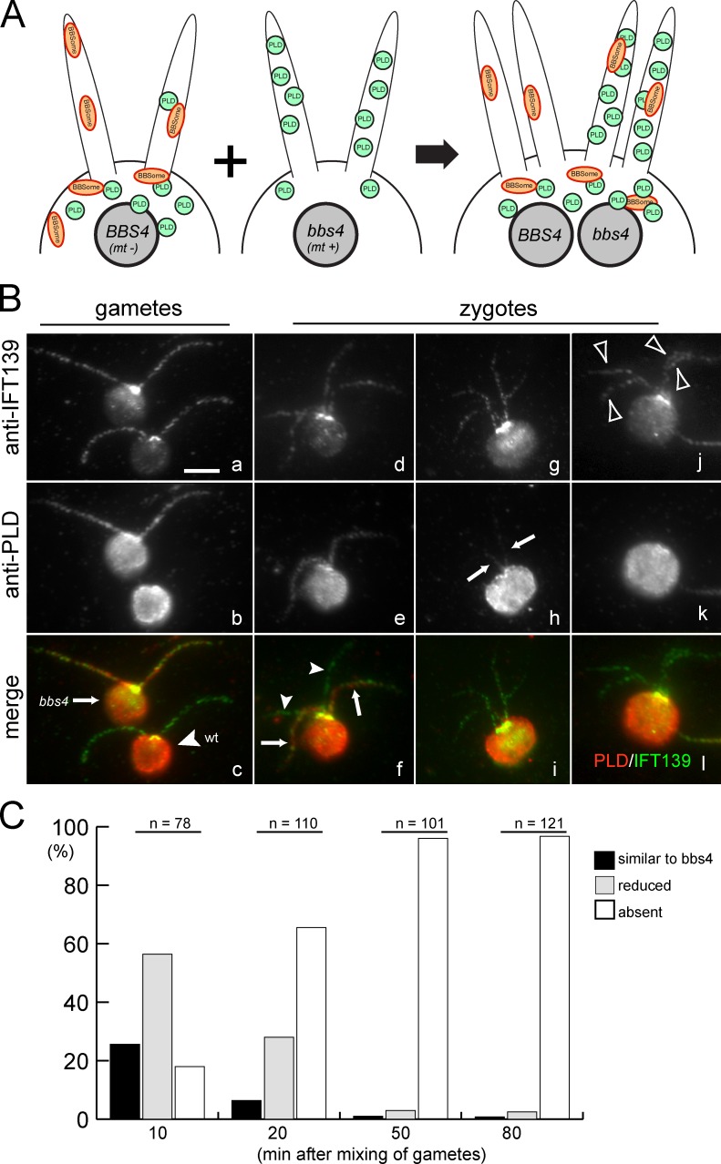
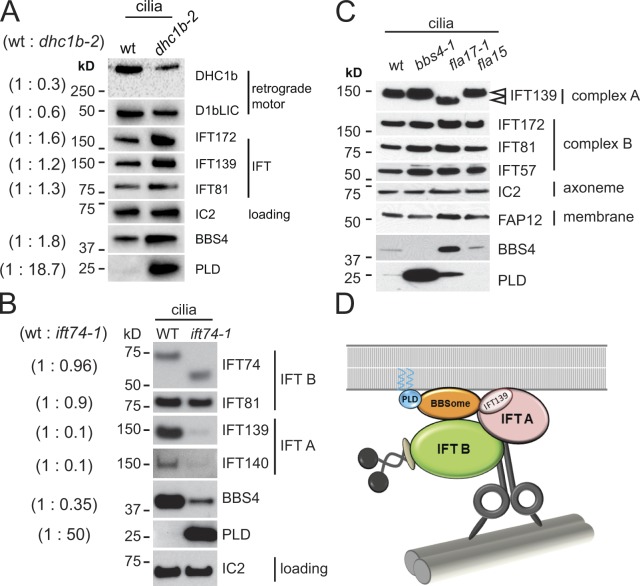
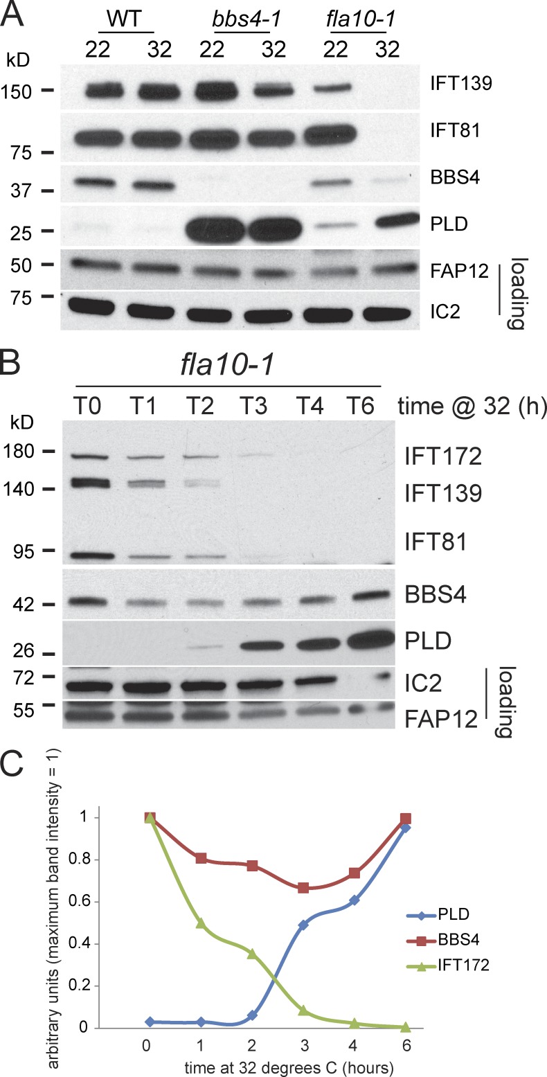
The BBSome is a complex of seven proteins, including BBS4, that is cycled through cilia by intraflagellar transport (IFT). Previous work has shown that the membrane-associated signaling protein phospholipase D (PLD) accumulates abnormally in cilia of Chlamydomonas reinhardtii bbs mutants. Here we show that PLD is a component of wild-type cilia but is enriched ∼150-fold in bbs4 cilia; this accumulation occurs progressively over time and results in altered ciliary lipid composition. When wild-type BBSomes were introduced into bbs cells, PLD was rapidly removed from the mutant cilia, indicating the presence of an efficient BBSome-dependent mechanism for exporting ciliary PLD. This export requires retrograde IFT. Importantly, entry of PLD into cilia is BBSome and IFT independent. Therefore, the BBSome is required only for the export phase of a process that continuously cycles PLD through cilia. Another protein, carbonic anhydrase 6, is initially imported normally into bbs4 cilia but lost with time, suggesting that its loss is a secondary effect of BBSome deficiency.


Figures

Figure 1.
Figure 1.
BBSome deficiency causes PLD redistribution from the cell body into cilia. (A) Western blot of isolated wild-type and bbs4-1 cilia probed with antibodies to PLD and, as loading controls, antibodies to the membrane-associated protein FAP12, the axonemal dynein subunit IC2, and the IFT complex B protein IFT172. (B) Western blot of isolated cilia from wild type (wt) and bbs1-1, bbs4-1, bbs7-1, and bbs8-1 probed with antibodies to BBS4 and PLD. IC2 was used as loading control. (C) Western blots of wild-type and bbs4-1 cilia (FL) and ciliary fractions (Ax, axonemes; AP and DP, aqueous and detergent phase from Triton X-114 phase partitioning, respectively) were probed with antibodies to PLD and BBS4, and with antibodies to the axonemal protein IC2 and the matrix protein IFT139 as loading controls. (D) Western blot of isolated ciliary membrane from wild type and bbs4-1 probed with anti-PLD. The bbs4-1 sample was diluted as indicated. IFT139, small amounts of which can be detected in the ciliary membrane, was used as a loading control. The signal strength obtained from the undiluted wild-type sample is between that of the 1:128 and 1:256 dilution of the bbs4-1 sample. (E) Western blots of whole cells (WC), deciliated cell bodies (CB), and isolated cilia (cilia) from wild type and bbs4-1 were probed with anti-PLD. Equivalent amounts of cells and cilia were loaded (i.e., one whole cell, one cell body, and two cilia). The IFT particle protein IFT139 was used as a loading control. (F) Wild-type and bbs4-1 gametes were stained with anti-IFT139 and anti-PLD. PLD was readily detected in cilia from bbs4-1 but not detected in wild-type cilia. Bar, 5 µm. (G) Two optical sections of a bbs4-1 vegetative cell stained with anti-PLD and anti-acetylated tubulin. Merged images are shown in color. The positions of the optical sections are indicated in the diagrams. Solid arrowheads, cilia; open arrowheads, microtubular roots. Bar, 5 µm. (H) Schematic presentation of PLD distribution in wild-type and bbs4-1 mutant cells.
Figure 2.
Figure 2.
The lipid composition of bbs4-1 cilia is altered. (A) Schematic presentation of the PLD substrate PE 18:0/18:3 and the site of PLD cleavage. (B) Schematic presentation of the enzymatic breakdown of PE into PA, ethanolamine, DAG, and downstream products. (C and D) Gas chromatography–mass spectrometry profiles showing the elution times for small metabolites from wild-type and bbs4-1 cilia. Ethanolamine (peaks marked by red arrows in C) is enriched ∼40× and free stearic acid (peaks marked by red arrows in D) is enriched ∼6× in bbs4-1 cilia. Accuracy of quantitation is ±10%.
Figure 3.
Figure 3.
The biochemical defects in bbs4-1 cilia increase with time. (A) Schematic presentation of the experimental design for obtaining age-sorted cilia from C. reinhardtii. (B) Western blots comparing regenerated wild-type and bbs4-1 cilia isolated at various time points after deciliation. (top) Western blots were probed with anti-FMG-1, anti-mastigoneme protein, anti-PKD2, anti-IFT57, anti-BBS4, anti-IC2 as a loading control, anti-PLD, and anti-CAH6. Ciliary length measurements for a similar experiment are shown in Fig. S3 E. (bottom) Long and short exposures of a Western blot of isolated ciliary membrane fractions from the same experiment probed with anti-PLD. (C) Graph showing the accumulation of PLD with time in bbs4-1 cilia; the dashed line corresponds to PLD levels in standard bbs4-1 cilia, i.e., cilia harvested without prior deciliation at least 6 h after the beginning of the light phase. (D) Silver-stained gel comparing matrix and membrane isolated from standard and 2-h-old regenerated wild-type and bbs4-1 cilia. Solid arrows, proteins altered in bbs4-1 ciliary membrane; open arrow, THB1. Note the differences in composition in the membrane of young and old cilia; no major differences were observed between young and old axonemal (not depicted) and matrix fractions, indicating that age-induced changes in ciliary protein composition are largely restricted to the ciliary membrane. (E) Graph showing the loss of CAH6 in bbs4-1 cilia as a percentage of that in wild-type cilia over time. The data shown in C and E are from a single representative experiment out of two repeats.
Figure 4.
Figure 4.
PLD is removed from cilia of bbs4-1 cells when BBSomes are introduced by fusion with wild-type cells. (A) Schematic presentation of the PLD ciliary export assay. In wild-type gametes BBSomes are present in both cytoplasm and cilia, and nearly all of the cell’s PLD is in the cell body; bbs4-1 gametes lack BBSomes and have undergone a massive redistribution of PLD from the cell body to the cilia. Wild-type (BBS4, mt−) and bbs4-1 (bbs4, mt+) gametes were mixed and allowed to fuse. The resulting zygotes have four cilia (two each from the wild-type and mutant gametes); BBSomes are present in the shared cytoplasm. (B) Gametes (a–c) and quadriciliated zygotes (d–l) were stained with antibodies to PLD and to IFT139 to visualize the cilia. Arrow in c, bbs4 gamete; arrowhead in c, wild-type gamete; arrows in f and h, cilia containing PLD derived from mutant gametes; arrowheads in f, cilia lacking PLD derived from a wild-type gamete; arrowheads in j, four cilia of a zygote without detectable PLD. Bar, 5 µm. (C) Quadriciliated zygotes were scored by eye for the amount of PLD in their cilia. Black bars, quadriciliated cells with two wild-type cilia and two cilia with levels of PLD characteristic of the bbs4-1 gamete; gray bars, quadriciliated cells with two wild-type cilia and two cilia with detectable but reduced amounts of PLD; white bars, quadriciliated cells without detectable PLD in their cilia.
Figure 5.
Figure 5.
Ciliary PLD is elevated in retrograde IFT mutants. (A) Western blot of isolated wild-type and dhc1b-2 cilia probed with antibodies to the proteins indicated. The amounts of the dynein 1b heavy chain, DHC1b, and light intermediate chain, D1bLIC, are reduced in the mutant cilia because of the instability of the dynein motor complex. BBS4 and in particular PLD are enriched in mutant cilia. IC2 was used as a loading control. For quantification of relative protein levels, values for band intensities were first normalized to the IC2 band intensity and the ratios (left, parentheses) were then calculated. (B) Western blot of cilia isolated from wild type and ift74-1 probed with antibodies to the proteins indicated. In ift74-1 the IFT complex B protein IFT74 is truncated. Note reduced amounts of IFT complex A proteins and BBS4 and increased levels of PLD in the mutant cilia. Relative protein levels (in parentheses) were determined as in A. (C) Western blot probing isolated cilia from wild type, bbs4-1, fla17-1 (expressing a truncated IFT139; arrowheads), and fla15-1. Note increase of BBS4 and PLD in fla17-1. (D) Summary diagram showing the IFT motors, IFT complexes A and B, the BBSome, and PLD. The BBSome functions as an IFT cargo adapter linking PLD and other cargoes to retrograde IFT particles. PLD is predicted to be myristoylated and palmitoylated (Fig. S4 C), offering a possible explanation for its membrane association. Dynein is shown linking the IFT particle to a doublet microtubule, which provides a track for IFT.
Figure 6.
Figure 6.
PLD accumulates in cilia after depletion of IFT proteins. (A) Western blot of cilia isolated from wild type, bbs4-1, and fla10-1 at 22°C and after 2.5 h at 32°C. The blot was probed with antibodies to the proteins indicated. Depletion of IFT proteins and accumulation of PLD in response to the temperature shift was observed only in cilia from fla10-1. (B) Western blot comparing fla10-1 cilia isolated at various time points from 0 to 6 h (T0–T6) after shifting the cells to 32°C. The blot was probed with antibodies to the proteins indicated. Note progressive depletion of IFT proteins and concomitant accumulation of PLD. (C) Relative amounts of PLD, BBS4, and IFT172 in the Western blot analysis shown in B. Maximum band intensity for each protein was set to 1. At later time points the mean length of fla10-1 cilia was reduced (Fig. S3 D). Because similar amounts of protein were loaded for each sample, the slight increase observed for BBS4 during the second half of the incubation time is likely to reflect the higher number of cilia loaded.

References

    1. Beales P.L. 2005. Lifting the lid on Pandora’s box: the Bardet-Biedl syndrome. Curr. Opin. Genet. Dev. 15:315–323. 10.1016/j.gde.2005.04.006 - DOI - PubMed
    1. Berbari N.F., Johnson A.D., Lewis J.S., Askwith C.C., Mykytyn K.. 2008a. Identification of ciliary localization sequences within the third intracellular loop of G protein-coupled receptors. Mol. Biol. Cell. 19:1540–1547. 10.1091/mbc.E07-09-0942 - DOI - PMC - PubMed
    1. Berbari N.F., Lewis J.S., Bishop G.A., Askwith C.C., Mykytyn K.. 2008b. Bardet-Biedl syndrome proteins are required for the localization of G protein-coupled receptors to primary cilia. Proc. Natl. Acad. Sci. USA. 105:4242–4246. 10.1073/pnas.0711027105 - DOI - PMC - PubMed
    1. Berthold P., Schmitt R., Mages W.. 2002. An engineered Streptomyces hygroscopicus aph 7″ gene mediates dominant resistance against hygromycin B in Chlamydomonas reinhardtii. Protist. 153:401–412. 10.1078/14344610260450136 - DOI - PubMed
    1. Blacque O.E., Leroux M.R.. 2006. Bardet-Biedl syndrome: an emerging pathomechanism of intracellular transport. Cell. Mol. Life Sci. 63:2145–2161. 10.1007/s00018-006-6180-x - DOI - PMC - PubMed

Publication types

LinkOut - more resources