. 2013 Jun;41(11):5784-98.
doi: 10.1093/nar/gkt231. Epub 2013 Apr 15.

HP1 promotes tumor suppressor BRCA1 functions during the DNA damage response

Affiliations

HP1 promotes tumor suppressor BRCA1 functions during the DNA damage response

Young-Ho Lee et al. Nucleic Acids Res. 2013 Jun.

Abstract

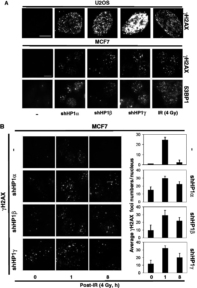
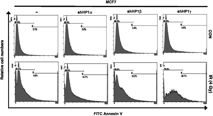
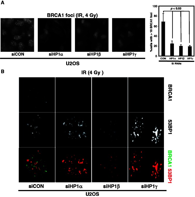
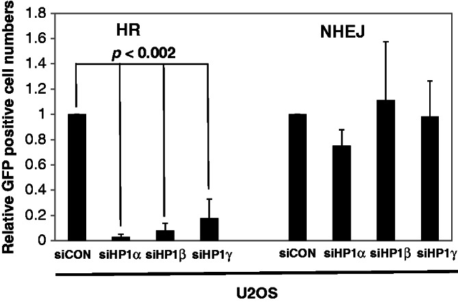
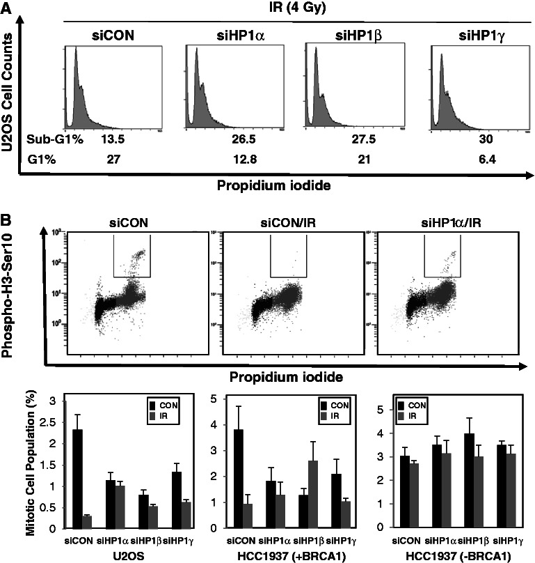
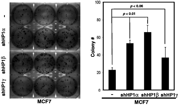
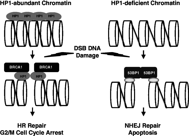
The DNA damage response (DDR) involves both the control of DNA damage repair and signaling to cell cycle checkpoints. Therefore, unraveling the underlying mechanisms of the DDR is important for understanding tumor suppression and cellular resistance to clastogenic cancer therapeutics. Because the DDR is likely to be influenced by chromatin regulation at the sites of DNA damage, we investigated the role of heterochromatin protein 1 (HP1) during the DDR process. We monitored double-strand breaks (DSBs) using the γH2AX foci marker and found that depleting cells of HP1 caused genotoxic stress, a delay in the repair of DSBs and elevated levels of apoptosis after irradiation. Furthermore, we found that these defects in repair were associated with impaired BRCA1 function. Depleting HP1 reduced recruitment of BRCA1 to DSBs and caused defects in two BRCA1-mediated DDR events: (i) the homologous recombination repair pathway and (ii) the arrest of cell cycle at the G2/M checkpoint. In contrast, depleting HP1 from cells did not affect the non-homologous end-joining (NHEJ) pathway: instead it elevated the recruitment of the 53BP1 NHEJ factor to DSBs. Notably, all three subtypes of HP1 seemed to be almost equally important for these DDR functions. We suggest that the dynamic interaction of HP1 with chromatin and other DDR factors could determine DNA repair choice and cell fate after DNA damage. We also suggest that compromising HP1 expression could promote tumorigenesis by impairing the function of the BRCA1 tumor suppressor.


Figures

Figure 1.
Figure 1.
HP1 depletion induces γH2AX and 53BP1 foci formation. (A) U2OS cells or HP1-depleted U2OS cells were stained with an anti-γ2AX antibody (Millipore). The γ2AX images from irradiated U2OS cells (4 Gy) are included as a positive control. MCF7 cell or HP1-depleted MCF7 cells were also stained with anti-γ2AX antibody and anti-53BP1 antibody (Millipore). The images of γH2AX foci and 53BP1 foci were captured on the same areas of the slide for comparison (200 ms by fluorescence microscopy). Scale Bar = 10 µm. (B) MCF7 cell or HP1-depleted MCF7 cells were irradiated (4 Gy), and foci formation was analyzed at 0, 1 and 8 h post-IR. Foci numbers in each nucleus were calculated by image-pro software. The histogram summarizes the average numbers of foci in the nuclei from at least 10 individual cells (right panel). P < 0.001 for MCF7; P < 0.04 for MCF7-shHP1α, β and γ.
Figure 2.
Figure 2.
HP1 is involved in IR-induced apoptosis. MCF7 cells and HP1-depleted MCF7 cells were untreated (upper panels) or irradiated (lower panels), then incubated for 20 h after IR-exposure. The cells were collected, stained with annexin V and then 1 × 10⁵ cells (or ∼0.5 × 10⁵ cells for the MCF7-shHP1γ post-IR) from each sample were analyzed by flow cytometry.
Figure 3.
Figure 3.
Depletion of HP1 reduces BRCA1 foci formation, but it enhances 53BP1 foci formation. U2OS cells were transfected with the indicated siRNAs and then irradiated (4 Gy). The immunofluorescence images of BRCA1 (A and B) and 53BP1 (B) were taken at 4 h after irradiation. The percentages of cells that have >10 BRCA1 foci are plotted on the graph. These were obtained from three independent experiments that analyzed >100 cells similar to those shown in (A). (B) Double immunofluorescence assay: in the lower panel, the green foci are the BRCA1 foci and red foci are the 53BP1 foci.
Figure 4.
Figure 4.
HP1-dependent recruitment of BRCA1 to DSB sites. AsiSI-ER-U2OS cells and HP1-depleted AsiSI-ER-U2OS cells were cultured and treated with 4-OHT (4-hydroxytamoxifen; 100 µM) for 4 h. Soluble chromatins were prepared from each culture and immunoprecipitated with specific antibodies (A: BRCA1, B: 53BP1, C: Rad51 and D: HP1). Immunoprecipitated chromatins were analyzed by quantitative PCR using specific primer pairs located at Chr 1: chr1_89231183, Chr 6: chr6_90404906, Chr 21: distal region of chr21_21292316. The results shown are relative signals from the individual precipitated DNA compared with input chromatin. siHP1 is siHP1 α, β and γ combined. Figure 4B includes two insets with enlarged scales showing the increased 53BP1 occupancy at DSB sites of Chr 1 and Chr 6 on 4-OHT treatment.
Figure 5.
Figure 5.
HP1 is required for HR repair. U2OS-NHEJ cells and U2OS-HR cells were transfected by individual siRNAs targeting the three respective HP1 subtypes on day one. The I-SecI expression construct was transfected at 24 h after siRNA transfection, and cells were incubated for additional 48 h. The numbers of GFP-positive cells were determined by fluorescence-activated cell sorting (FACS). GFP- and DSred-expression constructs were co-transfected to normalize for transfection efficiency in the FACS assays. The assays were repeated at least four times (n = 5), and the average relative numbers are presented as the mean ± SD.
Figure 6.
Figure 6.
HP1 plays an important role in apoptosis and cell cycle checkpoint control. (A) U2OS cells or HP1-depleted U2OS cells were cultured in low serum medium for 18 h and irradiated at 4 Gy. The cells were cultured for another 3 h, stained with PI and 5 × 10⁴ were analyzed by flow cytometry. (B) U2OS, HCC1937 (BRCA1-deficient or supplemented) cells and HP1-depleted MCF7 and HCC1937 cells were cultured in low serum medium for 18 h. Irradiated and sham-irradiated cells were cultured for 3 h before fixing, then stained with an anti-histone H3 phospho-serine 10 antibody (Millipore) and analyzed by FACS.
Figure 7.
Figure 7.
HP1 depletion enhances colony formation of MCF7 breast cancer cells. Equal numbers (100) of MCF7 cells or HP1-depleted MCF7 cells were cultured in each well of six-well plates. Colonies were stained with crystal violet on Day 10. A representative photo is shown (left panel), and the results were collected from three independent experiments, presented as mean ± SD (histogram, right panel). The plating efficiency was between ∼20 and 65%.
Figure 8.
Figure 8.
The roles of HP1 for BRCA1 function and the choice between HR and NHEJ repair. HP1 is a chromatin-associated protein, but the distribution of HP1 on chromatin is not even. Irradiation partially removes HP1 from chromatin, but the remaining HP1 molecules have active roles in recruiting BRCA1 to the damaged DNA sites. HP1 facilitates BRCA1 recruitment to chromatin after DNA damage, thereby activating G2/M cell cycle arrest and error-free HR repair. However, if the chromatin is HP1- or BRCA1-deficient, 53BP1 should be recruited resulting in error-prone NHEJ repair of the DNA damage. The loss of both the G2/M checkpoint control and HR repair pathway may increase the likelihood of apoptosis.

Similar articles

Cited by

References

    1. Goldberg AD, Allis CD, Bernstein E. Epigenetics: a landscape takes shape. Cell. 2007;128:635–638. - PubMed
    1. Cheung P, Allis CD, Sassone-Corsi P. Signaling to chromatin through histone modifications. Cell. 2000;103:263–271. - PubMed
    1. Hiragami K, Festenstein R. Heterochromatin protein 1: a pervasive controlling influence. Cell. Mol. Life Sci. 2005;62:2711–2726. - PMC - PubMed
    1. Zeng W, Ball AR, Jr, Yokomori K. HP1: heterochromatin binding proteins working the genome. Epigenetics. 2010;5:287–292. - PMC - PubMed
    1. Bannister AJ, Zegerman P, Partridge JF, Miska EA, Thomas JO, Allshire RC, Kouzarides T. Selective recognition of methylated lysine 9 on histone H3 by the HP1 chromo domain. Nature. 2001;410:120–124. - PubMed

Publication types

MeSH terms

Substances