

Review
doi: 10.1152/physrev.00019.2012.
Claudins and the modulation of tight junction permeability
Affiliations
- PMID: 23589827
- PMCID: PMC3768107
- DOI: 10.1152/physrev.00019.2012
Item in Clipboard
Review
Claudins and the modulation of tight junction permeability
Physiol Rev.
2013 Apr.
Abstract
Claudins are tight junction membrane proteins that are expressed in epithelia and endothelia and form paracellular barriers and pores that determine tight junction permeability. This review summarizes our current knowledge of this large protein family and discusses recent advances in our understanding of their structure and physiological functions.
Figures

Claudin family signature sequence in the predicted 1st extracellular loop. Top: Prosite pattern (accession no. PS01346), which is 89% specific for claudin family members (323). Ambiguities are indicated by listing acceptable amino acids at a given position between square parentheses, “x” is used for a position where any amino acid is accepted, with the range in the number of repetitions indicated in round parentheses. Bottom: updated version to include two other highly conserved residues located upstream and downstream of the classic signature. In addition to standard Prosite syntax, we use uppercase letters to indicate the more frequent residue at a position of ambiguity and lowercase to indicate the rarer one.
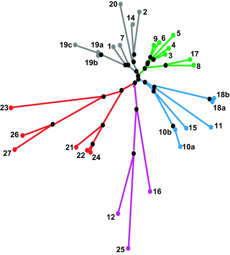
Phylogenetic tree of the 26 human claudin genes. [From Günzel and Fromm (120). Copyright 2012, with permission from John Wiley & Sons, Inc.]
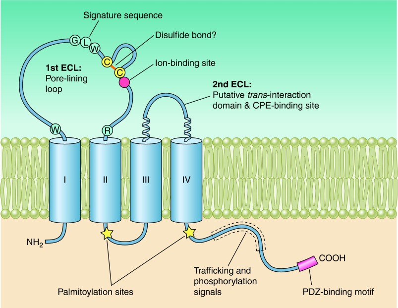
Model of claudin protein showing predicted topology and secondary structure as well as putative functional domains. Roman numerals indicate the predicted α-helical transmembrane domains.
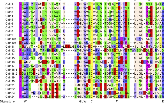
Alignment of the predicted first extracellular loop of human claudins. Residues within ECL1 were delineated using TMHMM (195). Sequence alignment was then performed with MUSCLE (76) and visualized in Jalview. Claudins 25–27 have lower homology and are not included in the alignment. Residues are color coded according to the following scheme: red, acidic; dark blue, basic; light blue, aromatic; green, polar; yellow, cysteine; purple, proline; cream, hydrophobic. Top: residue numbering for claudin-1 (identical to claudins 2, 4–9, 14, 17, 19, and 20). Bottom: consensus sequence of the claudin signature pattern.
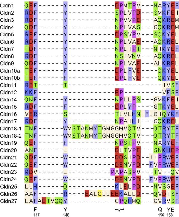
Alignment of the predicted second extracellular loop of human claudins. See legend to FIG. 4 for methods and color code. Bottom: letters indicate residues proposed to participate in trans-interactions, numbered according to the claudin-5 sequence. The bracket indicates the N-P motif proposed to bind to CPE.
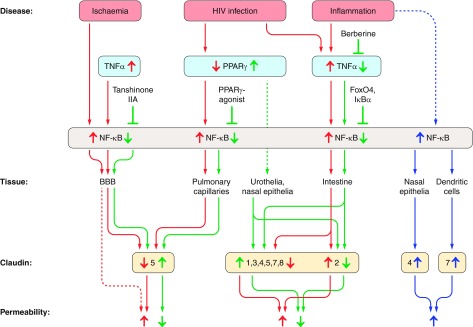
Summary of NF-κB-mediated TNF-α and PPAR-γ effects on claudin expression and epithelial permeability. In most epithelia, increases in NF-κB cause an increase in epithelial permeability, due to decreased expression in tightening tight junction proteins and/or increased expression in pore-forming claudin-2 (red arrows). Direct or indirect inhibition of NF-κB causes opposite effects (green arrows). In a few, exceptional cases, NF-κB was observed to upregulate tightening claudins, causing decreases in epithelial permeability (blue arrows). Figure is based on References , , , , , , , , , , , , , , , , . See text for detailed description.
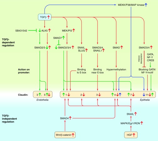
Summary of TGF-β-dependent and -independent SMAD/SNAIL/SLUG-signalling on claudin expression. Upregulation of SMAD2, SMAD3, SMAD4, SNAIL1 and/or SLUG by TGFβ (upper part), Wnt, β-catenin or HGF (lower part) causes a general downregulation of claudin expression, irrespective of claudin function (tightening or pore-forming, red arrows). Effects are transmitted by different effects on claudin promoters. Inhibition of SMAD2, SMAD3, and/or SMAD4 upregulation (e.g., by SMAD7), or interference with promoter binding, causes claudin upregulation (green arrows). TGF-β may also differentially affect certain claudins in a SMAD-independent way via the MEKK/p38/MAP kinase pathway (blue arrows). Based on References , , , , , , , , , , , , , , , , , . See text for detailed description.
References
-
- Acharya P, Beckel J, Ruiz WG, Wang E, Rojas R, Birder L, Apodaca G. Distribution of the tight junction proteins ZO-1, occludin, claudin-4, -8, and -12 in bladder epithelium. Am J Physiol Renal Physiol 287: F305–F318, 2004 - PubMed
-
- Aijaz S, Balda MS, Matter K. Tight junctions: molecular architecture and function. Int Rev Cytol 248: 261–298, 2006 - PubMed
Publication types
MeSH terms
Substances
Grants and funding
LinkOut - more resources
Full Text Sources
Other Literature Sources
Medical