Review
. 2013 Apr;93(2):653-80.
doi: 10.1152/physrev.00030.2012.

Urothelial signaling

Affiliations
Review

Urothelial signaling

Lori Birder et al. Physiol Rev. 2013 Apr.

Abstract

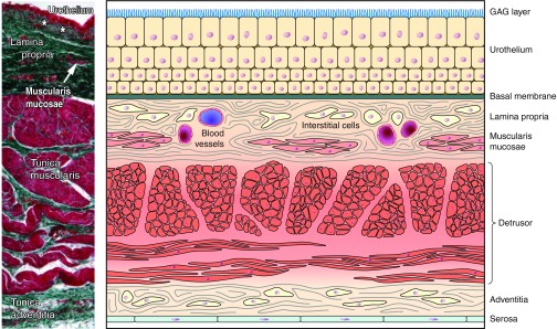
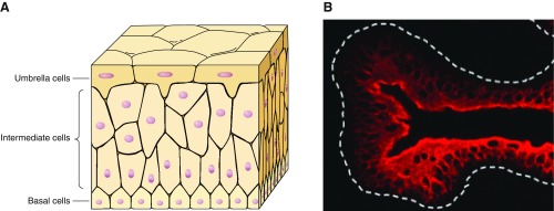
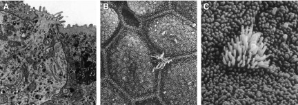
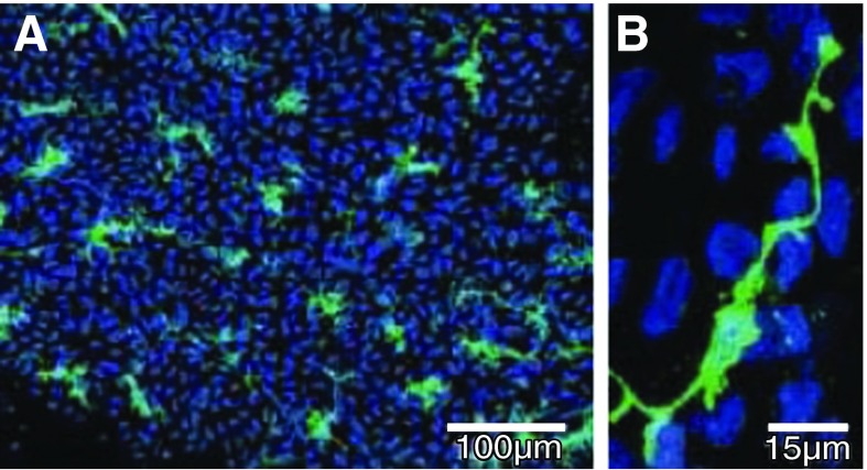
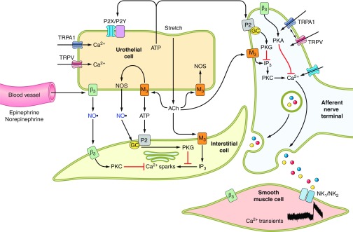
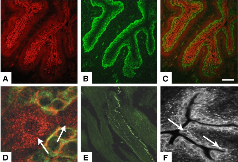
The urothelium, which lines the inner surface of the renal pelvis, the ureters, and the urinary bladder, not only forms a high-resistance barrier to ion, solute and water flux, and pathogens, but also functions as an integral part of a sensory web which receives, amplifies, and transmits information about its external milieu. Urothelial cells have the ability to sense changes in their extracellular environment, and respond to chemical, mechanical and thermal stimuli by releasing various factors such as ATP, nitric oxide, and acetylcholine. They express a variety of receptors and ion channels, including P2X3 purinergic receptors, nicotinic and muscarinic receptors, and TRP channels, which all have been implicated in urothelial-neuronal interactions, and involved in signals that via components in the underlying lamina propria, such as interstitial cells, can be amplified and conveyed to nerves, detrusor muscle cells, and ultimately the central nervous system. The specialized anatomy of the urothelium and underlying structures, and the possible communication mechanisms from urothelial cells to various cell types within the bladder wall are described. Changes in the urothelium/lamina propria ("mucosa") produced by different bladder disorders are discussed, as well as the mucosa as a target for therapeutic interventions.


Figures

Figure 1.
Figure 1.
Components of the bladder wall. Left: counterstained transverse section through normal human urinary bladder. [Modified from Neuhaus et al. (215), with permission from John Wiley and Sons, Inc.] Right: cartoon depicting bladder wall components (Modified from Andersson 2012.)
Figure 2.
Figure 2.
Urinary bladder urothelium and associated tight junctions. A: cartoon depicting multiple epithelial cell layers within the urothelium. B: immunohistochemical staining of mouse urothelium with an antibody to uroplakin III showing staining of superficial umbrella cells. [From Sun (266).]
Figure 3.
Figure 3.
Ultrastructural features of urethral epithelium. Right: images depict “open type” paraneurons within the dog urethra. A: paraneuron reaching the lumen. B and C: scanning EM identifies (arrows) microvillous cells among the epithelial cells. A: ×14,000. B: ×4,600. C: ×16,000. [From Hashimoto et al. (126).]
Figure 4.
Figure 4.
c-Kit-labeled interstitial cells (ICs) in the lamina propria. A: whole-mount preparation of guinea pig bladder mucosa labeled with anti-c-Kit (green) and nuclei counterstained with DAPI (blue). B: high magnification showing an IC with lateral branches. [From McCloskey (204), with permission from John Wiley and Sons, Inc.]
Figure 5.
Figure 5.
Images following application of liposomes within the urinary bladder. A: representative image showing a typical liposome (×4,000; Lipella, Inc.). B: liposomes instilled in a rat bladder forming a coating (in green) on urothelial surface. C: intravesical instillation of protamine sulfate (PS) in rat urinary bladder exhibit damaged urothelium. D: intravesical instillation of PS followed by liposomes in rat urinary bladder exhibit an intact urothelium, demonstrating a protective effect of liposomes on urothelial barrier. [From Kaufman et al. (154).]
Figure 6.
Figure 6.
Urothelial alterations following injury or inflammation. A: intact urothelium in rat urinary bladder (green pan-cytokeratin labeling epithelium; blue, DAP-I nuclear marker). B: area of damage (shown at arrow) to the rat urothelium following cyclophosphamide treatment in a rat. (Modified from Birder 2012.) C and D: scanning electron micrograph of apical surface of umbrella cell layer from normal rat (C) and 2 h after spinal cord injury (D). [Modified from Apodaca et al. (22).]
Figure 7.
Figure 7.
Urothelial alterations associated with bladder pain syndrome or senescence. Electron micrograph in control (A) and BPS (B) bladders. In A, parallel layers of upper lamina propria interstitial cells (ULP ICs, arrows) within a dense extracellular matrix. In B, arrows indicate fragmented layers of UPL ICs and presence of lymphocytes (L) and mast cells (M). [A and B from Gevaert et al. (110), with permission from John Wiley and Sons.] C and D: scanning electron micrographs of the urinary bladder of adult (C) and aging (D) female rats showing the luminal surface. In aging rats, the polygonal cell appearance and well-defined cell boundries (CB) appeared to be lost with focal areas of denuded urothelium (arrow). [C and D from Mahmoud et al. (196a).]
Figure 8.
Figure 8.
Hypothetical model depicting possible interactions between bladder nerves, urothelial cells, smooth muscle, interstitial cells, and blood vessels. Urothelial cells can also be targets for transmitters released from nerves or other cell types. Urothelial cells can be activated by either autocrine (i.e., autoregulation) or paracrine (release from nearby nerves or other cells) mechanisms. [Modified from Kanai et al. (153a), with permission from John Wiley and Sons, Inc.]
Figure 9.
Figure 9.
Expression of TRPs within the urinary bladder. A: cytokeratin-staining for bladder urothelium. B: TRPV4-positive immunoreactivity in mouse urothelium. C: a merged image of both cytokeratin staining and TRPV4 in mouse urothelium. D: confocal image of bladder urothelium in bladder whole mount stained for TRPV1 (cy3, red) and cytokeratin 17 (FITC, green), a marker for basal urothelial cells. Diffuse cytoplasmic pattern of TRPV1 staining can be seen in the apical and underlying urothelial layers. E: immunohistochemical localization of TRPA1 within the urinary bladder wall (scale, 100 μm). F: immunohistochemical localization of TRPM8 within the urinary bladder urothelium. [Modified from Chopra and Birder, unpublished observations; Birder et al. (39); Streng et al. (263); and Yamada et al. (300).]

Comment in

  • Re: Urothelial signaling.
    Wein AJ. Wein AJ. J Urol. 2013 Dec;190(6):2307-8. doi: 10.1016/j.juro.2013.08.108. Epub 2013 Sep 5. J Urol. 2013. PMID: 24209573 No abstract available.

References

    1. Abraham P, Rabi S, Selvakumar D. Protective effect of aminoguanidine against oxidative stress and bladder injury in cyclophosphamide-induced hemorrhagic cystitis in rat. Cell Biochem Funct 27: 56–62, 2009 - PubMed
    1. Abrams P, Amarenco G, Haab F. Urinary prostaglandin E2 levels are elevated in patients with overactive bladder and painful bladder syndrome and correlate with bladder diary symptoms. EAU Abstr 783: 2010
    1. Acharya P, Beckel J, Ruiz WG, Wang E, Rojas R, Birder L, Apodaca G. Distribution of the tight junction proteins ZO-1, occludin, and claudin-4, -8, and -12 in bladder epithelium. Am J Physiol Renal Physiol 287: F305–F318, 2004 - PubMed
    1. Aitken KJ, Bagli DJ. The bladder extracellular matrix. Part I: architecture, development and disease. Nat Rev Urol 6: 596–611, 2009 - PubMed
    1. Aizawa N, Igawa Y, Nishizawa O, Wyndaele JJ. Effects of CL316,243, a beta 3 adrenoceptor agonist, and intravesical prostaglandin E2 on the primary bladder afferent activity of the rat. Neurourol Urodyn 29: 771–776, 2010 - PubMed

Publication types

MeSH terms

Substances