. 2013 Apr 30;110(18):7306-11.
doi: 10.1073/pnas.1214441110. Epub 2013 Apr 15.

LDL receptor and its family members serve as the cellular receptors for vesicular stomatitis virus

Affiliations

LDL receptor and its family members serve as the cellular receptors for vesicular stomatitis virus

Danit Finkelshtein et al. Proc Natl Acad Sci U S A. .

Abstract

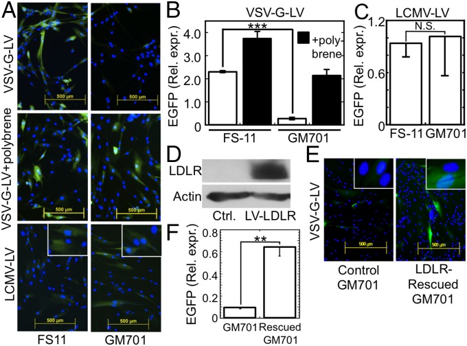
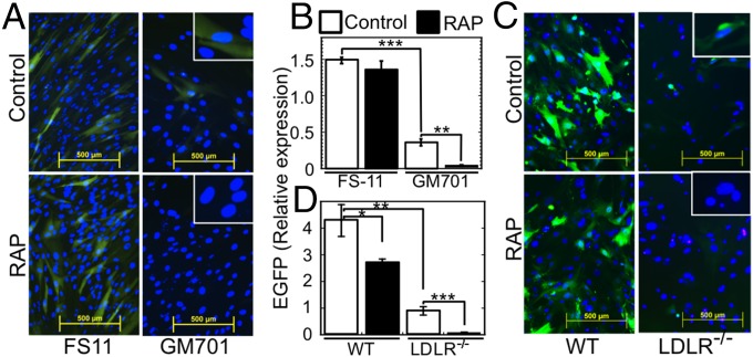
Vesicular stomatitis virus (VSV) exhibits a remarkably robust and pantropic infectivity, mediated by its coat protein, VSV-G. Using this property, recombinant forms of VSV and VSV-G-pseudotyped viral vectors are being developed for gene therapy, vaccination, and viral oncolysis and are extensively used for gene transduction in vivo and in vitro. The broad tropism of VSV suggests that it enters cells through a highly ubiquitous receptor, whose identity has so far remained elusive. Here we show that the LDL receptor (LDLR) serves as the major entry port of VSV and of VSV-G-pseudotyped lentiviral vectors in human and mouse cells, whereas other LDLR family members serve as alternative receptors. The widespread expression of LDLR family members accounts for the pantropism of VSV and for the broad applicability of VSV-G-pseudotyped viral vectors for gene transduction.


Conflict of interest statement

The authors declare no conflict of interest.

Figures

Fig. 1.
Fig. 1.
Soluble LDLR binds VSV and inhibits infection by VSV and transduction by a VSV-G-pseudotyped lentiviral vector. (A) Survival ± SD of WISH cells as determined by Neutral red staining after treatment with sLDLR and challenge by VSV at the indicated MOI. n = 3. (Inset) SDS/PAGE of sLDLR (10 µg). Molecular mass markers (kDa) are shown on the right lane. (B) Surviving WISH cells, bovine MDBK cells, and murine L cells after treatment with serially twofold-diluted sLDLR (starting at 8 µg/mL) followed by VSV (MOI = 1 for WISH and MDBK cells, MOI = 0.07 for L cells. C, no virus; V, VSV without sLDLR. (C) Surviving WISH cells after addition of sLDLR (1 µg/mL) at the indicated times relative to the time of VSV (MOI = 0.1) addition. In well R, sLDLR was added for 120 min and removed before VSV challenge. C and V are as in B. (D) Quantitative RT-PCR of VSV RNA after attachment of VSV (MOI = 10) at 4 °C for 4 h to WISH cells in the presence of the indicated sLDLR concentrations. VSV RNA ± SE is normalized to TATA binding protein mRNA; *P < 0.02, **P < 0.002, compared with the leftmost bar, n = 3. (Inset) RT-PCR products of VSV RNA, isolated after similar experiments, performed at 4 °C and at 37 °C. (E) Surface plasmon resonance analysis of VSV binding to immobilized sLDLR in PBS with or without CaCl2 (1 mM). (F) Surface plasmon resonance analysis of sLDLR binding to immobilized VSV-G-LV in PBS + 1 mM CaCl2. (G) (Upper) Immunoblotting of VSV-G after coimmunoprecipitation of a solubilized VSV-sLDLR complex with the following antibodies (lanes): 1, mAb 28.28 anti-LDLR; 2, mAb C7 anti-LDLR; 3, isotype control mAb; 4, no antibody. A VSV-G marker is shown in lane 5. (Lower) Reblotting of the membrane with anti-LDLR mAb 29.8. (H) EGFP expression (green) after transduction of FS-11 fibroblasts with either EGFP-encoding VSV-G-LV or EGFP-encoding LCMV-LV in the presence or absence of sLDLR (5 µg/mL). Nuclei were counterstained with Hoechst 33258 (blue). (Insets) Enlarged magnifications. (I) Average ± SD EGFP expression in cultures transfected as shown in H. ***P < 0.003, n = 4. N.S., not significant (P = 0.525), n = 4.
Fig. 2.
Fig. 2.
VSV and LDL share a common cell surface receptor. (A) Surviving WISH epithelial cells, pretreated with trypsin-EDTA or EDTA, washed and challenged with VSV (0.015 MOI, 15 min). Figure is representative of six replicates. VSV yield (Lower) was determined by a plaque assay of the culture supernatants. *P < 0.03, n = 3. (B) Internalized Dil-LDL (red) in FS-11 fibroblasts after binding (1.67 µg/mL, 4 h, 4 °C) in the presence of the indicated VSV MOI. The cultures were then washed, and bound Dil-LDL was allowed to internalize (1 h, 37 °C). (Insets) Higher magnifications. (C) (Upper) Immunoblot of LDLR in WT FS-11 fibroblasts and LDLR-deficient GM701 fibroblasts. (Lower) Lack of Dil-LDL uptake by LDLR-deficient GM701 fibroblasts. (D) Flow cytometry of FS-11 fibroblasts treated with Dil-LDL as in A in the absence or presence of VSV (MOI = 2000). n = 3. (E) LDLR-deficient GM701 fibroblasts untreated or treated with sLDLR (1 µg/mL) and challenged with VSV (MOI = 1).
Fig. 3.
Fig. 3.
LDLR and its family members are the major and the alternative VSV receptors, respectively. (A) Crystal violet-stained WISH cells, untreated (Ctrl.) or treated with anti-LDLR mAbs (30 min, 4 °C) and then subjected to limited infection by VSV (MOI = 0.05, 4 °C, 1 h). (B) Crystal violet-stained cultures of WT (FS-11) and LDLR-deficient (GM701) fibroblasts, either untreated (Control) or treated with isotype control mAb or anti-LDLR mAb 29.8 (12.5 µg/mL each), followed by VSV as in A. (C) Crystal violet-stained cultures of WT FS-11 fibroblasts and LDLR-deficient GM701 fibroblasts, treated with RAP (100 nM, 30 min, 37 °C) alone, VSV (MOI = 1) alone, or RAP followed by VSV. (D) Plaque assay of culture supernatants from WT FS-11 fibroblasts and LDLR-deficient GM701 fibroblasts (50,000 cells per well) preincubated (30 min, 37 °C) in DMEM-10 or in DMEM-10 + RAP (100 nM), then challenged with VSV (0.5 MOI, 30 min, 37 °C), washed three times, and incubated in DMEM-10 (0.1 mL, 37 °C, 7 h). ***P < 0.001, n = 4. (E) Crystal violet-stained WISH cells grown to confluence in 96-well plates, incubated (30 min, 37 °C) with the indicated combinations of RAP (200 nM), neutralizing anti-LDLR mAb 29.8, and nonneutralizing anti-LDLR mAb 28.28 (50 µg/mL each); cells were then challenged with VSV at the indicated MOI. Cell viability (bar plot) was determined by reading the OD540 of cultures treated with VSV at MOI = 0.06. ***P < 0.002, n = 4.
Fig. 4.
Fig. 4.
LDLR and its family members mediate VSV internalization by human fibroblasts. (A) Internalized VSV in WT FS-11 fibroblasts and LDLR-deficient GM701 fibroblasts after incubation with VSV (MOI = 500, 4 min, 37 °C) and washing three times with PBS. The cultures were then fixed and stained with anti-VSV-G (red). (B) Internalized VSV in WT FS-11 fibroblasts preincubated with the indicated combinations of RAP and anti-LDLR mAbs (30 min, 37 °C), followed by VSV as in A. (C) VSV foci in A and B were counted in fields containing at least 30 cells. **P < 0.01; *P < 0.05 (compared with FS-11 challenged with VSV only, leftmost bar); n = 3.
Fig. 5.
Fig. 5.
LDLR is the main entry port of VSV-G-LV. (A) EGFP expression in WT FS-11 fibroblasts and LDLR-deficient GM701 fibroblasts, 72 h posttransduction with either EGFP-encoding VSV-G-LV in the absence or presence of polybrene, or with EGFP-encoding LCMV-LV in the absence of polybrene. (Insets) Higher magnifications. (B) Average ± SD of the relative EGFP expression (Rel. expr.) after transduction with VSV-G-LV in the absence (open bars) or presence (filled bars) of polybrene. ***P < 0.0001, n = 3. (C) Average ± SD of the relative EGFP expression after transduction with LCMV-LV. N.S., not significant (P = 0.78), n = 3. (D) Immunoblot of LDLR after either mock transduction of GM701 fibroblasts with polybrene alone (Ctrl.) or their transduction with VSV-G-LV encoding LDLR in the presence of polybrene (LV-LDLR). (E) EGFP expression in cultures of LDLR-reconstituted or mock-transduced GM701 fibroblasts, transduced for 48 h with EGFP-encoding VSV-G-LV. (Insets) Higher magnifications. (F) Average ± SD of the relative EGFP expression shown in E. **P < 0.01, n = 3.
Fig. 6.
Fig. 6.
Other LDLR family members are alternative entry ports of VSV-G-LV in human and mouse cells. (A) EGFP expression in WT FS-11 fibroblasts and LDLR-deficient GM701 fibroblasts, transduced with EGFP-encoding VSV-G-LV in the absence (Control) or presence of RAP (100 nM). (Insets) Higher magnifications. (B) Average ± SD of EGFP expression shown in A. ***P < 0.0002, n = 3. *P < 0.03, n = 3. (C) EGFP expression in WT murine embryonic fibroblasts (WT) and LDLR-deficient MEFs, transduced with EGFP-encoding VSV-G-LV as in A. (Insets) Higher magnifications. (D) Average ± SD of EGFP expression shown in C. All fluorescence intensity values were normalized to the nuclei counts. *P < 0.05, **P < 0.007, ***P < 0.002, n = 3.

References

    1. Lichty BD, Power AT, Stojdl DF, Bell JC. Vesicular stomatitis virus: Re-inventing the bullet. Trends Mol Med. 2004;10(5):210–216. - PubMed
    1. Ge P, et al. Cryo-EM model of the bullet-shaped vesicular stomatitis virus. Science. 2010;327(5966):689–693. - PMC - PubMed
    1. Naldini L, et al. In vivo gene delivery and stable transduction of nondividing cells by a lentiviral vector. Science. 1996;272(5259):263–267. - PubMed
    1. Rose NF, et al. An effective AIDS vaccine based on live attenuated vesicular stomatitis virus recombinants. Cell. 2001;106(5):539–549. - PubMed
    1. Roche S, Albertini AA, Lepault J, Bressanelli S, Gaudin Y. Structures of vesicular stomatitis virus glycoprotein: membrane fusion revisited. Cell Mol Life Sci. 2008;65(11):1716–1728. - PMC - PubMed

Publication types