. 2013 Apr 30;110(18):7252-7.
doi: 10.1073/pnas.1303890110. Epub 2013 Apr 15.

Protein-DNA complexes are the primary sources of replication fork pausing in Escherichia coli

Affiliations

Protein-DNA complexes are the primary sources of replication fork pausing in Escherichia coli

Milind K Gupta et al. Proc Natl Acad Sci U S A. .

Abstract

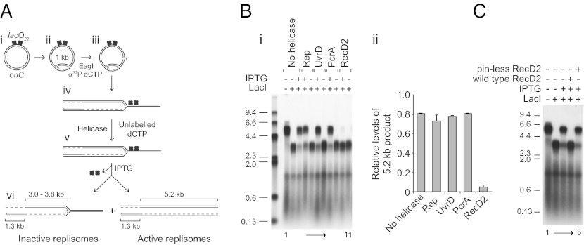
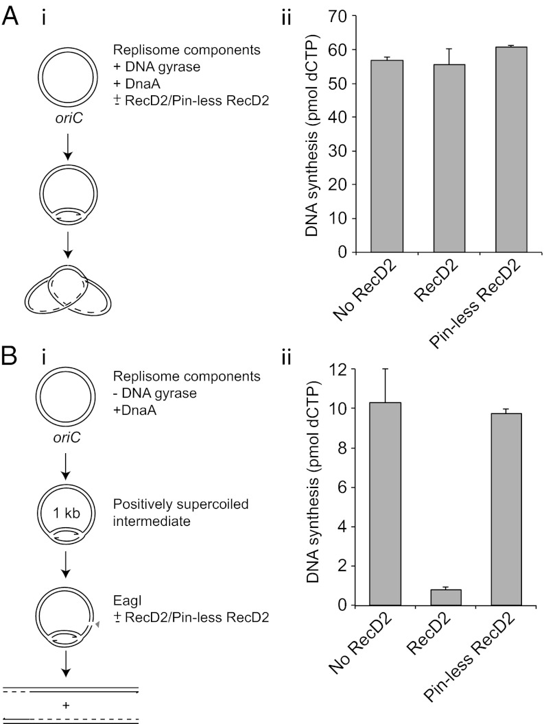
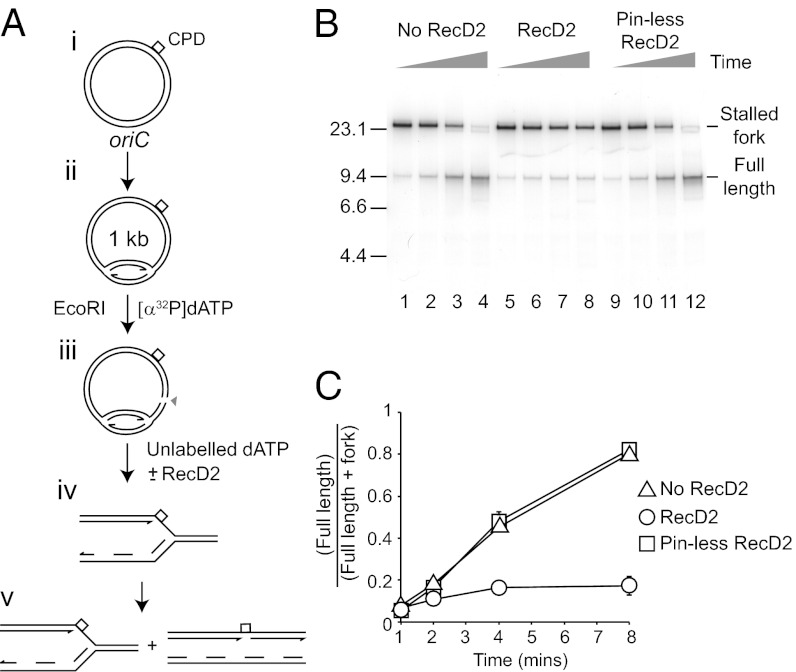
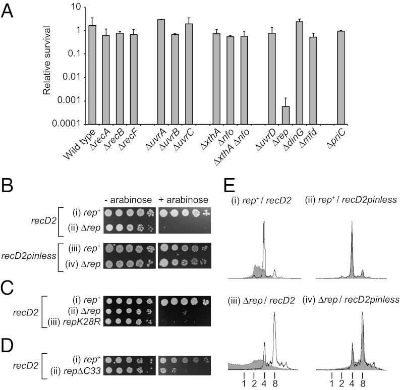
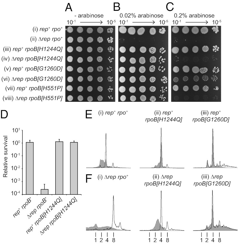
Replication fork pausing drives genome instability, because any loss of paused replisome activity creates a requirement for reloading of the replication machinery, a potentially mutagenic process. Despite this importance, the relative contributions to fork pausing of different replicative barriers remain unknown. We show here that Deinococcus radiodurans RecD2 helicase inactivates Escherichia coli replisomes that are paused but still functional in vitro, preventing continued fork movement upon barrier removal or bypass, but does not inactivate elongating forks. Using RecD2 to probe replisome pausing in vivo, we demonstrate that most pausing events do not lead to replisome inactivation, that transcription complexes are the primary sources of this pausing, and that an accessory replicative helicase is critical for minimizing the frequency and/or duration of replisome pauses. These findings reveal the hidden potential for replisome inactivation, and hence genome instability, inside cells. They also demonstrate that efficient chromosome duplication requires mechanisms that aid resumption of replication by paused replisomes, especially those halted by protein-DNA barriers such as transcription complexes.


Conflict of interest statement

The authors declare no conflict of interest.

Figures

Fig. 1.
Fig. 1.
RecD2 inactivates replication forks blocked by nucleoprotein complexes in vitro. (A) reaction scheme to monitor the ability of replisomes halted at LacI–lacO complexes to continue replication upon IPTG-induced dissociation of the barrier. (B, i) Denaturing agarose gel of replication products formed with pPM561 in the absence or the presence of 400 nM LacI with or without subsequent addition of 1 mM IPTG. Helicases added to 100 nM final concentration at step iv/v (A) are indicated. Sizes of markers in kilobases are indicated. (B, ii) Levels of the 5.2-kb leading strand products formed in the presence of repressor after subsequent addition of IPTG relative to those obtained in the absence of repressor (see lane 1 in B). (C) The effects of wild-type and pinless RecD2 on the ability of forks paused at LacI–lacO complexes to continue upon addition of IPTG.
Fig. 2.
Fig. 2.
RecD2 inactivates blocked but not elongating replication forks. (A, i) Method to analyze the impact of RecD2 on elongating replication forks. (A, ii) DNA synthesis in the presence of DNA gyrase upon simultaneous addition of DnaA with or without either wild-type or pinless RecD2, each present at 100 nM final concentration. (B, i) Method to monitor the impact of RecD2 on the ability of forks stalled by positive supercoiling to continue DNA synthesis upon relief of topological strain. (B, ii) Levels of DNA synthesis upon addition of a restriction enzyme with or without either wild-type or pinless RecD2 (each at 100 nM).
Fig. 3.
Fig. 3.
RecD2 inactivates forks paused by template lesions. (A) reaction scheme to monitor replication fork pausing at, and bypass of, a cyclobutane pyrimidine dimer (CPD) within the leading strand template. (B) Native agarose gel of replication products formed on plasmid DNA harboring a pyrimidine dimer within the leading strand template. Time points were taken 1, 2, 4, and 8 min after addition of restriction enzyme and radiolabel. Positions of replication forks stalled at the pyrimidine dimer and full-length products generated by bypass of the lesion are indicated. Wild-type and pinless RecD2 were present at 100 nM, as indicated. (C) Accumulation of full-length products as a function of time.
Fig. 4.
Fig. 4.
Expression of recD2 is toxic in strains lacking Rep. (A) Colony-forming ability of wild-type E. coli (BW25113) and otherwise isogenic strains bearing single gene deletions upon expression of recD2 using a plasmid-based arabinose-inducible system (pMG31). Survival is represented by the number of colonies formed upon induction of recD2 expression relative to the number of colonies formed in the absence of induction. Table S2 gives strain numbers. (B) Colony-forming ability of rep+ (TB28) and Δrep (N6577) strains harboring pBADrecD2 and pBADrecD2pinless. (C) Colony-forming ability of rep+ (MG1655), rep (N4982), and repK28R (SS1076) containing pBADrecD2. (D) Colony-forming ability of rep+ (MKG08) and repΔC33 (MKG10) upon recD2 overexpression (pMG31). (E) DNA content of (i and ii) wild-type (TB28) and (iii and iv) Δrep (N6577) cells without and with induction of expression (unshaded and shaded histograms, respectively) from pBADrecD2 and pBADrecD2pinless as monitored by flow cytometry. DNA content with respect to number of chromosome equivalents per cell is indicated below. Note that Δrep cells have a higher mean number of chromosomes per cell compared with rep+ (compare the uninduced samples in i and ii with iii and iv), as noted previously (39).
Fig. 5.
Fig. 5.
RecD2-directed lethality is associated with transcription complexes. (AC) Colony-forming ability of rep+ and Δrep strains bearing either wild-type or mutant rpoB alleles upon no, low-, or high-level induction from pBADrecD2 (0%, 0.02% and 0.2% arabinose, respectively). Strains (i–viii) are TB28, N6577, PM486, N7604, AM2158, HB278, N7616, and HB280. (D) Colony-forming ability of rep+ and Δrep strains bearing rpoB+ or rpoB[H1244Q] upon induction of recD2 expression with 0.2% arabinose. Strain numbers are as in AC. (E and F) DNA content of rep+ and Δrep strains bearing rpoB+, rpoB[H1244Q], or rpoB[G1260D] without and with induction of expression of recD2 with 0.2% arabinose (unshaded and shaded histograms, respectively). The number of chromosome equivalents per cell is indicated below. Note that both rpoB[H1244Q] and rpoB[G1260D] also reduced the median number of chromosome equivalents in Δrep cells in the absence of recD2 expression from eight to four, the same number as seen in rep+ cells (F, compare i with ii and iii). This may reflect suppression of the reduced rate of genome duplication in Δrep cells (30). Strain numbers are as in AC.

References

    1. French S. Consequences of replication fork movement through transcription units in vivo. Science. 1992;258(5086):1362–1365. - PubMed
    1. Lindahl T, Wood RD. Quality control by DNA repair. Science. 1999;286(5446):1897–1905. - PubMed
    1. Paeschke K, Capra JA, Zakian VA. DNA replication through G-quadruplex motifs is promoted by the Saccharomyces cerevisiae Pif1 DNA helicase. Cell. 2011;145(5):678–691. - PMC - PubMed
    1. Heller RC, Marians KJ. Replisome assembly and the direct restart of stalled replication forks. Nat Rev Mol Cell Biol. 2006;7(12):932–943. - PubMed
    1. Horiuchi T, Fujimura Y. Recombinational rescue of the stalled DNA replication fork: A model based on analysis of an Escherichia coli strain with a chromosome region difficult to replicate. J Bacteriol. 1995;177(3):783–791. - PMC - PubMed

Publication types

MeSH terms

LinkOut - more resources