. 2013 Apr 30;110(18):7193-8.
doi: 10.1073/pnas.1305286110. Epub 2013 Apr 15.

Phosphoinositides and membrane curvature switch the mode of actin polymerization via selective recruitment of toca-1 and Snx9

Affiliations

Phosphoinositides and membrane curvature switch the mode of actin polymerization via selective recruitment of toca-1 and Snx9

Jennifer L Gallop et al. Proc Natl Acad Sci U S A. .

Abstract

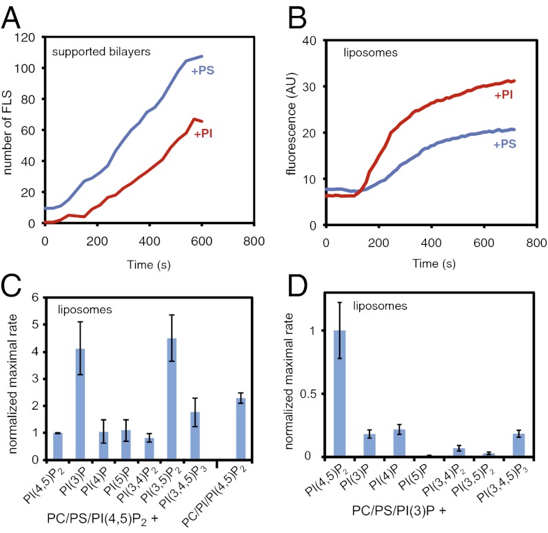
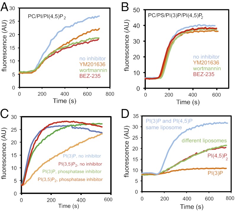
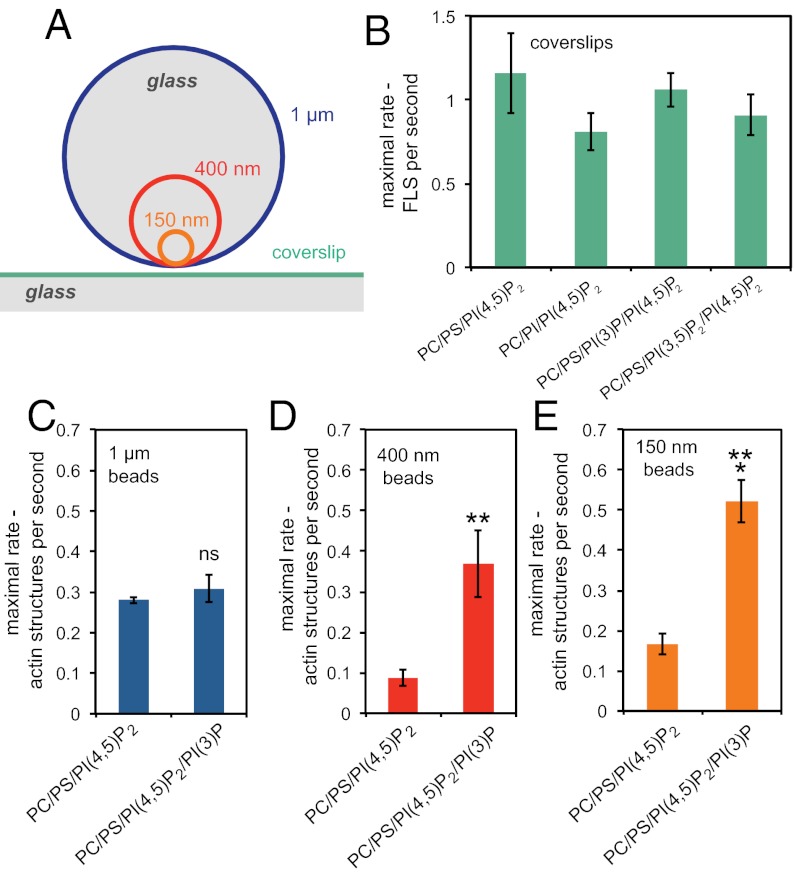
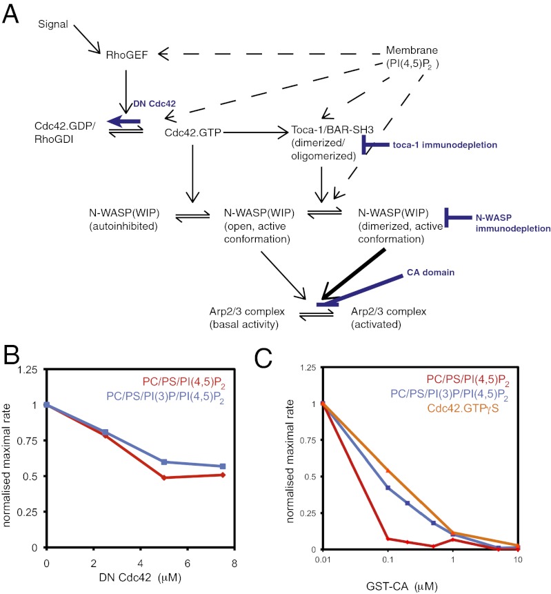
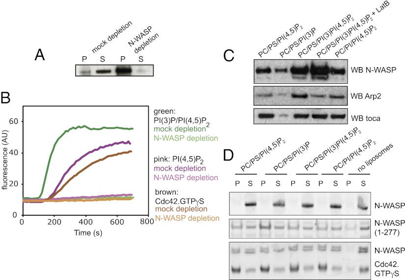
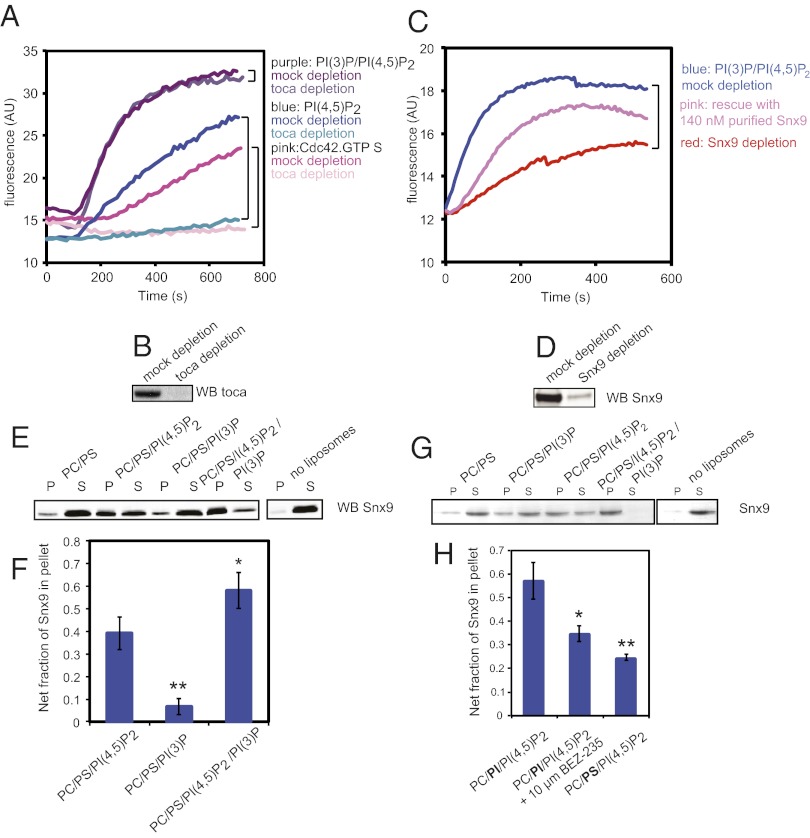
The membrane-cytosol interface is the major locus of control of actin polymerization. At this interface, phosphoinositides act as second messengers to recruit membrane-binding proteins. We show that curved membranes, but not flat ones, can use phosphatidylinositol 3-phosphate [PI(3)P] along with phosphatidylinositol 4,5-bisphosphate [PI(4,5)P2] to stimulate actin polymerization. In this case, actin polymerization requires the small GTPase cell cycle division 42 (Cdc42), the nucleation-promoting factor neural Wiskott-Aldrich syndrome protein (N-WASP) and the actin nucleator the actin-related protein (Arp) 2/3 complex. In liposomes containing PI(4,5)P2 as the sole phosphoinositide, actin polymerization requires transducer of Cdc42 activation-1 (toca-1). In the presence of phosphatidylinositol 3-phosphate, polymerization is both more efficient and independent of toca-1. Under these conditions, sorting nexin 9 (Snx9) can be implicated as a specific adaptor that replaces toca-1 to mobilize neural Wiskott-Aldrich syndrome protein and the Arp2/3 complex. This switch in phosphoinositide and adaptor specificity for actin polymerization from membranes has implications for how different types of actin structures are generated at precise times and locations in the cell.


Conflict of interest statement

The authors declare no conflict of interest.

Figures

Fig. 1.
Fig. 1.
PI and PI(3)P contribute to actin polymerization from liposomes. (A) Numbers of filopodia-like structures from supported lipid bilayers showing that filopodia-like structure formation is favored by PS and not PI. The lipid compositions were 48% PC, 48% PS or PI, and 4% PI(4,5)P2. (B) Pyrene actin assay of actin polymerization from liposomes showing that it is favored by PI rather than PS, with the same liposomes used in A. (C) Bar chart showing the maximal rate of actin polymerization (slope of a pyrene actin assay) with a lipid composition of 48% PC, 47% PS, 4% PI(4,5)P2, and 1% of the test phosphoinositide, or PC/PI/PI(4,5)P2. The values are normalized against PC/PS/PI(4,5)P2 liposomes. (D) Substituting PI(4,5)P2 and keeping 1% PI(3)P constant shows that PI(4,5)P2 is essential. Data are normalized to PC/PS/PI(3)P/PI(4,5)P2 liposomes. Data in A and B is representative of four independent experiments. Data in C and D is the average of three experiments, with error bars showing the SD.
Fig. 2.
Fig. 2.
Phosphorylation of PI by class I PI3 kinase is important, and PI(3)P and PI(4,5)P2 work together to stimulate actin polymerization. (A) Pyrene actin assay on addition of PC/PI/PI(4,5)P2 into extracts preincubated with 10 μM wortmannin, BEZ-235, or YM201636. (B) Pyrene actin assay on addition of 48% PC, 47% PI, 1% PI(3)P, and 4% PI(4,5)P2 liposomes in extracts that were preincubated with the drugs similarly to A showing that the 3-position stimulates actin polymerization. (C) Pyrene actin assay on addition of 48% PC, 47% PS, 1% PI(3)P or 1% PI(3,5)P2, and 4% PI(4,5)P2 in extract preincubated with a 1/100 dilution of a phosphatase inhibitor mixture. (D) Addition of liposomes where PI(3)P and PI(4,5)P2 are present in the same or distinct liposomes. Data is representative of two or three independent experiments.
Fig. 3.
Fig. 3.
Membrane curvature contributes to actin polymerization from PI(3)P/PI(4,5)P2 membranes. (A) Relative sizes of glass beads (to scale) compared with a coverslip. (B) Bar chart showing that the rate of appearance of filopodia-like structures is unaltered by the presence of PI(3)P or PI(3,5)P2. The lipid compositions were 48% PC, 47% PS, 1% PI(3)P or PI(3,5)P2, and 4% PI(4,5)P2. (C) Rate of appearance of actin structures growing on bilayers composed of 48% PC, 48% PS, 4% PI(4,5)P2 and 48% PC, 47% PS, 4% PI(4,5)P2, and 1% PI(3)P supported on 1-μm-diameter glass beads showing no significance difference between the compositions. (D) Rate of appearance of actin structures growing on bilayers supported on 400-nm-diameter glass beads showing a very significant increase in the presence of PI(3)P (**P = 0.008). (E) Number of actin structures growing on bilayers supported on 150-nm-diameter glass beads showing a significant increase in the presence of PI(3)P (***P = 0.0001). Data are the mean of six experiments; error bars show SEM.
Fig. 4.
Fig. 4.
Actin polymerization stimulated by PI(3)P and PI(4,5)P2 uses Cdc42 and is enhanced by GTPγS. (A) Schematic diagram of signaling from membranes through Cdc42, N-WASP, toca-1, and Arp2/3 complex and highlighting the steps that were inhibited. (B) Dose–response of actin polymerization in a pyrene actin assay in the presence of increasing concentrations of dominant negative Cdc42 with PI(4,5)P2 and PI(3)P/PI(4,5)P2 stimuli (composition used in Fig. 3). (C) Dose–response of actin polymerization in response to Cdc42.GTPγS, PI(4,5)P2, PI(3)P/PI(4,5)P2 stimuli with increasing concentrations of Arp2/3 complex-activation inhibitor, GST-CA domain. PI(3)P/PI(4,5)P2 and Cdc42.GTPγS are more resistant to the inhibitor.
Fig. 5.
Fig. 5.
Actin polymerization stimulated by PI(3)P and PI(4,5)P2 uses N-WASP; however, the specificity for PI(3)P/PI(4,5)P2 binding is not within N-WASP itself. (A) Western blot of an immunodepletion showing the bead pellet and extract in the supernatant with control IgG and anti–N-WASP antibodies. (B) Pyrene actin assays on mock and N-WASP–depleted extract shows that N-WASP is critical for actin polymerization with Cdc42.GTPγS, PI(3)P, and PI(3)P/PI(4,5)P2 liposomes. (C) Liposome sedimentation assay from addition of liposomes into extract showing that although Arp2/3 complex and N-WASP binding to the liposomes is increased by the presence of PI(3)P, toca-1 binding is not. Use of latrunculin shows that N-WASP sedimentation is not due to bulk actin polymerization. Arp2 recruitment is driven by both membrane and polymerized actin. (D) Liposome sedimentation assays with the indicated lipid compositions with purified N-WASP, active N-WASP fragment (1–277), or N-WASP and activated Cdc42. There is no specificity in binding for PI(3)P/PI(4,5)P2-containing liposomes, suggesting an external recruitment factor. P, pellet; S, supernatant.
Fig. 6.
Fig. 6.
PI(3)P/PI(4,5)P2-containing liposomes stimulate actin polymerization by recruiting additional N-WASP and Arp2/3 complex to the liposome surface via Snx9. (A) Pyrene actin assay in response to GTPγS.Cdc42, PI(4,5)P2, or PI(3)P/PI(4,5)P2 from mock-depleted or toca-1–depleted extracts showing that PI(3)P/PI(4,5)P2 stimulates actin polymerization independently of toca-1. (B) Western blot of toca-1 in mock- and toca-1–depleted extract used showing effective immunodepletion. (C) Pyrene actin assay in response to PI(3)P/PI(4,5)P2 from mock-depleted extract, Snx9-depleted extract, and Snx9-depleted extract supplemented with 140 nM purified Snx9. (D) Western blot of Snx9 from mock- and Snx9-depleted extract showing effective immunodepletion. (E) Sedimentation of Snx9 from extracts with indicated liposome compositions showing that it binds preferentially to liposomes containing both PI(3)P and PI(4,5)P2. (F) Quantification of Snx9 that is pelleted in the liposome compositions in E, adjusted for binding to control PC/PS liposomes alone, determined by Western blotting. *P = 0.037 and **P = 0.0022 are significant differences by t test. Data are representative of three independent experiments; error bars are SD. (G) Sedimentation of purified Snx9 with indicated liposome compositions showing that it binds preferentially to liposomes containing both PI(3)P and PI(4,5)P2. (H) Liposome sedimentation assay showing the quantification of Snx9 in extract pelleted in PC/PI/PI(4,5)P2 in the absence or presence of 10 μM BEZ-235, and in control PC/PS/PI(4,5)P2 liposomes. *P = 0.037 and **P = 0.0061 indicate a significant and very significant difference in binding relative to PC/PI/PI(4,5)P2 liposomes alone by t test. Data are representative of four experiments; error bars show SEM.

Similar articles

Cited by

References

    1. Ma L, Cantley LC, Janmey PA, Kirschner MW. Corequirement of specific phosphoinositides and small GTP-binding protein Cdc42 in inducing actin assembly in Xenopus egg extracts. J Cell Biol. 1998;140(5):1125–1136. - PMC - PubMed
    1. Papayannopoulos V, et al. A polybasic motif allows N-WASP to act as a sensor of PIP(2) density. Mol Cell. 2005;17(2):181–191. - PubMed
    1. Lebensohn AM, Kirschner MW. Activation of the WAVE complex by coincident signals controls actin assembly. Mol Cell. 2009;36(3):512–524. - PMC - PubMed
    1. Saarikangas J, Zhao H, Lappalainen P. Regulation of the actin cytoskeleton-plasma membrane interplay by phosphoinositides. Physiol Rev. 2010;90(1):259–289. - PubMed
    1. Zhao H, Hakala M, Lappalainen P. ADF/cofilin binds phosphoinositides in a multivalent manner to act as a PIP(2)-density sensor. Biophys J. 2010;98(10):2327–2336. - PMC - PubMed

Publication types

MeSH terms