. 2013 Apr 30;110(18):7312-7.
doi: 10.1073/pnas.1220998110. Epub 2013 Apr 15.

Exosomes reflect the hypoxic status of glioma cells and mediate hypoxia-dependent activation of vascular cells during tumor development

Affiliations

Exosomes reflect the hypoxic status of glioma cells and mediate hypoxia-dependent activation of vascular cells during tumor development

Paulina Kucharzewska et al. Proc Natl Acad Sci U S A. .

Abstract

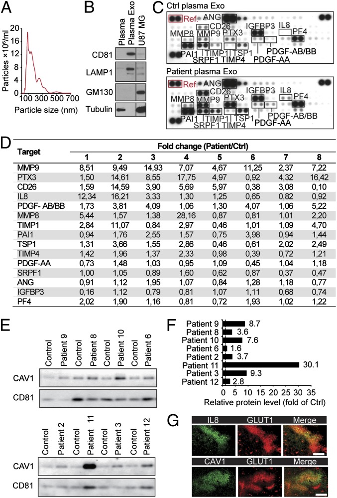
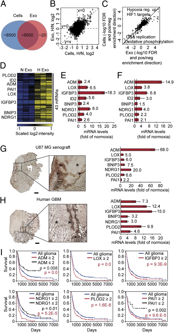
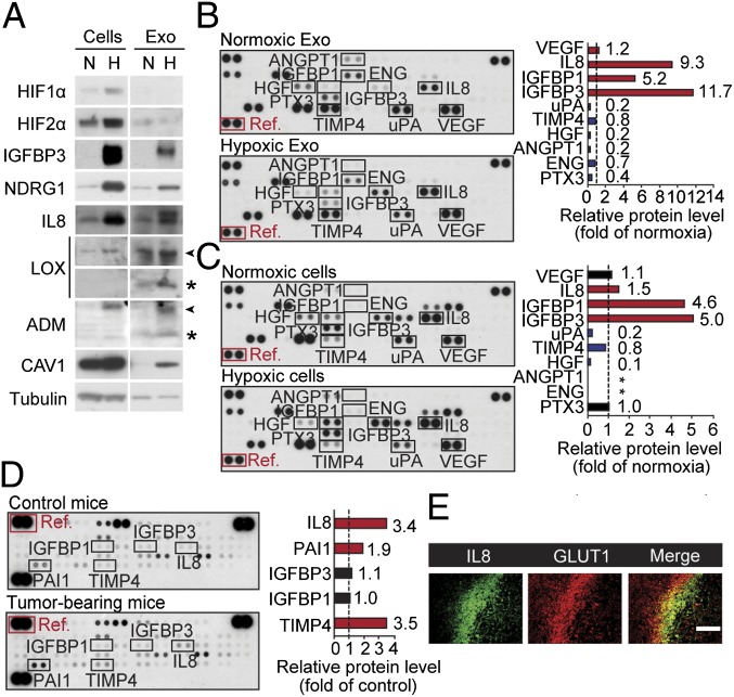
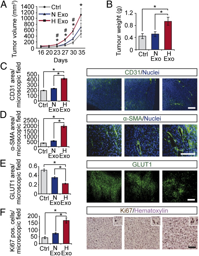
Hypoxia, or low oxygen tension, is a major regulator of tumor development and aggressiveness. However, how cancer cells adapt to hypoxia and communicate with their surrounding microenvironment during tumor development remain important questions. Here, we show that secreted vesicles with exosome characteristics mediate hypoxia-dependent intercellular signaling of the highly malignant brain tumor glioblastoma multiforme (GBM). In vitro hypoxia experiments with glioma cells and studies with patient materials reveal the enrichment in exosomes of hypoxia-regulated mRNAs and proteins (e.g., matrix metalloproteinases, IL-8, PDGFs, caveolin 1, and lysyl oxidase), several of which were associated with poor glioma patient prognosis. We show that exosomes derived from GBM cells grown at hypoxic compared with normoxic conditions are potent inducers of angiogenesis ex vivo and in vitro through phenotypic modulation of endothelial cells. Interestingly, endothelial cells were programmed by GBM cell-derived hypoxic exosomes to secrete several potent growth factors and cytokines and to stimulate pericyte PI3K/AKT signaling activation and migration. Moreover, exosomes derived from hypoxic compared with normoxic conditions showed increased autocrine, promigratory activation of GBM cells. These findings were correlated with significantly enhanced induction by hypoxic compared with normoxic exosomes of tumor vascularization, pericyte vessel coverage, GBM cell proliferation, as well as decreased tumor hypoxia in a mouse xenograft model. We conclude that the proteome and mRNA profiles of exosome vesicles closely reflect the oxygenation status of donor glioma cells and patient tumors, and that the exosomal pathway constitutes a potentially targetable driver of hypoxia-dependent intercellular signaling during tumor development.


Conflict of interest statement

The authors declare no conflict of interest.

Figures

Fig. 1.
Fig. 1.
GBM patient plasma exosomes have increased levels of hypoxia-regulated proteins involved in tumor development. (A) Nanoparticle-sized distribution profile of GBM patient-derived exosomes indicates an average diameter of 148 ± 79 nm. (B) GBM patient whole plasma, plasma exosomes (Plasma Exo), and U87 MG cell lysates were analyzed for the indicated proteins by Western blotting. (C) Antibody array analyses of Exo from GBM patients (Patient plasma Exo) and matched control subjects (Ctrl plasma Exo). Shown is array data from a representative patient-control experiment. (D) Fold change of relative protein levels (normalized to array reference marked with red box in C) in the respective patient-control pairs (–8). (E and F) Exo isolated from GBM patients and matched control subjects were analyzed for caveolin 1 (CAV1) by Western blotting (n = 8) with CD81 as loading control. (G) Hypoxic regions of patient GBM tumors display increased expression of IL8 and CAV1. (Scale bar: 100 μm.)
Fig. 2.
Fig. 2.
The hypoxic transcriptional profile of exosomes reflects the hypoxic signaling response of GBM cells and tumors. (A) Number of transcripts detected by microarray analysis in GBM cell-derived exosomes and GBM cells. (B) Ratios of hypoxia/normoxia intensities plotted as log2 scale for cells and exosomes. (C) False discovery rates (−log10 space) relating to gene set enrichment analysis. Positive value, up-regulation in hypoxia; negative value, down-regulation in hypoxia of a specific gene set. (D) Heat map of 43 transcripts with significantly higher expression levels in exosomes secreted by hypoxic compared with normoxic GBM cells. (E) Validation of hypoxic induction of indicated mRNAs in exosomes by qRT-PCR. Data are presented as fold increase in hypoxic compared with normoxic exosomes ± SD and are representative of three independent experiments. Values beside bars indicate fold change in hypoxic exosomes. (F) Similar experiment as in E performed with GBM cells. (G and H) Identification of the hypoxic exosome gene expression profile in U87 MG GBM xenografts and GBM patient tumors. Laser-capture microdissection of hypoxic (stars) and normoxic (arrowheads) tumor regions was performed as described in SI Materials and Methods and analyzed for the expression of indicated transcripts by qRT-PCR. (Scale bar: 2 mm.) (I) Kaplan–Meier survival curves of hypoxia-regulated transcripts, as indicated. Blue lines, median expression level of all gliomas (n = 343). Black lines, expression ≤ twofold compared with median. Red lines, expression ≥ twofold compared with median.
Fig. 3.
Fig. 3.
Hypoxic regulation of the GBM cell and exosome angiogenesis-related proteome. (A) Equal amount of total protein from normoxic (N) or hypoxic (H) GBM cells and corresponding exosomes (Exo) were analyzed for the indicated proteins and tubulin (loading control) by Western blotting. Arrowheads and stars denote proforms and mature forms of proteins, respectively. GBM Exo (B) and cells (C) from N and H conditions were subjected to human angiogenesis protein antibody array analyses. (B and C Left) Array data from a representative experiment. (B and C Right) Data are representative of at least three independent experiments and represent fold change of relative protein levels (normalized to array reference marked with red box) in H compared with N samples. Stars indicate protein expression below detection level. (D) Circulating Exo were isolated from plasma of tumor-free control mice and GBM tumor-bearing mice, and equal amounts of total protein were analyzed by angiogenesis antibody arrays. (Left) Array data from a representative experiment. (Right) Data are representative of at least three independent experiments and represent fold change of relative protein levels (normalized to array reference marked with red box) in Exo from GBM tumor-bearing mice compared with control mice. (E) Hypoxic regions of GBM xenografts display increased IL8 protein levels. (Scale bar: 100 μm.)
Fig. 4.
Fig. 4.
Role of exosomes in hypoxia-dependent cross-talk between malignant cells and vascular cells. (A) Mouse aortas were incubated in the absence (Ctrl) or presence of exosomes (Exo) (25 µg/mL) derived from normoxic (N) or hypoxic (H) GBM cells for 24 h and then embedded in Matrigel overlaid with medium supplemented with 2% mouse serum ± Exo. Shown are representative photomicrographs of microvessels at day 7. (B) Quantitative analysis of microvessel number (Left) and length (Right) presented as the mean ± SD, n = 6 aortas per group. *P < 0.05. (C) HUVECs were cultured for 24 h in the absence (Ctrl) or presence of Exo (10 µg/mL) and then grown on Matrigel for 20 h. Shown are representative photomicrographs of EC tubes from the different treatment groups. (Scale bar: 500 µm.) (D) Quantitative analysis of tubes/microscopic field presented as the mean ± SD, n = 6 per group. *P < 0.05. (E) HUVECs were cultured in the presence of varying concentrations of Exo derived from N or H GBM cells for a total period of 72 h and assessed for proliferation by [3H]-thymidine incorporation. (F) HUVECs were cultured in the presence of varying concentrations of Exo derived from N or H GBM cells for 48 h and assessed for cell death by 7-AAD staining. (G) Hypoxia potentiates autocrine stimulation by Exo. GBM cells were assessed for transwell migration over a period of 6 h in the absence (Ctrl) or presence of Exo (50 µg/mL) derived from of N or H GBM cells. (E–G) Data are presented as fold of untreated, control cells and are the mean ± SD, n ≥ 6 per group. *P < 0.05. (H) Primary HBVPs were assessed for transwell migration over a period of 6 h in serum-free medium (Ctrl) or in the different media as indicated. EC preconditioning with H GBM cell Exo significantly increased paracrine stimulation of pericyte migration (compare EC CM and EC + Exo CM). (I) Pericytes were analyzed for proliferative activity with the different treatments, as indicated. EC Exo preconditioning significantly increased pericyte proliferation, and the activity was in the soluble, vesicle-free CM fraction (compare EC + Exo CM and EC + Exo CM Sol). (J and K) Similar experiment as in H and I, respectively, with GBM cells; EC Exo preconditioning significantly increased paracrine stimulation of GBM cell migration (J) and proliferation (K). (HK) Data are presented as fold of untreated, control cells, and are the mean ± SD, n ≥ 6 per group. *P < 0.05.
Fig. 5.
Fig. 5.
Hypoxic induction of exosome-mediated stimulation of tumor development. Human GBM xenografts were established with or without exosomes (1 μg/mL) from normoxic (N Exo) or hypoxic (H Exo) GBM cells. Tumor volumes at the indicated time points (A) and final tumor weights (B) were determined. Data are presented as the mean ± SEM, *P < 0.05 compared with untreated Control; #P < 0.05 compared with N Exo tumors (n = 5). Tumors were analyzed by immunofluorescence microscopy for vascular density (C), pericyte coverage (D), hypoxic area (E), and proliferation (F). (Scale bars: 100 µm.) Results are the mean ± SEM, *P < 0.05.

References

    1. Harris AL. Hypoxia—a key regulatory factor in tumour growth. Nat Rev Cancer. 2002;2(1):38–47. - PubMed
    1. Pouysségur J, Dayan F, Mazure NM. Hypoxia signalling in cancer and approaches to enforce tumour regression. Nature. 2006;441(7092):437–443. - PubMed
    1. Bertout JA, Patel SA, Simon MC. The impact of O2 availability on human cancer. Nat Rev Cancer. 2008;8(12):967–975. - PMC - PubMed
    1. Evans SM, et al. Hypoxia is important in the biology and aggression of human glial brain tumors. Clin Cancer Res. 2004;10(24):8177–8184. - PubMed
    1. Finger EC, Giaccia AJ. Hypoxia, inflammation, and the tumor microenvironment in metastatic disease. Cancer Metastasis Rev. 2010;29(2):285–293. - PMC - PubMed

Publication types

MeSH terms

Associated data