. 2013 Apr 30;110(18):7217-22.
doi: 10.1073/pnas.1302377110. Epub 2013 Apr 15.

Crystal structure of a prokaryotic (6-4) photolyase with an Fe-S cluster and a 6,7-dimethyl-8-ribityllumazine antenna chromophore

Affiliations

Crystal structure of a prokaryotic (6-4) photolyase with an Fe-S cluster and a 6,7-dimethyl-8-ribityllumazine antenna chromophore

Fan Zhang et al. Proc Natl Acad Sci U S A. .

Abstract

The (6-4) photolyases use blue light to reverse UV-induced (6-4) photoproducts in DNA. This (6-4) photorepair was thought to be restricted to eukaryotes. Here we report a prokaryotic (6-4) photolyase, PhrB from Agrobacterium tumefaciens, and propose that (6-4) photolyases are broadly distributed in prokaryotes. The crystal structure of photolyase related protein B (PhrB) at 1.45 Å resolution suggests a DNA binding mode different from that of the eukaryotic counterparts. A His-His-X-X-Arg motif is located within the proposed DNA lesion contact site of PhrB. This motif is structurally conserved in eukaryotic (6-4) photolyases for which the second His is essential for the (6-4) photolyase function. The PhrB structure contains 6,7-dimethyl-8-ribityllumazine as an antenna chromophore and a [4Fe-4S] cluster bound to the catalytic domain. A significant part of the Fe-S fold strikingly resembles that of the large subunit of eukaryotic and archaeal primases, suggesting that the PhrB-like photolyases branched at the base of the evolution of the cryptochrome/photolyase family. Our study presents a unique prokaryotic (6-4) photolyase and proposes that the prokaryotic (6-4) photolyases are the ancestors of the cryptochrome/photolyase family.


Conflict of interest statement

The authors declare no conflict of interest.

Figures

Fig. 1.
Fig. 1.
Function and crystal structure of PhrB. (A) HPLC analysis of PhrB (6-4) photoproduct repair with single stranded DNA. The “standard” is a mixture of damaged DNA with a TT (6-4) lesion and undamaged DNA (black dotted line). In the other samples, damaged DNA was incubated with PhrB for 60 and 120 min under blue light (BL, purple and magenta lines, respectively) or 60 min in darkness (orange line). Thereafter, DNA was separated from the protein and subjected to HPLC. (B) HPLC analysis of PhrB (6-4) photoproduct repair with double stranded DNA. The standard is a mixture of damaged DNA with a TT (6-4) lesion and undamaged DNA (black dashed line). In the other samples, damaged DNA was incubated with PhrB for 5 and 15 min under blue light (BL, blue and red lines, respectively) or 15 min in darkness (green line). Thereafter, DNA was separated from the protein and subjected to HPLC. (C) Overall structure and cofactor arrangement with distances of PhrB. The ribbon representation shows the α/β-domain (N-terminal antenna binding domain; blue) and the α-helical domain (catalytic domain; green) connected by a long interdomain linker (orange). The cofactors DMRL, FAD, and the [4Fe-4S] cluster are illustrated in ball-and-sticks mode and as enlarged view (Lower).
Fig. 2.
Fig. 2.
DMRL binding site. (A) DMRL chromophore with σA-weighted 2Fo-Fc electron density map contoured at 1.0σ (blue mesh) and residues in potential hydrogen bonding or van der Waals contacts (Ile51 is omitted for clarity). Hydrogen bonds between DMRL and surrounding amino acids shown by black dotted lines, van der Waals contacts shown in red. (B) Antenna chromophores of photolyases superimposed onto the PhrB structure. Overall view and close-up view of antenna chromophores. PhrB is illustrated in ribbon representation and α-helices of PhrB are shown as cylinders (green). FAD and the DMRL antenna of PhrB are drawn as ball and sticks, the corresponding antenna chromophores are shown as sticks. Homologous binding pockets are used by 8-HDF in Anacystis nidulans CPD photolyase (PDB code 1TEZ), 8-HDF in Thermus thermophilus CPD photolyase (PDB code 2J07), 8-HDF in Drosophila melanogaster (6-4) photolyase (soaked chromophore, PDB code 3CVV), or the isoalloxazine ring of FAD in Sulfolobus tokodaii CPD photolyase (PDB code 2E0I). A different binding pocket is used by methenyltetrahydrofolate in Escherichia coli CPD photolyase (PDB code 1DNP) or MTHF in Arabidopsis thaliana Cry3 (PDB code 2VTB).
Fig. 3.
Fig. 3.
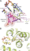
Catalytic center. (A) The active site of PhrB. Sections of Drome (6-4) PL in complex with photodamaged DNA (PDB code 3CVU, DNA in magenta, amino acids in purple, and FAD in blue) and of PhrB (amino acids in green and FAD in yellow) are shown after superposition of the FAD molecules. Amino acid residues in Drome (6-4) PL, which interact with the DNA lesion and the homologous residues in PhrB, are shown. Potential hydrogen bonds are represented by dashed lines. (B) Water W731 of PhrB (green) forms a potential hydrogen bond with the N5 atom of FAD (C atoms in yellow), replacing the role of Asn378 of the class I CPD photolyase from E. coli (PDB code 1DNP), drawn in purple. Its PhrB homolog, Glu403, faces away from FAD and interacts with His388.
Fig. 4.
Fig. 4.
Potential role of the PhrB α7–α8 loop region in binding of the (6-4) DNA lesion in comparison with eukaryotic Drome (6-4) PL. PhrB and Drome (6-4) PL are illustrated in ribbon representation and α-helices are shown as cylinders in green and brown, respectively. The α7–α8 loop is drawn in black; the (6-4) lesion stabilizing loop of Drome (6-4) PL is drawn in yellow. This loop in Drome (6-4) PL is located between helices that correspond to α17 and α18 in PhrB and stabilizes the flip out of the (6-4) photoproduct by inserting a pair of amino acid side chains (Gln418 and Arg421) into the resulting gap in the double stranded DNA oligomer. (A) The DNA fragment with the (6-4) lesion (shown as ball and sticks) from the Drome (6-4) PL–DNA complex (PDB code 3CVU) superimposed onto the PhrB structure. (B) A close-up view of A. Two side chain conformations shown for Arg183 (as sticks), the original one as seen in the crystal structure (yellow) and a second one (brown) being a rotamer that is in close contact (<4 Å) with the central phosphate as indicated by the dotted lines. (C) The Drome (6-4) PL complex with photodamaged DNA oligomer (PDB code 3CVU). DNA is drawn as sticks and the (6-4) lesion highlighted as ball and sticks. Only a portion of the DNA oligomer is shown for clarity. (D) A close-up view of C. Arg421 forms a salt bridge with the DNA (6-4) lesion.
Fig. 5.
Fig. 5.
Structural comparison between PhrB and the primase S. cerevisiae large subunit PriL. Overall structural comparison of PhrB and the PriL carboxyl-terminal domain (PriL-CTD) are drawn in green and blue, respectively; coordinating Cys residues and Fe-S clusters in orange/yellow and black/red, respectively. Numbers of Cys residues and α-helices refer to PhrB. (A) Superposition of PhrB and PriL–CTD (Sacce PriL, PDB Code 3LGB) based on Cα atoms, yielding an rmsd of 3.1 Å for 112 Cα atoms. The homologous regions are drawn in ribbon representation; the rest of the protein is transparent. (B) Close-up view of [4Fe-4S] clusters, coordinating Cys residues and relevant portions of the protein backbones.
Fig. 6.
Fig. 6.
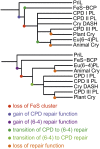
Two possible evolutionary pathways from the ancestor of primase PriL and CPFs to present PriL and CPF group members. Proposed major functional changes are indicated by colored dots. The Lower tree has fewer functional changes. In this preferred scenario, the first photolyase was a (6-4) photolyases with an iron-sulfur cluster. Eu(6-4)PL: eukaryotic (6-4) photolyases; Cry: cryptochromes; CPD PL: CPD photolyases; CryDASH: DASH-type cryptochromes.

References

    1. Sancar A. Structure and function of DNA photolyase and cryptochrome blue-light photoreceptors. Chem Rev. 2003;103(6):2203–2237. - PubMed
    1. Cashmore AR. Cryptochromes: Enabling plants and animals to determine circadian time. Cell. 2003;114(5):537–543. - PubMed
    1. Chaves I, et al. The cryptochromes: Blue light photoreceptors in plants and animals. Annu Rev Plant Biol. 2011;62:335–364. - PubMed
    1. Essen LO. Photolyases and cryptochromes: Common mechanisms of DNA repair and light-driven signaling? Curr Opin Struct Biol. 2006;16(1):51–59. - PubMed
    1. Müller M, Carell T. Structural biology of DNA photolyases and cryptochromes. Curr Opin Struct Biol. 2009;19(3):277–285. - PubMed

Publication types

MeSH terms

Associated data

LinkOut - more resources