. 2013 Apr;8(4):771-82.
doi: 10.1038/nprot.2013.039.

A rapid and reproducible assay for modeling myelination by oligodendrocytes using engineered nanofibers

Affiliations

A rapid and reproducible assay for modeling myelination by oligodendrocytes using engineered nanofibers

Seonok Lee et al. Nat Protoc. 2013 Apr.

Abstract

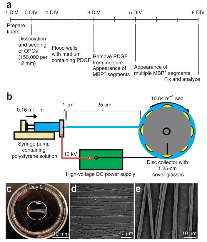
Current methods for studying oligodendrocyte myelination using primary neurons are limited by the time, cost and reproducibility of myelination in vitro. Nanofibers with diameters of >0.4 μm fabricated from electrospinning of liquid polystyrene are suitable scaffolds for concentric membrane wrapping by oligodendrocytes. With the advent of aligned electrospinning technology, nanofibers can be rapidly fabricated, standardized, and configured into various densities and patterns as desired. Notably, the minimally permissive culture environment of fibers provides investigators with an opportunity to explore the autonomous oligodendrocyte cellular processes underlying differentiation and myelination. The simplicity of the system is conducive to monitoring oligodendrocyte proliferation, migration, differentiation and membrane wrapping in the absence of neuronal signals. Here we describe protocols for the fabrication and preparation of nanofibers aligned on glass coverslips for the study of membrane wrapping by rodent oligodendrocytes. The entire protocol can be completed within 2 weeks.


Conflict of interest statement

The authors declare no competing financial interests.

Figures

Figure 1
Figure 1
Fabrication and preparation of the fibers for assaying oligodendroglial membrane wrapping. (a) Timeline for fiber fabrication and OPC culture for analysis of myelin-like segment formation using immunocytochemistry. (b) Diagram of an electrospinning setup consisting of a syringe pump (pale yellow), a high-voltage DC power supply (green) and a rapidly rotating wheel collector (dark gray). A polystyrene solution (cyan) is dispensed from the syringe pump to the collector that is positioned 25 cm from the tip of the syringe (in the figure, the 1-cm length refers to the distance from the tip of the syringe to the tip of the needle, before ejection of the polystyrene liquid starts). A high-voltage DC power supply (green) grounds a rapidly rotating wheel collector containing taped 12-mm coverslips onto which aligned nanofibers are collected. (c) Phase image of the fibers aligned on a coverslip at low magnification. (d) Phase image of the fibers aligned on a coverslip at high magnification. (e) SEM image of 2–4-μm diameter fibers spaced 10–15 μm apart.
Figure 2
Figure 2
Temporal and spatial distribution of OPCs cultured on fibers: proliferation, migration, differentiation and formation of myelin-like segments. (a) Phase image of the fibers seeded with rat cortical OPCs in 100 μl of culture medium containing PDGF, promoting association with the fibers. (b) Immunostaining of OPC-fiber culture reveals that at day 1, OPCs (green, positive for PDGFRα) are spatially organized in culture either ensheathing or contacting several fibers with their processes. They maintain close association with the fibers during proliferation and migration. At this early stage of the culture, very few contaminating GFAP-positive astrocytes are present. (c) At day 3, OPCs start to differentiate into oligodendrocytes and can be identified by MBP. A small number of GFAP-positive astrocytes can be identified at this stage (white arrows). (d) At day 5, more mature MBP-positive oligodendrocytes start to appear among OPCs that are spatially organized around the fibers. (e,f) At day 8, the majority of cells are MBP-positive oligodendrocytes that form longer and mature myelin-like segments shown at low magnification (e) and at high magnification (f). (g) Electron micrographs of the fibers ensheathed by OPCs. (h) A high-magnification image of a cross-section illustrating an oligodendrocyte wrapping multiple layers of membrane around a large-diameter fiber. Arrows indicate dark electron-dense lines.

References

    1. Colello RJ, Pott U. Signals that initiate myelination in the developing mammalian nervous system. Mol Neurobiol. 1997;15:83–100. - PubMed
    1. Ravikumar M, Jain S, Miller RH, Capadona JR, Selkirk SM. An organotypic spinal cord slice culture model to quantify neurodegeneration. J Neurosci Methods. 2012;211:280–288. - PubMed
    1. Yang Y, Lewis R, Miller RH. Interactions between oligodendrocyte precursors control the onset of CNS myelination. Dev Biol. 2011;350:127–138. - PMC - PubMed
    1. Lubetzki C, et al. Even in culture, oligodendrocytes myelinate solely axons. Proc Natl Acad Sci USA. 1993;90:6820–6824. - PMC - PubMed
    1. Nash B, et al. Functional duality of astrocytes in myelination. J Neurosci. 2011;31:13028–13038. - PMC - PubMed

Publication types