

PET imaging in prostate cancer: focus on prostate-specific membrane antigen
- PMID: 23590171
- PMCID: PMC4067736
- DOI: 10.2174/1568026611313080008
PET imaging in prostate cancer: focus on prostate-specific membrane antigen
Abstract
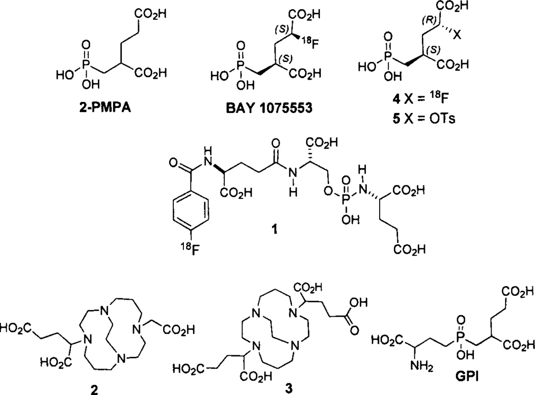
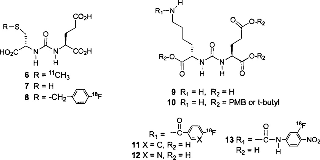
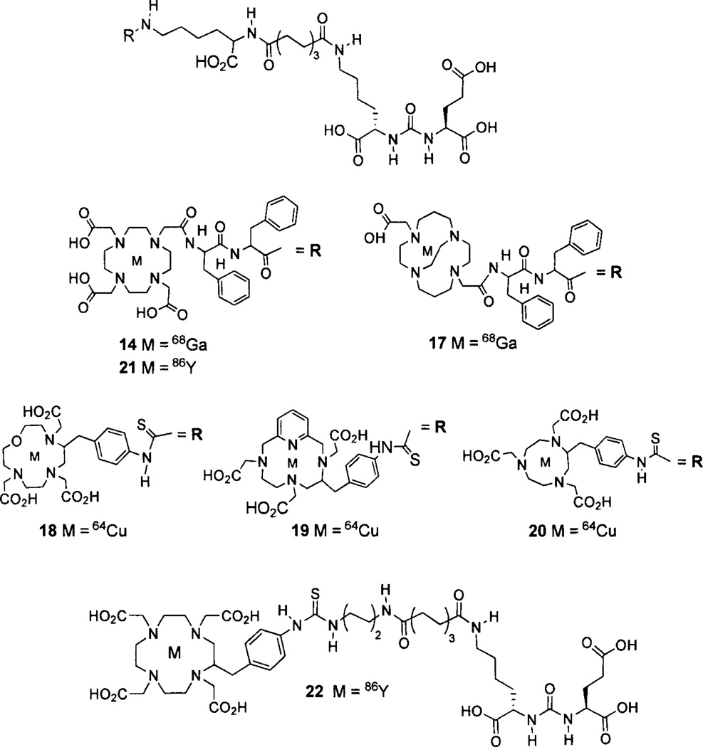
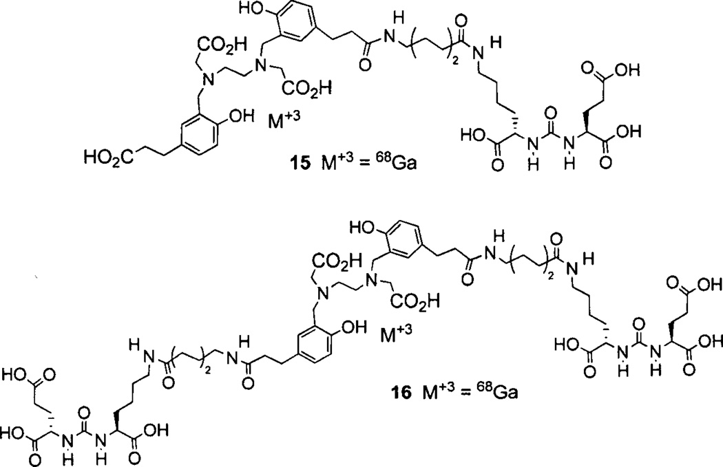
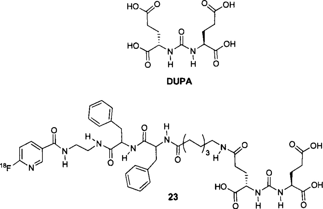
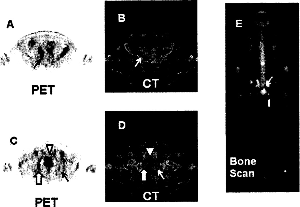
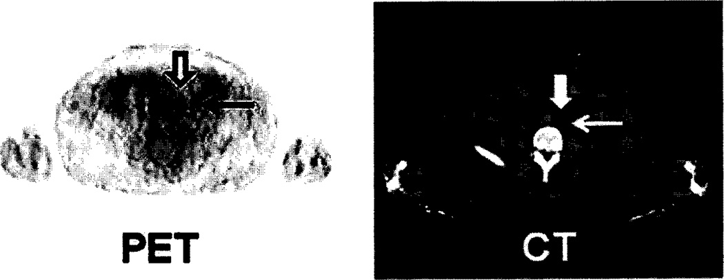
Prostate cancer (PCa) is the second leading cause of cancer-related death in American men. Positron emission tomography/computed tomography (PET/CT) with emerging radiopharmaceuticals promises accurate staging of primary disease, restaging of recurrent disease, detection of metastatic lesions and, ultimately, for predicting the aggressiveness of disease. Prostate-specific membrane antigen (PSMA) is a well-characterized imaging biomarker of PCa. Because PSMA levels are directly related to androgen independence, metastasis and progression, PSMA could prove an important target for the development of new radiopharmaceuticals for PET. Preclinical data for new PSMA-based radiotracers are discussed and include new (89)Zr- and (64)Cu-labeled anti-PSMA antibodies and antibody fragments, (64)Cu-labeled aptamers, and (11)C-, (18)F-, (68)Ga-, (64)Cu-, and (86)Y-labeled low molecular weight inhibitors of PSMA. Several of these agents, namely (68)Ga- HBED-CC conjugate 15, (18)F-DCFBC 8, and BAY1075553 are particularly promising, each having detected sites of PCa in initial clinical studies. These early clinical results suggest that PET/CT using PSMA-targeted agents, especially with compounds of low molecular weight, will make valuable contributions to the management of PCa.
Conflict of interest statement
The authors confirm that this article content has no conflicts of interest.
Figures
References
Publication types
MeSH terms
Substances
Grants and funding
LinkOut - more resources
Full Text Sources
Other Literature Sources
Medical
Miscellaneous
