

Mechanism of the interaction of plant alkaloid vincristine with DNA and chromatin: spectroscopic study
- PMID: 23590199
- PMCID: PMC3651685
- DOI: 10.1089/dna.2012.1886
Mechanism of the interaction of plant alkaloid vincristine with DNA and chromatin: spectroscopic study
Abstract
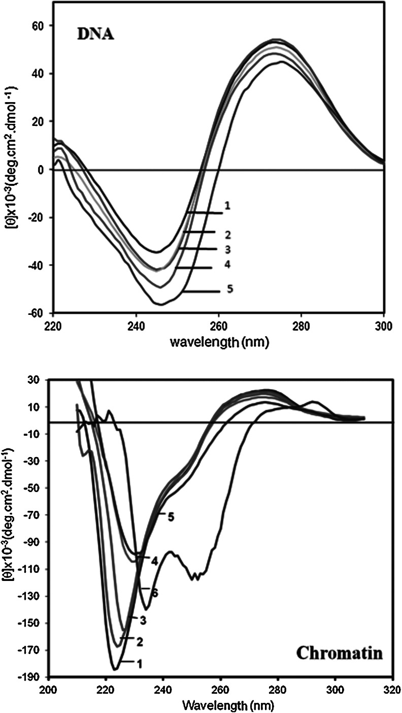
Chromatin has been successfully used as a tool for the study of genome function in cancers. Vincristine as a vinca alkaloid anticancer drug exerts its action by binding to tubulins. In this study the effect of vincristine on DNA and chromatin was investigated employing various spectroscopy techniques as well as thermal denaturation, equilibrium dialysis and DNA-cellulose affinity. The results showed that the binding of vincristine to DNA and chromatin reduced absorbance at both 260 and 210 nm with different extent. Chromopheres of chromatin quenched with the drug and fluorescence emission intensity decreased in a dose-dependent manner. Chromatin exhibited higher emission intensity changes compared to DNA. Upon addition of vincristine, Tm of DNA and chromatin exhibited hypochromicity without any shift in Tm. The binding of the drug induced structural changes in both positive and negative extremes of circular dichroism spectra and exhibited a cooperative binding pattern as illustrated by a positive slope observed in low r values of the binding isotherm. Vincristine showed higher binding affinity to double stranded DNA compared to single stranded one. The results suggest that vincristine binds with higher affinity to chromatin compared to DNA. The interaction is through intercalation along with binding to phosphate sugar backbone and histone proteins play fundamental role in this process. The binding of the drug to chromatin opens a new insight into vincristine action in the cell nucleus.
Figures


References
-
- Alberts B. Amodio F. Jenkins M. Gutman E. Ferris F. Studies with DNA-cellulose chromatography. I. DNA-binding proteins from escherichia coli. Cold Spring Harb Symp Quant Biol. 1968;33:289–305. - PubMed
-
- Arni P. Hertner T. Chromosomal aberrations in vitro induced by aneugens. Mutat Res. 1997;379:83–93. - PubMed
-
- Bradbury E.M. Van Holde K.E. Chromatin structure and dynamics: A historical perspective. In: Zlatanova J., editor; Leuba S.H., editor. Chromatin Structure and Dynamics: State of the Art. Elsevier, Amesterdam; The Netherlands Elsevier: 2004.
-
- Cera C. Dalumbo M. Anticancer activity of anthracycline antibiotics and DNA condensation. Anticancer Drugs. 1990;5:265–271. - PubMed
-
- Chaires J.B. Dattagupta N. Crothes D.M. Binding of daunomycin to calf thymus nucleosomes. Biochemistry. 1983;22:284–292. - PubMed
Publication types
MeSH terms
Substances
LinkOut - more resources
Full Text Sources
Other Literature Sources
