Intramedullary dermoid cyst with relatively atypical symptoms: a case report and review of the literature
- PMID: 23590721
- PMCID: PMC3639845
- DOI: 10.1186/1752-1947-7-104
Intramedullary dermoid cyst with relatively atypical symptoms: a case report and review of the literature
Abstract
Background: Intraspinal dermoid cysts are rare and benign tumors that occur primarily due to the defective closure of the neural tube, an ectodermal derivative, during the process of development. They are slow-growing tumors manifesting in the second and third decades of life.
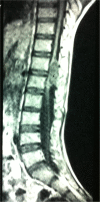
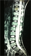
Case presentation: We present here a case of a 14-year-old Sindhi boy with a six-month history of paraparesis of the lower limbs and a progressive loss of power of grade 3/5, and hypoesthesia in the L4/L5 dermatomes of his right lower limb. A plain magnetic resonance imaging scan revealed a well-demarcated intraspinal intramedullary cyst containing an abscess at the level of T12 and L1 causing localized cord compression, which was producing the symptoms. Near total excision of the cyst was successfully performed and was sent for biopsy, which revealed keratinocytes and keratin flakes. With one month of follow-up, along with physiotherapeutic management, the patient gradually improved and was able to walk without support.
Conclusions: Critical evaluation of every case with aggravating symptoms should be carried out, and neurological and radiological examinations should be conducted to ensure the well-being of patients.
Figures
References
-
- Moin H, Mohagheghzadeh P. A case of intracranial dermoid cyst presenting like an epidermoid cyst in Ethmoidal region. J Res Med Sci. 2004;6:312–314.
-
- Love JG, Kennohan JW. Dermoid and epidermoid tumors (cholesteatomas) of central nervous system. JAMA. 1936;107:1876–1883. doi: 10.1001/jama.1936.02770490030009. - DOI
LinkOut - more resources
Full Text Sources
Other Literature Sources


