

Mantle cell lymphoma presenting as a pelvi-ureteric junction obstruction: a case report
- PMID: 23590763
- PMCID: PMC3637467
- DOI: 10.1186/1752-1947-7-105
Mantle cell lymphoma presenting as a pelvi-ureteric junction obstruction: a case report
Abstract
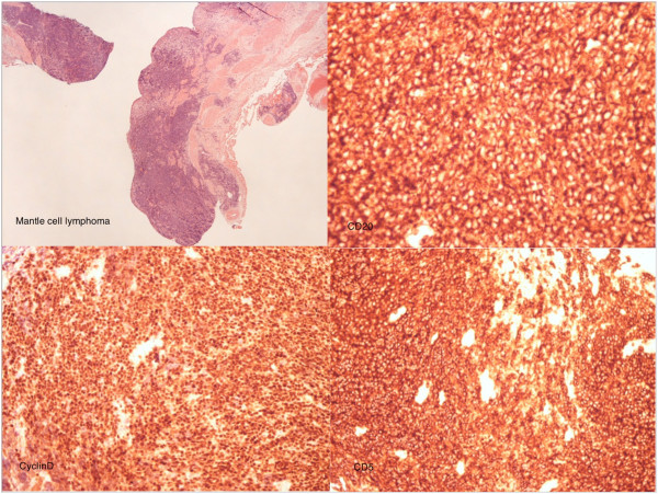
Introduction: Mantle cell lymphoma is one of the several subtypes of non-Hodgkin's lymphoma. Mantle cell lymphoma is the rarest of the subtypes, accounting for about 6% of all non-Hodgkin's lymphoma cases in the United States and Europe. Lymphoid neoplasms of the urinary tract and male genital organs are relatively rare, accounting for less than 5% of extranodal lymphomas. We present a rare case of mantle cell lymphoma infiltrating the ureter causing pelvi-ureteric junction obstruction on tissue diagnosis.
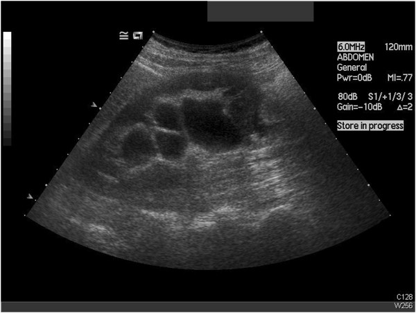
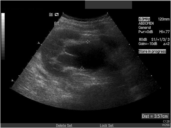
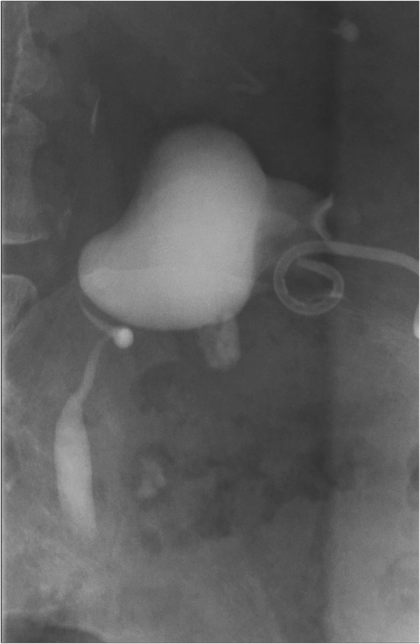
Case presentation: A 78-year-old Caucasian woman was referred to our department with right flank pain, pyrexia and features of a urinary tract infection. A nephrostogram revealed a grossly distended right pelvicalyceal system in a pelvi-ureteric junction obstruction pattern. She underwent an elective pyeloplasty after her acute management and the results of histological examination revealed mantle cell lymphoma.
Conclusion: We describe a rare presentation of mantle cell lymphoma as a pelvi-ureteric junction obstruction. To the best of our knowledge, there has not been any previously published report of the above finding. Our patient had a history of a previous lymphoma but the aim of this manuscript is to highlight a possible presentation rather than determining whether the mantle cell lymphoma was de novo or a transformation from her previous splenic lymphoma with villous lymphocytes.
Figures


References
-
- Greer JP, Foerster J, Rodgers GM, Paraskevas F, Glader B, Arber DA, Means RT. Wintrobe’s Clinical Hematology. 12. Philadelphia, PA: Lippincott Williams and Wilkins; 2009.
-
- Marcus R, Sweetenham JW, Williams ME. Lymphoma – Pathology, Diagnosis and Treatment. Cambridge: Cambridge University Press; 2007.
-
- Lee HJ, Seo JW, Cho HS, Kang Y, Bae EJ, Lee DW, Jeon DH, Lee JS, Chang SH, Park DJ. Renal involvement of mantle cell lymphoma leading to end stage renal disease. Hemodial Int. 2012;16(1):104–108. - PubMed
-
- Huret JL. t(11;14)(q13;q32). Atlas Genet Cytogenet Oncol Haematol 1998. http://AtlasGeneticsOncology.org/Anomalies/t1114ID2021.html.
LinkOut - more resources
Full Text Sources
Other Literature Sources
