

Autophagy and heterophagy dysregulation leads to retinal pigment epithelium dysfunction and development of age-related macular degeneration
- PMID: 23590900
- PMCID: PMC3722332
- DOI: 10.4161/auto.24546
Autophagy and heterophagy dysregulation leads to retinal pigment epithelium dysfunction and development of age-related macular degeneration
Abstract
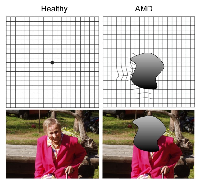
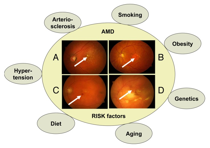
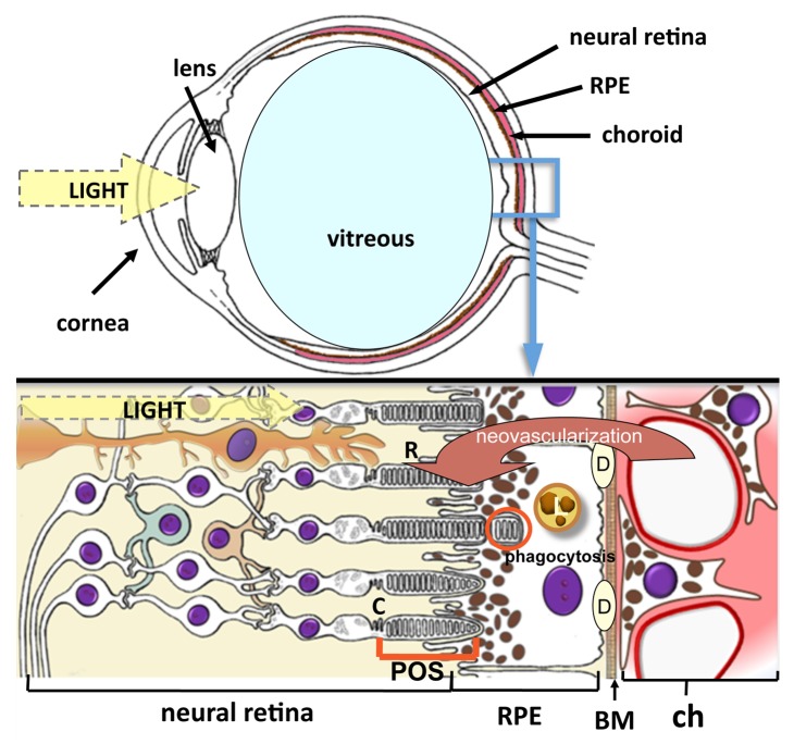
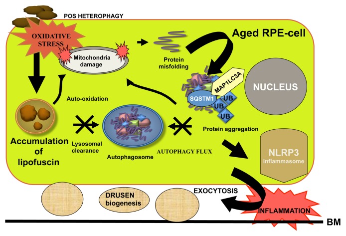
Age-related macular degeneration (AMD) is a complex, degenerative and progressive eye disease that usually does not lead to complete blindness, but can result in severe loss of central vision. Risk factors for AMD include age, genetics, diet, smoking, oxidative stress and many cardiovascular-associated risk factors. Autophagy is a cellular housekeeping process that removes damaged organelles and protein aggregates, whereas heterophagy, in the case of the retinal pigment epithelium (RPE), is the phagocytosis of exogenous photoreceptor outer segments. Numerous studies have demonstrated that both autophagy and heterophagy are highly active in the RPE. To date, there is increasing evidence that constant oxidative stress impairs autophagy and heterophagy, as well as increases protein aggregation and causes inflammasome activation leading to the pathological phenotype of AMD. This review ties together these crucial pathological topics and reflects upon autophagy as a potential therapeutic target in AMD.
Keywords: AMD; RPE; autophagy; heterophagy; inflammasome; lysosome; oxidative stress; phagocytosis; proteasome.
Figures
Similar articles
-
A novel melano-lysosome in the retinal epithelium of rhesus monkeys.Exp Eye Res. 2011 Dec;93(6):937-46. doi: 10.1016/j.exer.2011.10.011. Epub 2011 Nov 2. Exp Eye Res. 2011. PMID: 22056912 Free PMC article.
-
Loss of NRF-2 and PGC-1α genes leads to retinal pigment epithelium damage resembling dry age-related macular degeneration.Redox Biol. 2019 Jan;20:1-12. doi: 10.1016/j.redox.2018.09.011. Epub 2018 Sep 14. Redox Biol. 2019. PMID: 30253279 Free PMC article.
-
Defects in retinal pigment epithelial cell proteolysis and the pathology associated with age-related macular degeneration.Prog Retin Eye Res. 2016 Mar;51:69-89. doi: 10.1016/j.preteyeres.2015.09.002. Epub 2015 Sep 4. Prog Retin Eye Res. 2016. PMID: 26344735 Free PMC article. Review.
-
Heat shock proteins as gatekeepers of proteolytic pathways-Implications for age-related macular degeneration (AMD).Ageing Res Rev. 2009 Apr;8(2):128-39. doi: 10.1016/j.arr.2009.01.001. Ageing Res Rev. 2009. PMID: 19274853 Review.
-
Crosstalk of protein clearance, inflammasome, and Ca2+ channels in retinal pigment epithelium derived from age-related macular degeneration patients.J Biol Chem. 2023 Jun;299(6):104770. doi: 10.1016/j.jbc.2023.104770. Epub 2023 May 1. J Biol Chem. 2023. PMID: 37137441 Free PMC article.
Cited by
-
[Atrophy of the macula in the context of its wet, age-related degeneration : An inescapable consequence of anti-VEGF therapy?].Ophthalmologe. 2016 Dec;113(12):1036-1045. doi: 10.1007/s00347-016-0306-9. Ophthalmologe. 2016. PMID: 27364637 Review. German.
-
17β-estradiol ameliorates oxidative stress and blue light-emitting diode-induced retinal degeneration by decreasing apoptosis and enhancing autophagy.Drug Des Devel Ther. 2018 Sep 4;12:2715-2730. doi: 10.2147/DDDT.S176349. eCollection 2018. Drug Des Devel Ther. 2018. PMID: 30233136 Free PMC article.
-
Over-expression of CNTF in bone marrow mesenchymal stem cells protects RPE cells from short-wavelength, blue-light injury.In Vitro Cell Dev Biol Anim. 2018 May;54(5):355-365. doi: 10.1007/s11626-018-0243-9. Epub 2018 Mar 21. In Vitro Cell Dev Biol Anim. 2018. PMID: 29564604
-
In-silico simulated prototype-patients using TPMS technology to study a potential adverse effect of sacubitril and valsartan.PLoS One. 2020 Feb 13;15(2):e0228926. doi: 10.1371/journal.pone.0228926. eCollection 2020. PLoS One. 2020. PMID: 32053711 Free PMC article.
-
Antioxidants and Mechanistic Insights for Managing Dry Age-Related Macular Degeneration.Antioxidants (Basel). 2024 May 4;13(5):568. doi: 10.3390/antiox13050568. Antioxidants (Basel). 2024. PMID: 38790673 Free PMC article. Review.
References
-
- Kaarniranta K, Salminen A, Haapasalo A, Soininen H, Hiltunen M. Age-related macular degeneration (AMD): Alzheimer’s disease in the eye? J Alzheimers Dis. 2011;24:615–31. - PubMed
-
- Vision 2020. Right to sight. Blindness and visual impairment: Global facts. Available at: http://vision2020.org/main.cfm?type=FACTS Accessed August 22, 2011.
Publication types
MeSH terms
Substances
Grants and funding
LinkOut - more resources
Full Text Sources
Other Literature Sources
Medical