. 2013 Jul:60:97-106.
doi: 10.1016/j.yjmcc.2013.04.004. Epub 2013 Apr 13.

Optimization of direct fibroblast reprogramming to cardiomyocytes using calcium activity as a functional measure of success

Affiliations

Optimization of direct fibroblast reprogramming to cardiomyocytes using calcium activity as a functional measure of success

Russell C Addis et al. J Mol Cell Cardiol. 2013 Jul.

Abstract

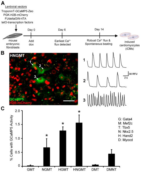
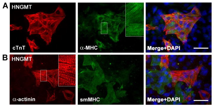
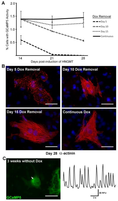
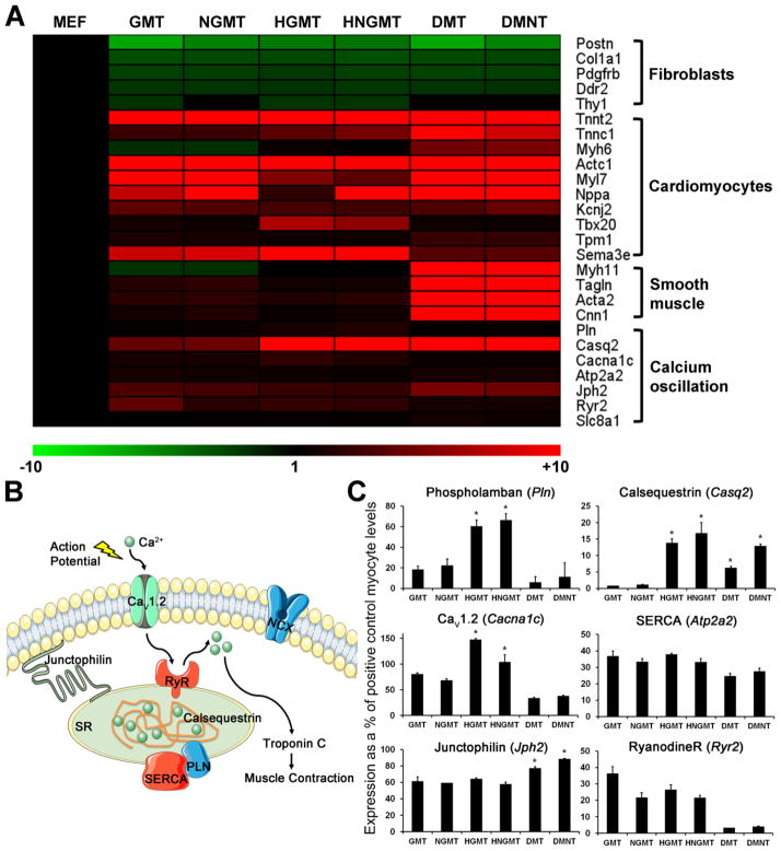
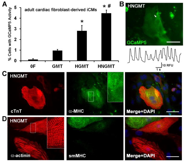
Direct conversion of fibroblasts to induced cardiomyocytes (iCMs) has great potential for regenerative medicine. Recent publications have reported significant progress, but the evaluation of reprogramming has relied upon non-functional measures such as flow cytometry for cardiomyocyte markers or GFP expression driven by a cardiomyocyte-specific promoter. The issue is one of practicality: the most stringent measures - electrophysiology to detect cell excitation and the presence of spontaneously contracting myocytes - are not readily quantifiable in the large numbers of cells screened in reprogramming experiments. However, excitation and contraction are linked by a third functional characteristic of cardiomyocytes: the rhythmic oscillation of intracellular calcium levels. We set out to optimize direct conversion of fibroblasts to iCMs with a quantifiable calcium reporter to rapidly assess functional transdifferentiation. We constructed a reporter system in which the calcium indicator GCaMP is driven by the cardiomyocyte-specific Troponin T promoter. Using calcium activity as our primary outcome measure, we compared several published combinations of transcription factors along with novel combinations in mouse embryonic fibroblasts. The most effective combination consisted of Hand2, Nkx2.5, Gata4, Mef2c, and Tbx5 (HNGMT). This combination is >50-fold more efficient than GMT alone and produces iCMs with cardiomyocyte marker expression, robust calcium oscillation, and spontaneous beating that persist for weeks following inactivation of reprogramming factors. HNGMT is also significantly more effective than previously published factor combinations for the transdifferentiation of adult mouse cardiac fibroblasts to iCMs. Quantification of calcium function is a convenient and effective means for the identification and evaluation of cardiomyocytes generated by direct reprogramming. Using this stringent outcome measure, we conclude that HNGMT produces iCMs more efficiently than previously published methods.


Figures

Figure 1
Figure 1. The TroponinT-GCaMP5 calcium reporter
A) The Troponin T (TNNT2) gene promoter drives expression of the genetically-encoded calcium indicator, GCaMP5, and a zeocin resistance cassette. The GCaMP protein consists of a Ca2+-sensitive calmodulin domain fused to GFP. When intracellular Ca2+ is low, the calmodulin domain blocks proper folding of GFP, resulting in a low level of GFP fluorescence. When intracellular Ca2+ is high, the calmodulin domain shifts to permit GFP folding and bright fluorescence. B) Validation of the TroponinT-GCaMP5 reporter in embryonic cardiomyocytes. Ventricular cardiomyocytes were isolated from mouse embryos at 14.5 dpc and transduced with reporter lentivirus. GCaMP activity was recorded 48 hours post-transduction. The left panel corresponds to a single frame of supplemental Movie S1. White arrows indicate regions examined for oscillating fluorescence, shown in the right panel. RFU, Relative Fluorescence Units calculated using the “Intensity v Time Monitor” in ImageJ software.
Figure 2
Figure 2. Functional quantification of direct fibroblast reprogramming to induced cardiomyocytes
A) Schematic of direct fibroblast reprogramming to induced cardiomyocytes (iCMs). B) GCaMP activity in beating iCMs, 14 days post-induction. The left panel corresponds to a single frame of supplemental Movie S4, which shows Ca2+ flux in spontaneously beating iCMs generated by HNGMT treatment, overlaid with a red fluorescent image to show the PGK-H2B-mCherry vector that was used to quantify total cells. The right panel traces oscillations in GCaMP fluorescence, corresponding to oscillations in intracellular Ca2+, at the regions marked by the white arrows. Scale bar 50μm. RFU, Relative Fluorescence Units calculated using the “Intensity v Time Monitor” in ImageJ software. C) Quantification of functional iCMs using GCaMP activity. Functional iCMs were identified on the basis of repeated GCaMP oscillation in 10s movies made at 14 days post-induction. Activity is expressed as the percentage of cells displaying repeated GCaMP oscillation in 10s movies divided by the total number of cells per field. Data are presented as means ± SEM. *denotes p<0.05 in comparison to GMT.
Figure 3
Figure 3. Immunocytochemical analysis of Day 14 iCMs
Cardiomyocyte markers cardiac Troponin T (cTnT), α-myosin heavy chain (α-MHC), and sarcomeric α-actinin are expressed in HNGMT-iCMs. Regions in white boxes are enlarged in insets to show sarcomere structure. Putative smooth muscle cells were also detected by staining for smooth muscle myosin heavy chain (smMHC, Myh11). DAPI stain (blue) marks nuclei. Scale bars 50μm.
Figure 4
Figure 4. Stability of the reprogrammed iCM phenotype
A) GCaMP activity was quantified at 14, 21, and 28 days post-induction, comparing HNGMT-treated cells with doxycycline applied continuously or withdrawn at 5, 10, or 15 days post-induction. Data are presented as means ± SEM. Beating, GCaMP+ iCMs could be detected on Day 28 for all conditions excepting Day 5 Dox removal. B) Immunostaining for sarcomeric α-actinin (red) in representative HNGMT-iCMs, 28 days post-induction. DAPI stain (blue) marks nuclei. Scale bars 50μm. C) GCaMP activity recorded on Day 36 post-induction, with doxycycline removed on Day 15 (thus 3 weeks without exogenous transcription factor induction). Right panel shows oscillation in GCaMP fluorescence at the region indicated by the white arrow, left. Left panel is a single frame of supplemental Movie S6, which shows both GCaMP activity and robust cell contraction. Scale bar 50μm.
Figure 5
Figure 5. iCM gene expression analysis and assessment of components of excitation-contraction coupling
A) Heat map showing expression data for selected genes in microarray analysis. Microarrays were performed on RNA collected from putative iCMs on Day 21 post-induction. Expression is normalized to untreated MEFs, with positive fold changes indicated in red and negative fold changes in green. B) Key components of the excitation-contraction system: Action potential firing causes an inward Ca2+ current to enter the myocyte via CaV1.2 channels. The influx of Ca2+ ions stimulates Ryanodine receptors (RyR) to release additional Ca2+ into the cytosol from the calsequestrin stores of the sarcoplasmic reticulum (SR), causing the activation of Troponin C and the troponin-tropomyosin complex that mediates contraction. Cytosolic Ca2+ levels are then rapidly reduced by a combination of mechanisms including Na+/Ca2+ exchange by NCX and pumping into the SR by the Phospoholamban (PLN)-regulated sarcoendoplasmic reticulum calcium ATPase (SERCA). Junctophilin helps to tether the SR to the sarcolemma. Diagram was made with Servier Medical Art, with permission. C) Expression of excitation-contraction genes in iCMs. Gene expression data obtained via microarray analysis was confirmed using quantitative RT-PCR and normalized to expression levels present in embryonic cardiomyocytes (E14.5). Calsequestrin, CaV1.2, and Phospholamban are most highly expressed in iCMs generated by HGMT and HNGMT, the factor combinations that produced the most GCaMP+ cells. Data are presented as means ± SEM. * denotes p<0.05 for increase in expression compared to GMT.
Figure 6
Figure 6. iCMs derived from adult cardiac fibroblasts
A) Quantification of functional cardiac fibroblast-derived iCMs using GCaMP activity. Functional iCMs were identified on the basis of repeated GCaMP oscillation in 10s movies made at 14 days post-induction. Activity is expressed as the percentage of cells displaying repeated GCaMP oscillation in 10s movies divided by the total number of cells per field. Data are presented as means ± SEM. * denotes p<0.05 in comparison to GMT. # denotes p<0.05 in comparison to HGMT. B) GCaMP activity recorded in a typical cardiac fibroblast-derived iCM on Day 14 post-induction. Top panel is a single frame of supplemental Movie S7, which shows both GCaMP activity and spontaneous contraction. Lower panel shows oscillation in GCaMP fluorescence at the region indicated by the white arrow, above. Scale bar 50μm. C) Immunocytochemical analysis of cardiac fibroblast-derived iCMs, 14 days post-induction. Cardiomyocyte markers cardiac Troponin T (cTnT), α-myosin heavy chain (α-MHC), and sarcomeric α-actinin are expressed in HNGMT-iCMs. Regions in white boxes are enlarged in insets to show sarcomere structure. Putative smooth muscle cells were also detected by staining for smooth muscle myosin heavy chain (smMHC, Myh11). DAPI stain (blue) marks nuclei. Scale bars 50μm.

Similar articles

Cited by

References

    1. Ieda M, Fu JD, Delgado-Olguin P, Vedantham V, Hayashi Y, Bruneau BG, et al. Direct reprogramming of fibroblasts into functional cardiomyocytes by defined factors. Cell. 2010;142:375–86. - PMC - PubMed
    1. Jayawardena TM, Egemnazarov B, Finch EA, Zhang L, Payne JA, Pandya K, et al. MicroRNA-mediated in vitro and in vivo direct reprogramming of cardiac fibroblasts to cardiomyocytes. Circ Res. 2012;110:1465–73. - PMC - PubMed
    1. Protze S, Khattak S, Poulet C, Lindemann D, Tanaka EM, Ravens U. A new approach to transcription factor screening for reprogramming of fibroblasts to cardiomyocyte-like cells. J Mol Cell Cardiol. 2012 - PubMed
    1. Qian L, Huang Y, Spencer CI, Foley A, Vedantham V, Liu L, et al. In vivo reprogramming of murine cardiac fibroblasts into induced cardiomyocytes. Nature. 2012;485:593–8. - PMC - PubMed
    1. Song K, Nam YJ, Luo X, Qi X, Tan W, Huang GN, et al. Heart repair by reprogramming non-myocytes with cardiac transcription factors. Nature. 2012;485:599–604. - PMC - PubMed

Publication types