. 2013:3:1673.
doi: 10.1038/srep01673.

Thrombospondin-1 signaling through CD47 inhibits self-renewal by regulating c-Myc and other stem cell transcription factors

Affiliations

Thrombospondin-1 signaling through CD47 inhibits self-renewal by regulating c-Myc and other stem cell transcription factors

Sukhbir Kaur et al. Sci Rep. 2013.

Abstract

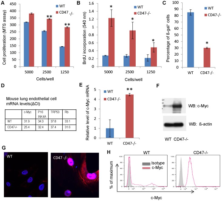
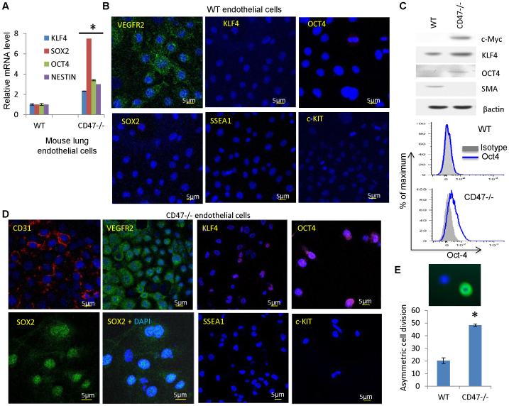
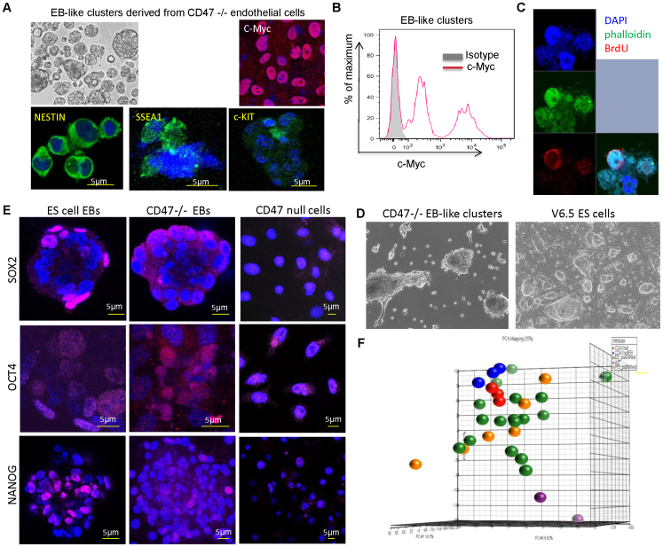
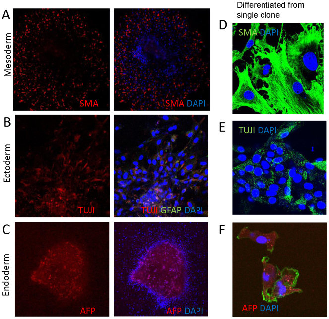
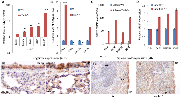
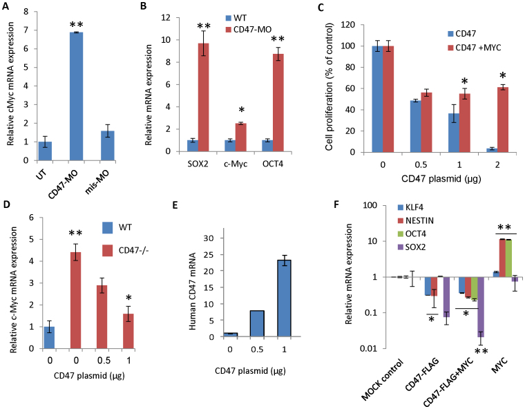
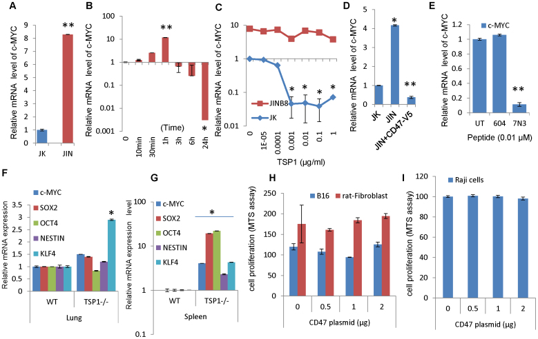
Signaling through the thrombospondin-1 receptor CD47 broadly limits cell and tissue survival of stress, but the molecular mechanisms are incompletely understood. We now show that loss of CD47 permits sustained proliferation of primary murine endothelial cells, increases asymmetric division, and enables these cells to spontaneously reprogram to form multipotent embryoid body-like clusters. c-Myc, Klf4, Oct4, and Sox2 expression is elevated in CD47-null endothelial cells, in several tissues of CD47- and thrombospondin-1-null mice, and in a human T cell line lacking CD47. CD47 knockdown acutely increases mRNA levels of c-Myc and other stem cell transcription factors in cells and in vivo, whereas CD47 ligation by thrombospondin-1 suppresses c-Myc expression. The inhibitory effects of increasing CD47 levels can be overcome by maintaining c-Myc expression and are absent in cells with dysregulated c-Myc. Thus, CD47 antagonists enable cell self-renewal and reprogramming by overcoming negative regulation of c-Myc and other stem cell transcription factors.


Figures

Figure 1
Figure 1. Enhanced proliferation and decreased senescence of CD47-null murine endothelial cells is associated with increased expression of c-Myc.
(A) MTS assay for cell survival and growth over 72 h expressed as % of day 0 values at the indicated plating densities of first passage WT and CD47 null cells. (B) BrdU assay for DNA synthesis. (C) Percentage of senescence-associated β-galactosidase expression at passage 3. (D) Expression of genes associated with cell immortalization in WT and CD47 null cells. (E) c-Myc mRNA levels in lung endothelial cells of WT and CD47 null mice. (F) CD47 limits c-Myc protein levels. (G) c-Myc expression (red) in WT and CD47−/− endothelial cells. Blue = DAPI nuclear stain. (H) Flow cytometric analysis of c-Myc expression in WT and CD47−/− endothelial cells. (*p < 0.05, **p < 0.01).
Figure 2
Figure 2. Stem cell and differentiation marker expression in WT and CD47-null endothelial cells.
(A) mRNA expression levels of stem cell transcription factors in WT and CD47 null lung endothelial cells. (B, D) Stem cell and differentiation marker expression in WT (B) and CD47 null mouse endothelial cells (D). The endothelial cells were stained using the indicated antibodies and DAPI (blue). Scale bars = 5 μm. (C) Protein expression of stem cell transcription factors and smooth muscle actin (SMA) assessed by western blotting and flow cytometry (Oct4) in cultured WT or CD47 null endothelial cells in EGM2 medium. (E) Asymmetric cell division frequencies in second passage WT and CD47−/− endothelial cells equilibrium labeled with BrdU and chased for one cell division. Asymmetric division was scored by counting BrdU+(green)/DAPI+ nuclei adjacent to BrdU/DAPI+ nuclei.
Figure 3
Figure 3. Stem cell marker expression in CD47 null EB-like clusters.
(A) Top left: Typical appearance of EB-like clusters photographed under phase contrast. Top right: Staining with c-Myc antibody (red) and DAPI (blue). Bottom panels: EB-like clusters or cells dissociated from them were stained using the indicated antibodies (green) and DAPI (blue). Scale bars = 5 μm. (B) Flow cytometric analysis of c-Myc expression in CD47-null cells dissociated from EB-like clusters. (C) Detection of asymmetric cell division in cells from CD47-null EB-like clusters equilibrium labeled with BrdU and then chased for two cell divisions without BrdU and stained with anti-BrdU (red) and phalloidin to visualize actin (green). Blue = DAPI. (D) Morphologies of CD47 null EB-like clusters and V6.5 ES cells growing in ES medium with LIF. The V6.5 culture also contains a MEF feeder layer. (E) CD47 null EB-like clusters and V6.5 ES cells cultured as in D and CD47 null endothelial cells in endothelial growth medium (right panels) were stained using the indicated antibodies (magenta) and DAPI (blue). Scale bars = 5 μm. (F) Principal component analysis of global gene expression data for WT (purple) and CD47 null endothelial cells (red), and CD47 null EB-like clusters (blue) compared with published expression data for iPS (gold) and ES cells (green).
Figure 4
Figure 4. Differentiation of CD47-null EB-like clusters.
(A) EB-like clusters were cultured in the presence of serum for 6 days to induce substrate adhesion and then transferred in to the indicated lineage specific media for 36 h and stained with smooth muscle actin antibody (red) to detect mesodermal cells. (B) Differentiated EB-like clusters were stained with the ectoderm neural markers glial fibrillary acidic protein (GFAP, green), neuron-specific beta III tubulin (TUJI, red). (C) Differentiated EB-like clusters were stained with anti-a-fetoprotein (red) to detect ectodermal cells. In all panels DAPI was used to visualize nuclei (blue). (D–F) A single clone isolated from a CD47-null EB-like cluster in serum-free medium was expanded and then differentiated in the indicated lineage-specific medium for 7 days and stained for the indicated markers. Blue = DAPI, green in (F) = actin.
Figure 5
Figure 5. Loss of CD47 is associated with up-regulation of stem cell transcription factors in vivo.
(A) Relative c-Myc mRNA expression in lung, kidney, liver, brain and spleen of CD47-null versus WT mice. (B) c-Myc mRNA levels in purified splenic cell populations from WT and CD47 null mice. (C) mRNA expression levels in lung from WT and CD47-null mice. (D) mRNA expression levels in spleen from WT and CD47-null mice. (*p < 0.05, **p < 0.01). (E–H) Increased frequency of Sox2 expressing cells in tissues from CD47-null mice. The alveolar (Alv) regions of lung tissues from WT (E) generally lack Sox2-positive cells (brown stain), whereas CD47-null lung shows more positive cells (F). In contrast, similar uniform Sox2 staining was observed in bronchiolar epithelium (BrEp) from WT and null mice, consistent with its previously reported expression in Clara cells. (G–H) Paraffin embedded sections of representative spleen tissues from WT (G) and CD47−/− (H) mice were stained with a specific antibody to Sox2. Sections were examined under light microscopy showing subcapsular (CP), red pulp (RP) and white pulp (WP) staining.
Figure 6
Figure 6. CD47 expression regulates c-Myc and stem cell transcription factor expression.
(A) Morpholino knockdown of CD47 in lung endothelial cells increases c-Myc mRNA expression. (B) In vivo morpholino knockdown of CD47 elevates c-Myc, Oct4, and Sox2 mRNA at 48 h in mouse spleen. (C) CD47 re-expression in CD47-null murine endothelial cells suppresses cell growth unless c-Myc expression is sustained. (D) CD47 re-expression in CD47 null endothelial cells alters expression c-Myc compared with WT (E) Expression level of transfected human CD47. (F) Re-expression of CD47 and c-Myc alters mRNA expression of stem cell transcription factors. (*p < 0.05, **p < 0.01).
Figure 7
Figure 7. Regulation of c-Myc and stem cell transcription factors by CD47 ligation.
(A) c-Myc mRNA in Jurkat (JK) and CD47-deficient JinB8 Jurkat T cells (JIN). (B) Time-dependence for regulation of c-Myc mRNA expression by the CD47 ligand thrombospondin-1. Jurkat cells were treated with 2.2 nM thrombospondin-1 for the indicated times before isolating RNA and assessing c-Myc mRNA by real time PCR normalized to β2m mRNA and expressed as a ratio to normalized c-Myc levels in control cells at the corresponding time points. (C) Thrombospondin-1 effects on c-Myc mRNA in WT Jurkat and CD47-deficient (JinB8) T lymphoma cells. (D) CD47 re-expression in JinB8 cells alters expression c-Myc compared with WT Jurkat cells. (E) Effects of CD47-binding TSP1 peptide 7N3 and control peptide 604 on c-Myc mRNA expression in Jurkat T cells. (F and G) mRNA levels in thrombospondin-1-null vs. WT lung and spleen. (H) CD47 over-expression in Rat1 fibroblasts and B16 melanoma cells does not suppress growth. (I) Deregulation of translocated c-Myc in Raji Burkitt's lymphoma cells prevents growth regulation by CD47 over-expression. (*p < 0.05, **p < 0.01).

References

    1. Matozaki T., Murata Y., Okazawa H. & Ohnishi H. Functions and molecular mechanisms of the CD47-SIRPalpha signalling pathway. Trends Cell Biol 19, 72–80 (2009). - PubMed
    1. Roberts D. D., Miller T. W., Rogers N. M., Yao M. & Isenberg J. S. The matricellular protein thrombospondin-1 globally regulates cardiovascular function and responses to stress. Matrix Biol. 31, 162–169 (2012). - PMC - PubMed
    1. Frazier W. A., Isenberg J. S., Kaur S. & Roberts D. D. in UCSD Nature Molecule Pages (2010). doi:10.1038/mp.a002870.01.
    1. Isenberg J. S. et al. Thrombospondin-1 limits ischemic tissue survival by inhibiting nitric oxide-mediated vascular smooth muscle relaxation. Blood 109, 1945–1952 (2007). - PMC - PubMed
    1. Thakar C. V. et al. Identification of thrombospondin 1 (TSP-1) as a novel mediator of cell injury in kidney ischemia. J Clin Invest 115, 3451–3459 (2005). - PMC - PubMed

Publication types