. 2013:4:1724.
doi: 10.1038/ncomms2720.

Molecular mechanics of mineralized collagen fibrils in bone

Affiliations
Free PMC article

Molecular mechanics of mineralized collagen fibrils in bone

Arun K Nair et al. Nat Commun. 2013.
Free PMC article

Abstract

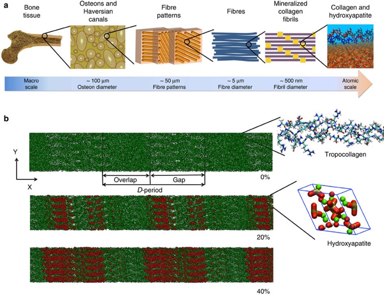
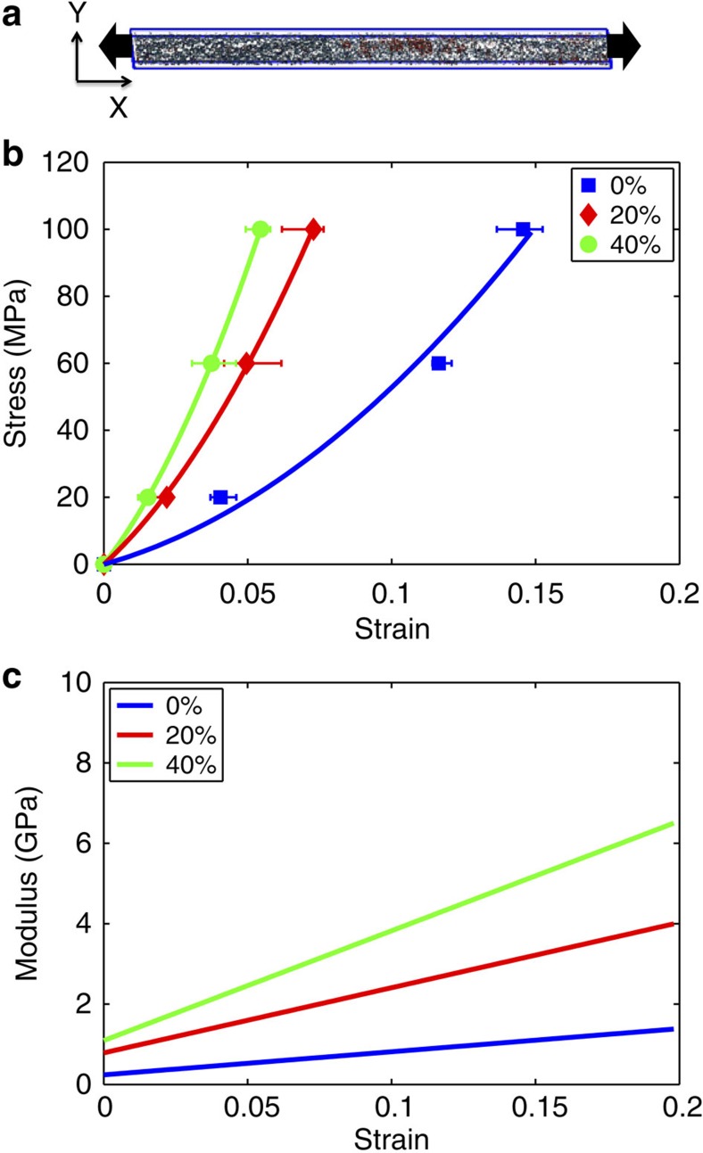
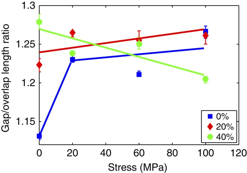
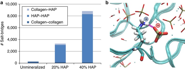
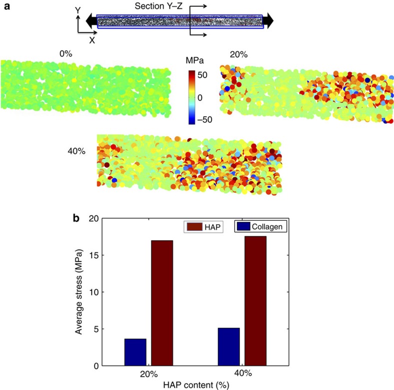
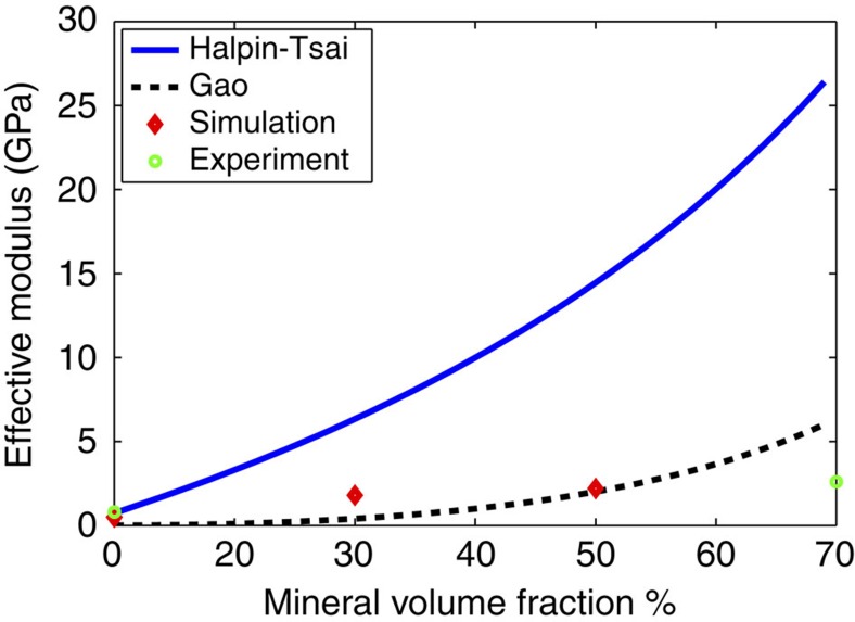
Bone is a natural composite of collagen protein and the mineral hydroxyapatite. The structure of bone is known to be important to its load-bearing characteristics, but relatively little is known about this structure or the mechanism that govern deformation at the molecular scale. Here we perform full-atomistic calculations of the three-dimensional molecular structure of a mineralized collagen protein matrix to try to better understand its mechanical characteristics under tensile loading at various mineral densities. We find that as the mineral density increases, the tensile modulus of the network increases monotonically and well beyond that of pure collagen fibrils. Our results suggest that the mineral crystals within this network bears up to four times the stress of the collagen fibrils, whereas the collagen is predominantly responsible for the material's deformation response. These findings reveal the mechanism by which bone is able to achieve superior energy dissipation and fracture resistance characteristics beyond its individual constituents.


Figures

Figure 1
Figure 1. Bone structure and model development.
(a) Hierarchical structure of bone ranging from the macroscale skeleton to nanoscale collagen (green) and HAP (red). (b) Collagen microfibril model with 0% mineralization (inset shows the collagen triple-helix structure), 20% mineral content (inset shows a HAP unit cell) and 40% mineral content. The HAP crystals are arranged such that the c axis of crystal aligns with the fibril axis. Ca atoms plotted in green, OH groups plotted in red and white, and the tetrahedron structure visualizes the PO4 group. The left three images in panel a taken from Launey et al.
Figure 2
Figure 2. Mineral distribution in the collagen microfibril at different mineralization stages.
(a) Distribution of HAP along the collagen fibril axis. The data shows that the maximum amount of HAP is found in the gap region (between 30 and 50 nm). (b) Spatial distribution of HAP in the unit cell for 20 and 40% mineral density. (c) HAP density distribution along the fibril axis for the 40% case normalized (same data as depicted in panel a) compared with experimental data. The comparison confirms that maximum deposition is found in the gap region.
Figure 3
Figure 3. Mechanical properties of collagen fibrils at different mineralization stages.
(a) Fibril unit cell with mineral content used to perform tensile test by measuring stress versus strain. (b) Stress–strain plots for non-mineralized collagen fibril (0%), 20% mineral density and 40% mineral-density cases. (c) Modulus versus strain for 0, 20 and 40% mineral density showing an increase in modulus as the mineral content increases. The error bars in b are computed from the maximum and minimum values of the periodic box length along the x direction at equilibrium.
Figure 4
Figure 4. Variation of the gap-to-overlap length ratio as the applied stress increases for different HAP contents.
The deformation mechanism of non-mineralized collagen fibrils is molecular straightening in the crimped and loosely packed gap region (at small deformations), followed by molecular stretching (across the full D-period) at larger deformations. These mechanisms result in an initial increase in the gap/overlap ratio, which then remains constant, as is observed in the non-mineralized model reported here (where the gap/overlap ratio increases from 0–20 MPa stress). In contrast, in the mineralized cases the deformation mechanism is radically changed. Indeed, in the lower mineralized model (20% HAP) we do not observe any significant change in the gap/overlap ratio, suggesting that the D-period deforms rather uniformly, due to the stiffening effect of HAP mainly in the gap region. However, in the highly mineralized model (40% HAP), the trend is inverted, where the gap/overlap region decreases, suggesting that in this case the gap region is stiffer and that deformation takes place primarily in the overlap region. The error bars are obtained from the maximum and minimum values of the periodic box length along the x direction obtained after equilibration of each sample, which is utilized to compute the gap and overlap lengths.
Figure 5
Figure 5. Salt bridges in mineralized and non-mineralized collagen fibrils.
(a) Number of salt bridges per unit cell. In the non-mineralized model, the number of salt bridges is ~260, due to interactions between charged side chains of collagen: lysine (+), arginine (+), glutamic acid (−) and aspartic acid (−). In the mineralized models, there is a high number of mineral–mineral salt bridges, due to the fact that the moieties forming HAP are highly charged: Ca2+, PO43− and OH. Salt bridges are also found relevant between collagen and mineral phase (about 220 and 520 in 20% HAP and 40% HAP case, respectively), contributing to the increase in mechanical properties of mineralized fibrils. Panel b shows an example of salt bridge between a lysine (+) side chain and mineral PO43− group.
Figure 6
Figure 6. Analysis of stress fields for different mineral contents.
(a) View of the cross-section of the y–z plane of the unit cell used to perform the stress analysis. The principal stress contours for an applied stress of 100 MPa in collagen microfibril show a nearly uniform stress distribution, whereas the data for 20 and 40% mineral-density cases clearly reveals significantly higher stress regions in the HAP mineral. (b) Quantification of the overall average stress in the collagen and mineral phases at 100 MPa applied stress, showing that the mineral phase features about four times the stress level compared with collagen.
Figure 7
Figure 7. Variation of effective modulus with respect to mineral volume fraction comparing simulation, experiment and analytical models.
Comparison of analytical models by Halpin–Tsai and Gao et al. with our simulation data and experimental results. The analytical model by Halpin–Tsai (equations (1) and (2)) predicts a higher modulus as the mineral volume fraction increases compared with Gao’s model, simulation and experimental modulus. The Gao model (equation (3)) agrees better overall, but predicts a lower modulus for very low mineral content and deviates from the atomistic and experimental predictions for higher mineral content.

References

    1. Fratzl P. (ed.). Collagen: Structure and Mechanics (Springer (2008)).
    1. Fratzl P., Gupta H. S., Paschalis E. P. & Roschger P. Structure and mechanical quality of the collagen-mineral nano-composite in bone. J. Mater. Chem. 14, 2115–2123 (2004).
    1. Fratzl P. & Weinkamer R. Nature’s hierarchical materials. Prog. Mater. Sci. 52, 1263–1334 (2007).
    1. Gupta H. S. et al.. Cooperative deformation of mineral and collagen in bone at the nanoscale. Proc. Natl Acad. Sci. USA 103, 17741–17746 (2006). - PMC - PubMed
    1. Jager I. & Fratzl P. Mineralized collagen fibrils: a mechanical model with a staggered arrangement of mineral particles. Biophys. J. 79, 1737–1746 (2000). - PMC - PubMed

Publication types