. 2013 Jun;4(3):331-8.
doi: 10.1007/s13244-013-0243-y. Epub 2013 Apr 17.

Hepatobiliary anomalies associated with ABCB4/MDR3 deficiency in adults: a pictorial essay

Affiliations

Hepatobiliary anomalies associated with ABCB4/MDR3 deficiency in adults: a pictorial essay

Julie Benzimra et al. Insights Imaging. 2013 Jun.

Abstract

Background: ABCB4/MDR3 gene variants are mostly associated with a peculiar form of cholelithiasis in European adults, currently referred to as low phospholipid-associated cholelithiasis (LPAC) syndrome.

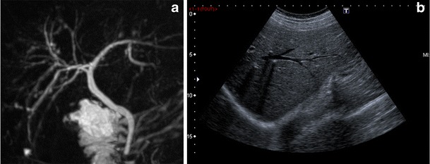
Methods: LPAC syndrome is a rare genetic disorder, characterised by the following clinical features: biliary symptoms before the age of 40, recurrence of the symptoms after cholecystectomy, and intrahepatic microlithiasis or intrahepatic hyperechogenic foci.

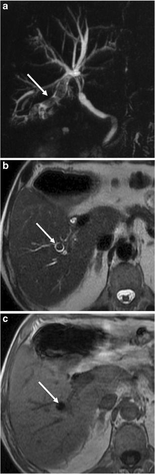
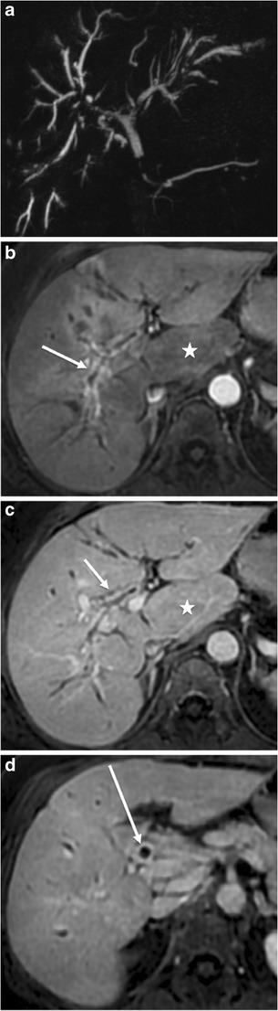
Results: Imaging features associated with ABCB4/MDR3 mutations are not specific and correspond to a wide spectrum of biliary abnormalities. The main feature is the presence of intrahepatic lithiasis. Other uncommon presentations have been described, such as uni- or multifocal spindle-shaped dilatations of the intrahepatic bile ducts filled with gallstones, secondary sclerosing cholangitis, biliary cirrhosis, and intrahepatic cholangiocarcinoma.

Conclusion: This review focuses on MR features related to ABCB4/MDR3 mutations.

Main messages: • LPAC syndrome is characterised by intrahepatic microlithiasis or intrahepatic hyperechogenic foci. • Ultrasound examination is very accurate in detecting intrahepatic stones. • At MR imaging, LPAC syndrome is associated with various presentations.


Figures

Fig. 1
Fig. 1
Ultrasound/MRCP discrepancy in a 44-year-old man with LPAC syndrome. a Transverse ultrasound showing typical comet-tail artefacts in the left lobe. b The MRCP shows no sign of biliary stone
Fig. 2
Fig. 2
Unifocal mild biliary dilatation with biliary stone in a 40-year-old man with LPAC syndrome. The dilatation is located in the segment V on T2-weighted acquisitions (white arrow in a) and 3D MRCP (b) and contains a small signal void corresponding to an endoluminal stone (white arrow in b). The gallbladder and the common bile duct show no abnormalities
Fig. 3
Fig. 3
Severe unisegmental dilatation with endoluminal stones in a 44-year-old woman with LPAC syndrome. Several dilated bile ducts in segment VIII containing macroscopic signal voids corresponding to biliary stones (white arrows). a Transverse T2-weighted acquisition with fat saturation and (b) 3D MCRP
Fig. 4
Fig. 4
Bisegmental dilatation in a 36-year-old woman with LPAC syndrome. Transverse T1-weighted acquisition with fat saturation after injection of a gadolinium chelate at portal phase (a and b), and 3D MCRP (c) show a mild biliary dilatation of both segment III (white arrow in a) and VI (white arrow in b). The MRCP shows a round signal void in the dilated bile ducts of the segment III (white arrow in c) corresponding to a biliary stone
Fig. 5
Fig. 5
Diffuse global bile duct abnormalities in a 48-year-old man with LPAC syndrome. Transverse T2-weighted acquisition (a), transverse T1-weighted acquisition with fat saturation (b), and coronal maximum intensity projection MCRP (c) show a biliary dilatation in both right and left lobes containing biliary stones depicted as T2 hypointense and T1 hyperintense endoluminal formations (white arrow in a and b). The MRCP shows large oval shape signal voids in the dilated bile ducts (white arrow in c) corresponding to biliary stones
Fig. 6
Fig. 6
Segmental dilatation of segment VI bile duct filled with several stones in a 69-year-old man with LPAC syndrome. Coronal maximum intensity projection MCRP (a), transverse T2-weighted acquisition (b), and transverse in-phase T1-weighted acquisition (c) show a dilatation of segment VI bile ducts filled with several stones (white arrow in a). The stones appear as endoluminal signal voids on T2-weighted acquisition (white arrow in b) and hypointensities on T1-weighted acquisitions (white arrow in c)
Fig. 7
Fig. 7
Severe LPAC syndrome in a 55-year-old man. Transverse T2-weighted acquisition (a), and coronal maximum intensity projection MCRP (b) show a rare presentation of severe LPAC syndrome consisting in diffuse biliary dilatation containing multiple biliary stones
Fig. 8
Fig. 8
Biliary irregularities in a 54-year-old man. Three-dimensional MRCP (a) and sagittal ultrasound of the right lobe (b) show right biliary abnormalities (a). These mild irregular calibre intrahepatic bile ducts were not demonstrated with ultrasound; on the other hand, small bile stones were easily depicted as hyperechoic formations with posterior attenuation
Fig. 9
Fig. 9
Diffuse and severe cholangitis in a 64-year-old woman. Maximum intensity projection coronal MRCP (a), T1-weighted transverse acquisitions with fat saturation after gadolinium chelate injection obtained at arterial (b), portal (c) and delayed phase (d) show biliary irregularities and stenoses (a) associated with intense biliary contrast uptake of the thickened biliary walls at the arterial (white arrows in b) and portal phase (white arrow in c). The common bile duct presents with the same abnormalities (white arrow in d). Note the segment I hypertrophy (white star in b and c)
Fig. 10
Fig. 10
Severe LPAC syndrome with secondary intrahepatic cholangiocarcinoma formation in a 55-year-old woman. Maximum intensity projection coronal MRCP (a), and transverse T2-weighted acquisition show right biliary irregularities and dilated left bile ducts filled with several small intrahepatic stones (white arrow in b). Two years later, T1-weighted transverse acquisitions with fat saturation after gadolinium chelate injection obtained at portal phase (c) and transverse T2-weighted acquisition (d) show an intrahepatic large mass with irregular contrast enhancement (white star). Liver biopsy confirmed the diagnosis of intrahepatic cholangiocarcioma
Fig. 11
Fig. 11
Biliary abscess formation in a 30-year-old woman. Transverse T1-weighted contrast enhanced acquisitions (a and b) show small round lesions with peripheral enhancement (arrows) corresponding to abscesses in segment II (a) and IV (b). The patient previously underwent right hepatectomy for multiple and diffuse bile duct stones

References

    1. Deleuze JF, Jacquemin E, Dubuisson C, et al. Defect of multidrug-resistance 3 gene expression in a subtype of progressive familial intrahepatic cholestasis. Hepatology. 1996;23:904–908. doi: 10.1002/hep.510230435. - DOI - PubMed
    1. Davit-Spraul A, Gonzales E, Baussan C, et al. The spectrum of liver diseases related to ABCB4 gene mutations: pathophysiology and clinical aspects. Semin Liver Dis. 2010;30:134–146. doi: 10.1055/s-0030-1253223. - DOI - PubMed
    1. Enjoji M, Yada R, Fujino T, et al. The state of cholesterol metabolism in the liver of patients with primary biliary cirrhosis: the role of MDR3 expression. Hepatol Int. 2009;3:490–496. doi: 10.1007/s12072-009-9137-y. - DOI - PMC - PubMed
    1. Wadsworth CA, Dixon PH, Wong JH, et al. Genetic factors in the pathogenesis of cholangiocarcinoma. Dig Dis. 2011;201(29):93–97. doi: 10.1159/000324688. - DOI - PMC - PubMed
    1. De Vree JM, Jacquemin E, Sturm E, et al. Mutations in the MDR3 gene cause progressive familial intrahepatic cholestasis. Proc Natl Acad Sci USA. 1998;95:282–287. doi: 10.1073/pnas.95.1.282. - DOI - PMC - PubMed

LinkOut - more resources